. 2022 Jan 26;14(629):eabb8975.
doi: 10.1126/scitranslmed.abb8975. Epub 2022 Jan 26.

Platelet-mimicking procoagulant nanoparticles augment hemostasis in animal models of bleeding

Affiliations

Platelet-mimicking procoagulant nanoparticles augment hemostasis in animal models of bleeding

Ujjal Didar Singh Sekhon et al. Sci Transl Med. .

Abstract

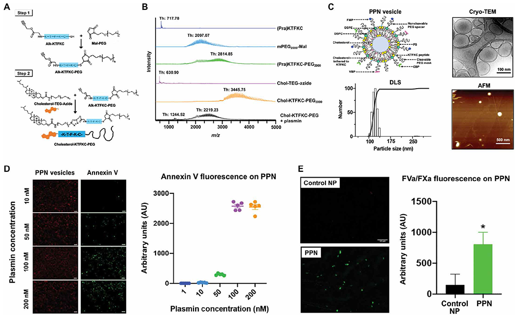
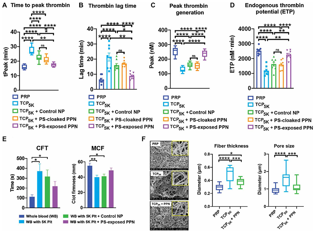
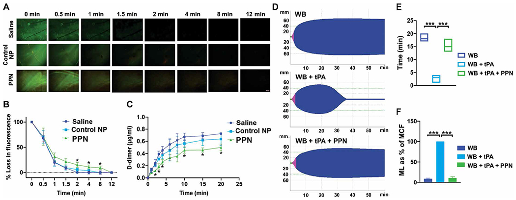
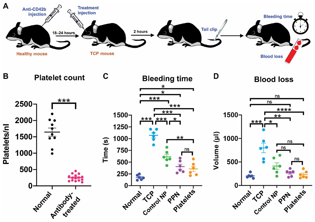
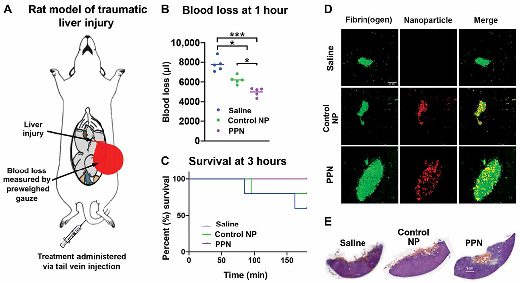
Treatment of bleeding disorders using transfusion of donor-derived platelets faces logistical challenges due to their limited availability, high risk of contamination, and short (5 to 7 days) shelf life. These challenges could be potentially addressed by designing platelet mimetics that emulate the adhesion, aggregation, and procoagulant functions of platelets. To this end, we created liposome-based platelet-mimicking procoagulant nanoparticles (PPNs) that can expose the phospholipid phosphatidylserine on their surface in response to plasmin. First, we tested PPNs in vitro using human plasma and demonstrated plasmin-triggered exposure of phosphatidylserine and the resultant assembly of coagulation factors on the PPN surface. We also showed that this phosphatidylserine exposed on the PPN surface could restore and enhance thrombin generation and fibrin formation in human plasma depleted of platelets. In human plasma and whole blood in vitro, PPNs improved fibrin stability and clot robustness in a fibrinolytic environment. We then tested PPNs in vivo in a mouse model of thrombocytopenia where treatment with PPNs reduced blood loss in a manner comparable to treatment with syngeneic platelets. Furthermore, in rat and mouse models of traumatic hemorrhage, treatment with PPNs substantially reduced bleeding and improved survival. No sign of systemic or off-target thrombotic risks was observed in the animal studies. These findings demonstrate the potential of PPNs as a platelet surrogate that should be further investigated for the management of bleeding.


Conflict of interest statement

Competing interests: A.S.G. is a cofounder of Haima Therapeutics LLC, which is focused on development of platelet-inspired hemostat technologies. A.S.G. and M.D.N. serve on the scientific advisory board of Haima. A.S.G. is coinventor on U.S. patent 9107845 entitled “Synthetic platelets” that is licensed to Haima. U.D.S.S. and A.S.G. are coinventors on a provisional patent application no. 63/147411 entitled “Bioresponsive nanoparticles for targeted amplification of coagulation and apoptosis signals,” filed by Case Western Reserve University. The other authors declare that they have no competing interests.

Figures

Fig. 1.
Fig. 1.. Design and characterization of PPNs.
(A) Shown is the two-step process for synthesis of cholesterol-KTFKC-PEG using alkyne-terminated plasmin-cleavable peptide (Alk-KTFKC), maleimide-terminated polyethylene glycol (Mal-PEG), and azide-terminated cholesterol-triethylene glycol (cholesterol-TEG-azide). Cholesterol-KTFKC-PEG was incorporated into PPNs providing a PEG cloak that could be cleaved by plasmin. (B) Matrix-assisted laser desorption ionization–time-of-flight mass spectrometry was used to characterize synthesized cholesterol-KTFKC-PEG and its plasmin-induced degradation (Th, theoretical mass). (C) Shown is the design of PPN vesicles and their size characterization by dynamic light scattering (DLS), cryo–transmission electron microscopy (cryo-TEM), and atomic force microscopy (AFM). The PPN diameter ranged from 100 to 150 nm. (D) Shown are representative images and fluorescence intensity quantification of fluorescently labeled annexin V that was bound to exposed phosphatidylserine at the surface of immobilized PPNs after exposure to plasmin. (E) Shown are representative images and fluorescence intensity quantification of Alexa Fluor 488–labeled antibody bound to factors FVa and FXa (green fluorescence) of the prothrombinase complex that was assembled at the surface of PPNs with exposed phosphatidylserine but not on control nanoparticles (Control NP) without exposed phosphatidylserine. *P ≤ 0.05, two tailed t test.
Fig. 2.
Fig. 2.. PPNs rescue thrombin generation and clot quality in platelet-depleted plasma.
(A to D) Shown are thrombin generation studies in human thrombocytopenic plasma with ~5000 platelets/μl (TCP5K) compared to platelet-rich human plasma (PRP). Time to peak thrombin (tPeak), thrombin lag time, peak thrombin generation, and endogenous thrombin potential are shown after the addition of control nanoparticles (Control NP) or PPNs with exposed phosphatidylserine (PS-exposed) or without exposed phosphatidylserine (PS-cloaked). (E) Rotational thromboelastometry analysis of human whole blood (WB) or human whole blood with ~5000 platelets /μl (WB with 5K Plt) was conducted after addition of Control NP or PPNs with exposed phosphatidylserine (PS-exposed), and clot formation time (CFT) and maximum clot firmness (MCF) were measured. (F) Shown are representative scanning electron microscopy (SEM) images of fibrin clots generated in human TCP5K compared to PRP before and after the addition of PPNs with exposed phosphatidylserine (PS-exposed). Insets show a magnified view (×5) of the main image. *P ≤ 0.05, **P ≤ 0.01, ***P ≤ 0.001, and ****P ≤ 0.0001, one way analysis of variance (ANOVA) with Tukey’s multiple comparisons test.
Fig. 3.
Fig. 3.. PPNs enhance human plasma and whole blood clot stability under fibrinolytic conditions.
(A to C) Shown is microfluidic analysis of human plasma clots exposed to tissue plasminogen activator (tPA) to create a fibrinolytic environment. Addition of PPNs, with exposed phosphatidylserine in response to plasmin (PPN) in human plasma, delayed clot lysis as indicated by fibrin green fluorescence (A and B) and reduced D-dimers in the clot lysate (C) compared to control nanoparticles (Control NP). (D to F) Shown is rotational thromboelastometry analysis of human whole blood in the presence of tPA. Addition of PPNs, with exposed phosphatidylserine in response to plasmin (PPN), enhanced clot stability as demonstrated by the MCF maintenance time (E) and reduced maximum lysis (ML) (ML as % of MCF) (F). *P ≤ 0.05, **P ≤ 0.01, and ***P ≤ 0.001, two-way ANOVA with Tukey’s multiple comparisons for microfluidic data and one-way ANOVA with Tukey’s multiple comparisons for rotational thromboelastometry data.
Fig. 4.
Fig. 4.. PPNs reduce tail bleeding in thrombocytopenic mice.
(A) In the tail transection bleeding mouse model, mice were rendered thrombocytopenic through intraperitoneal injection of anti-CD42b (anti-GPIb) antibody (0.5 μg/kg) to deplete platelets. Mice were administered by retro-orbital injection either Control NP or PPNs, ~18 to 24 hours later. Two hours after treatment, mouse tails were clipped 1 mm from the tip and bleeding time and blood loss were measured. (B) Shown are platelet counts in normal (wild-type) mice and antibody-treated thrombocytopenic mice. (C) Shown are bleeding times in normal (wild-type) mice and antibody-treated thrombocytopenic (TCP) mice before and after addition of control nanoparticles or PPNs. Also shown are bleeding times when mice were transfused with syngeneic platelets. (D) Shown is blood loss analysis using a hemoglobin assay in normal (wild-type) mice and antibody-treated thrombocytopenic (TCP) mice before and after addition of control nanoparticles or PPNs. Also shown is blood loss when mice were transfused with syngeneic platelets. n = 6 animals per group. *P ≤ 0.05, **P ≤ 0.01, ***P ≤ 0.001, and ****P ≤ 0.0001, one-way ANOVA with Tukey’s multiple comparisons test. ns, not significant.
Fig. 5.
Fig. 5.. PPNs reduce blood loss and improve survival in rats with hemorrhagic liver injury.
(A) In the rat liver injury hemorrhagic model, >30% of the liver was resected to cause intra-peritoneal hemorrhage and treatment after injury was administered via tail vein. (B) Shown is blood loss at 1 hour after treatment with PPNs compared to control nanoparticles (Control NP) or saline. (C) Shown is animal survival 3 hours after treatment with PPNs compared to control nanoparticles (Control NP) or saline. (D) Shown are representative immunofluorescence images of the liver injury site indicating greater fibrin fluorescence in hemostatic clots after PPN treatment compared to either control nanoparticles (Control NP) or saline. (E) Shown is Carstairs’ staining of representative hemostatic clots at the liver injury site indicating higher fibrin content (orange-red staining) in the clots of PPN-treated rats compared to Control NP– or saline-treated rats. n = 5 animals per group. *P ≤ 0.05 and ***P ≤ 0.001, one-way ANOVA with Tukey’s multiple comparisons test.

References

    1. Hoffman M, Monroe DM III, A cell-based model of hemostasis. Thromb. Haemost 85, 958–965 (2001). - PubMed
    1. Versteeg HH, Heemskerk JWM, Levi M, Reitsma PH, New fundamentals in hemostasis. Physiol. Rev 93, 327–358 (2013). - PubMed
    1. Etchill EW, Myers SP, Raval JS, Hassoune A, Sen Gupta A, Neal MD, Platelet transfusion in critical care and surgery: Evidence-based review of contemporary practice and future directions. Shock 47, 537–549 (2017). - PubMed
    1. Kumar A, Mhaskar R, Grossman BJ, Kaufman RM, Tobian AA, Kleinman S, Gernsheimer T, Tinmouth AT, Djubegovic B; AABB Platelet Transfusion Guidelines Panel, Platelet transfusion: A systematic review of the clinical evidence. Transfusion 55, 1116–1127 (2015). - PubMed
    1. Cohen MJ, Kutcher M, Redick B, Nelson M, Call M, Knudson MM, Schreiber MA, Bulger EM, Muskat P, Alarcon LH, Myers JG, Rahbar MH, Brasel KJ, Phelan HA, del Junco DJ, Fox EE, Wade CE, Holcomb JB, Cotton BA, Matijevic N; PROMMTT Study Group, Clinical and mechanistic drivers of acute traumatic coagulopathy. J. Trauma Acute Care Surg 75, S40–S47 (2013). - PMC - PubMed

Publication types