. 2022 Jan 26;18(1):e1009784.
doi: 10.1371/journal.ppat.1009784. eCollection 2022 Jan.

New insights into the role of endosomal proteins for African swine fever virus infection

Affiliations

New insights into the role of endosomal proteins for African swine fever virus infection

Miguel Ángel Cuesta-Geijo et al. PLoS Pathog. .

Abstract

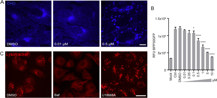
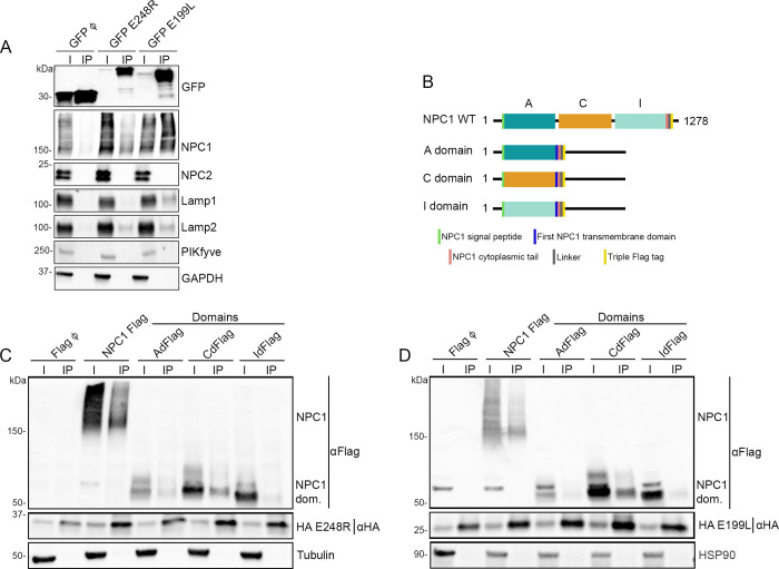
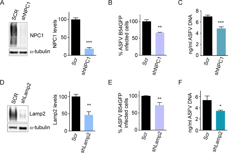
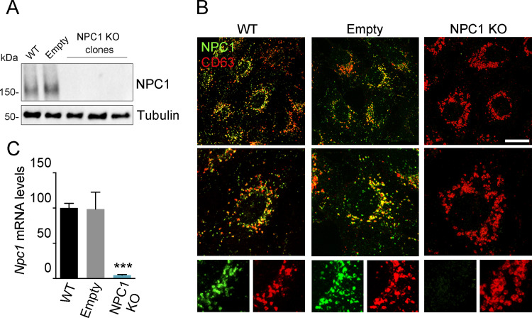
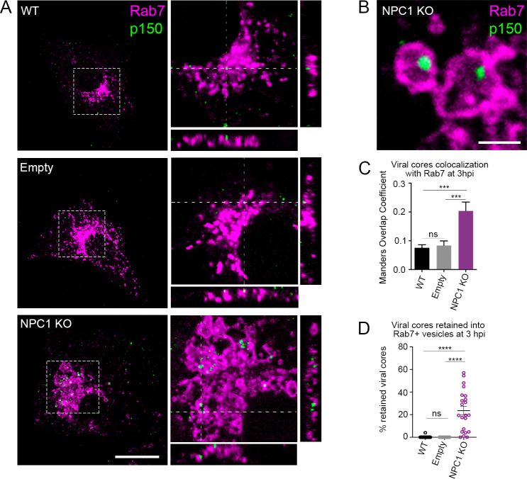
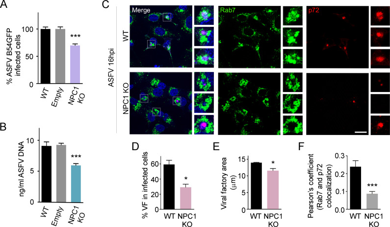
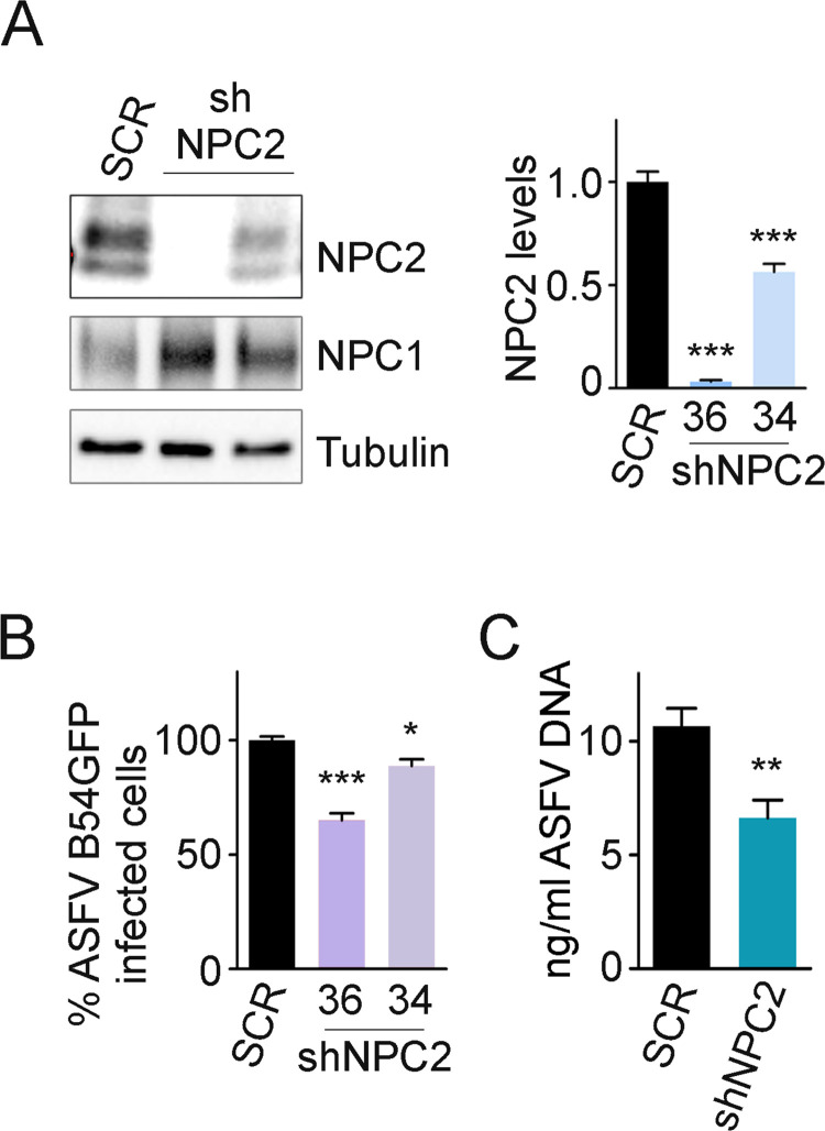
African swine fever virus (ASFV) infectious cycle starts with the viral adsorption and entry into the host cell. Then, the virus is internalized via clathrin/dynamin mediated endocytosis and macropinocytosis. Similar to other viruses, ASF virion is then internalized and incorporated into the endocytic pathway. While the endosomal maturation entails luminal acidification, the decrease in pH acts on the multilayer structure of the virion dissolving the outer capsid. Upon decapsidation, the inner viral membrane is exposed to interact with the limiting membrane of the late endosome for fusion. Viral fusion is then necessary for the egress of incoming virions from endosomes into the cytoplasm, however this remains an intriguing and yet essential process for infection, specifically for the egress of viral nucleic acid into the cytoplasm for replication. ASFV proteins E248R and E199L, located at the exposed inner viral membrane, might be implicated in the fusion step. An interaction between these viral proteins and cellular endosomal proteins such as the Niemann-Pick C type 1 (NPC1) and lysosomal membrane proteins (Lamp-1 and -2) was shown. Furthermore, the silencing of these proteins impaired ASFV infection. It was also observed that NPC1 knock-out cells using CRISPR jeopardized ASFV infection and that the progression and endosomal exit of viral cores was arrested within endosomes at viral entry. These results suggest that the interactions of ASFV proteins with some endosomal proteins might be important for the membrane fusion step. In addition to this, reductions on ASFV infectivity and replication in NPC1 KO cells were accompanied by fewer and smaller viral factories. Our findings pave the way to understanding the role of proteins of the endosomal membrane in ASFV infection.


Conflict of interest statement

The authors have declared that no competing interests exist.

Figures

Fig 1
Fig 1. Cholesterol efflux arrest and inhibition of ASFV infectivity.
(A) Treatment with increasing concentrations of U18666A chemical compound altered the distribution of unesterified cholesterol (CHO) in Vero cells (filipin; blue). Scale bar: 10 μM. (B) Cells pre-treated with increasing concentrations of U18666A compound and infected with recombinant ASFV BPP30GFP for 16 hpi. GFP fluorescence intensity was measured using a plate reader. Graph represents mean±sem of 3 independent experiments. Statistically significant differences are indicated by asterisks (****p<0.0001, *p<0.05). (C) Visualization of acidic vesicles with lysotracker (red) in Vero cells treated with U18666A drug or controls. These acidic vesicles were absent after treatment with lysosomotropic drugs (Scale bar: 25 μM).
Fig 2
Fig 2. E248R and E199L interactions with endosomal proteins NPC1, Lamp 1 and Lamp2.
(A) GFP was immunoprecipitated in lysates from HEK293T cells transfected with EGFP, EGFP E248R or EGFP E199L. Representative immunoblot analysis of cell lysates (I) and GFP-immunoprecipitates (IP) using GFP, NPC1, NPC2, Lamp1, Lamp2 and PIKfyve antibodies. GAPDH was used as control. (n  =  4 independent experiments, S2 and S3 Figs). (B) Schematic representation of NPC1 WT and specific recombinant constructions of individual domains shown in C and D. (C, D) HA was immunoprecipitated in lysates from HEK293T cells co-transfected with HA E248R (C) or HA E199L (D) with NPC1 Flag and FLAG NPC1 domains. Representative immunoblot analysis of total lysates (I) and HA-immunoprecipitates (IP) using Flag and HA antibody. Alpha-tubulin (C) or HSP90 (D) were used as control. “NPC1 dom.” refers to individual domains of NPC1 (n  =  3 independent experiments, S5 and S6 Figs).
Fig 3
Fig 3. Silencing of NPC1 and Lamp2 proteins affected ASFV infection.
(A, D) Representative immunoblots and densitometric analysis of NPC1 (A) and Lamp2 (D) in Vero cells transduced with lentiviral particles encoding NPC1 and Lamp2 shRNAs or Scrambled shRNA (SCR) as controls. Αlpha-tubulin was used as loading control. (B, E) Flow cytometry of cells shown in (A, D) and infected with fluorescent recombinant virus B54GFP at a MOI of 1pfu/ml for 16 hpi. (C, F) Quantification of ASFV DNA by qPCR in cells shown in (A, D). Graphs represent mean±sem of three independent experiments. Statistically significant differences are indicated by asterisks (***p < 0.001, **p < 0.01, *p < 0.05).
Fig 4
Fig 4. Validation of CRISPR KO NPC1 cells.
(A) Immunoblot of NPC1 and tubulin (loading control) in samples from WT, Empty and clones of NPC1 KO cells. (B) Visualization NPC1 (green) in late endosomes (CD63, red) in WT, Empty and NPC1 Vero KO cells. Scale bar: 25 μm. Zoom images are also shown. (C) Npc1 mRNA levels in WT, Empty and NPC1 KO cells as detected by qPCR. Graph represents mean±sem of three independent experiments. Statistically significant differences are indicated by asterisks (***p < 0.001).
Fig 5
Fig 5. Viral cores visualized inside dilated late endosomes in ASFV infected NPC1 KO cells.
(A) Representative micrographs of control cells (WT and Empty) and NPC1 KO cells infected at a MOI of 50 pfu/ml for 3hpi. Viral cores and late endosomes were detected with an antibody against p150 (green) and Rab7 (magenta), respectively. Orthogonal projections showed viral cores and Rab7 positive membranes in the Z stack. Note that numbers of retained viral cores were higher in NPC1 KO cells compared to controls. Scale bar: 10μm. (B) Insets depict endosomes containing viral cores shown in detail. Scale bar: 2 μm. (C) Overlapping Manders coefficient (mean±sem) from 2 independent experiments (n = ca. 25). (D) Percentages of viral cores inside endosomes (mean±sem) from 2 independent experiments (n = ca. 25). Statistically significant differences are indicated by asterisks (****p < 0.0001, ***p < 0.001, ns-not significative).
Fig 6
Fig 6. ASFV infection and replication in NPC1 KO cells.
Percentage of B54GFP infected cells (1 pfu/ml) at 16hpi in WT, Empty and NPC1 KO Vero cells detected by flow cytometry. Percentages were normalized to values in WT cells. (B) ASFV replication in WT, Empty and NPC1 KO ASFV infected cells quantified by real-time PCR. (C) Representative confocal images of WT and NPC1 KO cells stained for Rab7 (green), ASFV p72 (red) and DNA (Topro3, blue). Scale bar: 20 μm. Zoom images of ASFV viral factories (boxed regions) are also shown. (D) Percentages of viral factories in WT and NPC1 KO in infected cells shown in C. (E) Quantification of the cellular area occupied by viral factories stained with ASFV p72 in WT versus NPC1 KO infected cells shown in C. (F) Quantification of Rab7 and p72 colocalization in WT and NPC1 KO ASFV infected cells using ImageJ software. Graphs represent mean±sem from three independent experiments. Statistically significant differences are indicated by asterisks (***p < 0.001, **p < 0.01, *p < 0.05).
Fig 7
Fig 7. NPC2 downregulation reduced ASFV infectivity.
(A) NPC1, NPC2 and tubulin (loading control) were detected by western blot in Vero cells transduced with SCR (scramble) and shNPC2 (34 and 36) lentiviral particles. Quantification of NPC1 and NPC2 bands were corrected to tubulin data and then normalized to SCR values. (B) Percentage of B54GFP ASFV infected cells (1 pfu/ml) at 16 hpi in Vero cells shown in (A). Percentages were normalized to values in SCR cells. (C) Quantification of ASFV replication in SCR and shNPC2-36 transduced Vero cells by qPCR. Graphs represent mean±sem from three independent experiments. Statistically significant differences are indicated by asterisks (***p < 0.001, **p < 0.01, *p < 0.05).

References

    1. Andres G, Charro D, Matamoros T, Dillard RS, Abrescia NGA. The cryo-EM structure of African swine fever virus unravels a unique architecture comprising two icosahedral protein capsids and two lipoprotein membranes. J Biol Chem. 2020;295(1):1–12. Epub 2019/10/28. doi: 10.1074/jbc.AC119.011196 ; PubMed Central PMCID: PMC6952596. - DOI - PMC - PubMed
    1. Liu S, Luo Y, Wang Y, Li S, Zhao Z, Bi Y, et al.. Cryo-EM Structure of the African Swine Fever Virus. Cell Host Microbe. 2019;26(6):836–43 e3. Epub 2019/12/04. doi: 10.1016/j.chom.2019.11.004 . - DOI - PubMed
    1. Wang N, Zhao D, Wang J, Zhang Y, Wang M, Gao Y, et al.. Architecture of African swine fever virus and implications for viral assembly. Science. 2019;366(6465):640–4. Epub 2019/10/19. doi: 10.1126/science.aaz1439 . - DOI - PubMed
    1. Rowlands RJ, Michaud V, Heath L, Hutchings G, Oura C, Vosloo W, et al.. African swine fever virus isolate, Georgia, 2007. Emerg Infect Dis. 2008;14(12):1870–4. Epub 2008/12/03. doi: 10.3201/eid1412.080591 ; PubMed Central PMCID: PMC2634662. - DOI - PMC - PubMed
    1. Brookes V, Barrett T, Ward M, Roby J, Hernandez-Jover M, Cross E, et al.. A scoping review of African swine fever virus spread between domestic and free-living pigs. Transboundary and Emerging Diseases. 2020. - PubMed

Publication types