. 2022 Jun;87(6):2922-2932.
doi: 10.1002/mrm.29167. Epub 2022 Jan 26.

Model-based dynamic off-resonance correction for improved accelerated fMRI in awake behaving nonhuman primates

Affiliations

Model-based dynamic off-resonance correction for improved accelerated fMRI in awake behaving nonhuman primates

Mo Shahdloo et al. Magn Reson Med. 2022 Jun.

Abstract

Purpose: To estimate dynamic off-resonance due to vigorous body motion in accelerated fMRI of awake behaving nonhuman primates (NHPs) using the echo-planar imaging reference navigator, in order to attenuate the effects of time-varying off-resonance on the reconstruction.

Methods: In NHP fMRI, the animal's head is usually head-posted, and the dynamic off-resonance is mainly caused by motion in body parts that are distant from the brain and have low spatial frequency. Hence, off-resonance at each frame can be approximated as a spatially linear perturbation of the off-resonance at a reference frame, and is manifested as a relative linear shift in k-space. Using GRAPPA operators, we estimated these shifts by comparing the navigator at each time frame with that at the reference frame. Estimated shifts were then used to correct the data at each frame. The proposed method was evaluated in phantom scans, simulations, and in vivo data.

Results: The proposed method is shown to successfully estimate spatially low-order dynamic off-resonance perturbations, including induced linear off-resonance perturbations in phantoms, and is able to correct retrospectively corrupted data in simulations. Finally, it is shown to reduce ghosting artifacts and geometric distortions by up to 20% in simultaneous multislice in vivo acquisitions in awake-behaving NHPs.

Conclusion: A method is proposed that does not need sequence modification or extra acquisitions and makes accelerated awake behaving NHP imaging more robust and reliable, reducing the gap between what is possible with NHP protocols and state-of-the-art human imaging.

Keywords: EPI; dynamic off-resonance; fMRI; nonhuman primates; simultaneous multislice.


Figures

FIGURE 1
FIGURE 1
Ghosting artifacts and geometric distortion due to off‐resonance. Simultaneous multislice accelerated data (MB = 2, with CAIPI shifts 33 ) were simulated using a single data‐frame from fully sampled in vivo macaque data. Simulated spatially linear off‐resonance was added. (A) The fully sampled data were used to train split‐slice GRAPPA kernels. (B) In the absence of off‐resonance perturbations, these trained kernels could be used to completely separate the aliased slices. (C) Off‐resonance perturbation, however, results in inconsistency of the data to the trained kernels which leads to aliasing artifacts manifested as Nyquist ghosts as well as geometric distortion. (D) Increasing the off‐resonance level (indicated by numbers) enhances the level of ghosting artifacts and geometric distortion. Here, the display range is saturated to enhance the visibility of artifacts (indicated by white arrows). (E) Artifact power, taken as the 2 norm of the background values, versus off‐resonance level. Values corresponding to off‐resonance levels in (D) are marked by empty circles. Artifact power monotonically increases by increasing the levels of off‐resonance
FIGURE 2
FIGURE 2
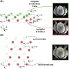
Dynamic off‐resonance estimation and correction. Spatially linear off‐resonance perturbations can be cast as linear shifts in k‐space data. (A) To estimate the off‐resonance perturbation, the EPI reference navigator data at each time frame were compared to the navigator data from a reference frame. Linear shifts between corresponding navigator lines were estimated using GRAPPA operators, accounting for the different echo times of the consecutive navigator lines. (B) The estimated linear shift coefficients that reflect the off‐resonance perturbation were used to correct the EPI data at each time frame. This procedure reduces the ghosting artifacts and geometric distortion (red outline), yielding an improved reconstruction (green outline)
FIGURE 3
FIGURE 3
Estimation of linear shim changes in phantom. Dynamic off‐resonance changes were induced in a bottle phantom by manually modifying the linear shim terms across acquisitions. The EPI reference navigator data were then used to estimate the linear shim term changes across (A) x, (B) y, and (C) z directions in the range ±20μ T/m. The estimated shim term changes (filled markers) are in good agreement with the ground truth (empty markers)
FIGURE 4
FIGURE 4
Reconstruction of the in vivo EPI acquisition. (A) SMS accelerated in vivo data were simulated using the single‐band k‐space data by assuming dynamic off‐resonance perturbations. Calibration inconsistency due to off‐resonance perturbation causes ghosting artifacts and geometric distortions. EPI reference navigator data were used to estimate and correct the simulated dynamic off‐resonance. Standard Nyquist ghost correction and dynamic zeroth‐order B0 correction were applied on both corrected and uncorrected image series prior to off‐resonance estimation and correction. Images from four different slices are shown in columns. The display window in the top and bottom quarter of images are saturated to better show the ghosting artifacts. The red outline shows the object boundary in the undistorted single‐band reference image. (B) Prospectively SMS accelerated in vivo acquisition was corrected and reconstructed using the proposed method. Formatting is identical to panel A. Estimating and accounting for the dynamic off‐resonance yields significantly reduced ghosting artifacts and geometric distortion
FIGURE 5
FIGURE 5
Estimated parameters and quantitative measurements for the in vivo EPI acquisition. (A) The six estimated off‐resonance model parameters are shown at each time time frame of the prospectively SMS accelerated in vivo acquisition. The display scaling of the offset terms cx, cy, and cz is magnified for easier visibility. Shaded areas show the standard error of the mean across slices. (B) Image entropy is shown at each time frame. Shaded areas show the standard error of the mean across slices. The proposed method decreases the mean entropy, indicating the decrease in ghosting artifacts. (C) Normalized root mean squared error compared to the single‐band reference image is shown. Shaded areas show the standard error of the mean across slices. Decreased geometric distortion achieved using the proposed method yields reduced nRMSE. (D) Histograms of tSNR is compared between the reconstructions. Only the tail of the histogram is shown to compare the distribution in pixels with highest tSNRs. The proposed method yields a histogram that is skewed to the right, indicating higher number of pixels with high tSNR

References

    1. Orban GA. Functional MRI in the awake monkey: the missing link. J Cogn Neurosci. 2002;14:965‐969. - PubMed
    1. Donahue CJ, Glasser MF, Preuss TM, et al. Quantitative assessment of prefrontal cortex in humans relative to nonhuman primates. Proc Nat Acad Sci. 2018;115:E5183‐E5192. - PMC - PubMed
    1. Moeller S, Yacoub E, Olman CA, et al. Multiband multislice GE‐EPI at 7 T, with 16‐fold acceleration using partial parallel imaging with application to high spatial and temporal whole‐brain FMRI. Magn Reson Med. 2010;63:1144‐1153. - PMC - PubMed
    1. Setsompop K, Gagoski BA, Polimeni JR, et al. Blipped‐controlled aliasing in parallel imaging for simultaneous multislice echo planar imaging with reduced g‐factor penalty. Magn Reson Med. 2012;67:1210‐1224. - PMC - PubMed
    1. Autio JA, Glasser MF, Ose T, et al. Towards HCP‐style macaque connectomes: 24‐channel 3T multi‐array coil. MRI sequences and preprocessing. NeuroImage. 2020;215:116800. - PMC - PubMed

Publication types