. 2022 Jan 27;8(1):2.
doi: 10.1186/s42234-021-00083-7.

Focused ultrasound neuromodulation on a multiwell MEA

Affiliations

Focused ultrasound neuromodulation on a multiwell MEA

Marta Saccher et al. Bioelectron Med. .

Abstract

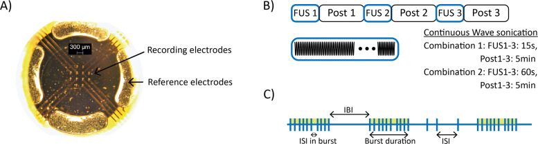
Background: Microelectrode arrays (MEA) enable the measurement and stimulation of the electrical activity of cultured cells. The integration of other neuromodulation methods will significantly enhance the application range of MEAs to study their effects on neurons. A neuromodulation method that is recently gaining more attention is focused ultrasound neuromodulation (FUS), which has the potential to treat neurological disorders reversibly and precisely.

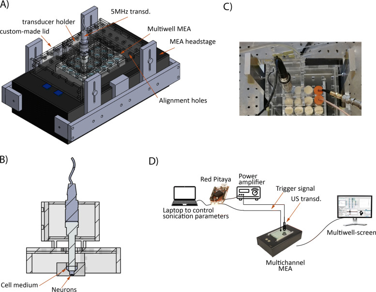
Methods: In this work, we present the integration of a focused ultrasound delivery system with a multiwell MEA plate.

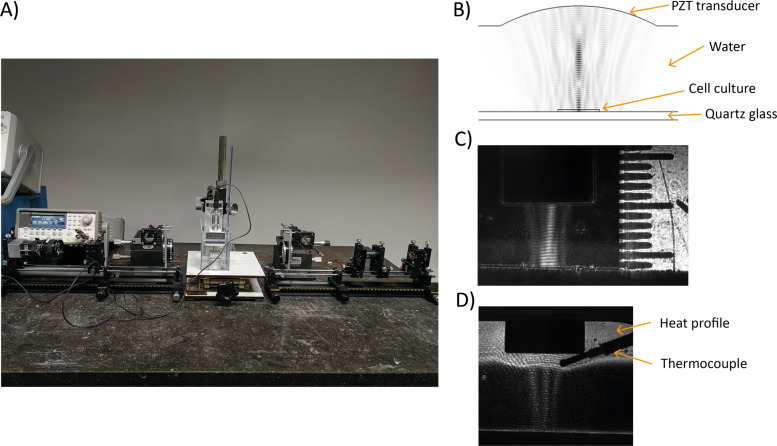
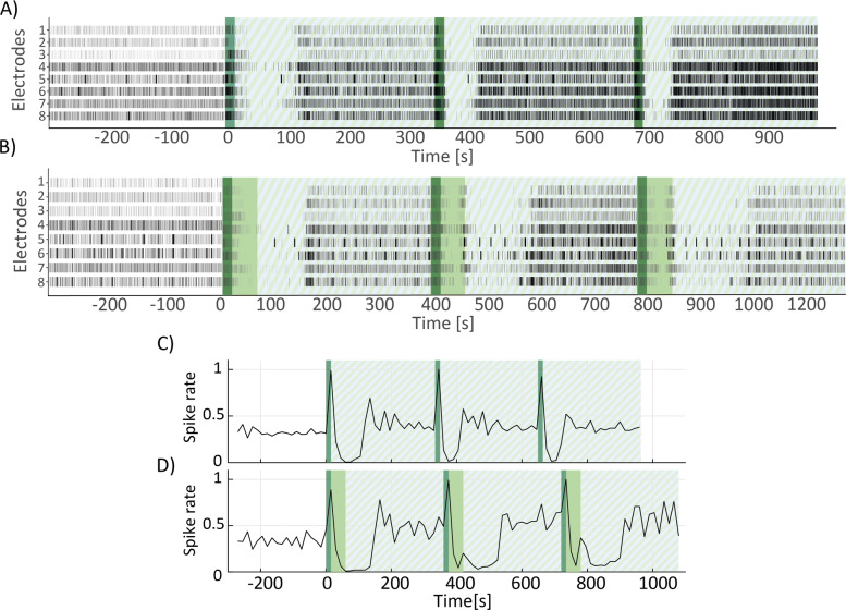
Results: The ultrasound delivery system was characterised by ultrasound pressure measurements, and the integration with the MEA plate was modelled with finite-element simulations of acoustic field parameters. The results of the simulations were validated with experimental visualisation of the ultrasound field with Schlieren imaging. In addition, the system was tested on a murine primary hippocampal neuron culture, showing that ultrasound can influence the activity of the neurons.

Conclusions: Our system was demonstrated to be suitable for studying the effect of focused ultrasound on neuronal cultures. The system allows reproducible experiments across the wells due to its robustness and simplicity of operation.

Keywords: Bioelectronics; Focused ultrasound; Multielectrode arrays; Neuromodulation; Ultrasound.


Conflict of interest statement

The authors declare that they have no competing interests.

Figures

Fig. 1
Fig. 1
Experiment features. A) Microscope view of MEA electrodes. In the centre the 12 recording electrodes and at the sides, the 4 reference electrodes. B) Sonication protocol. C) Spike train features
Fig. 2
Fig. 2
Experimental setup. A) 3D model of the experimental setup. The black box at the bottom is the MEA headstage, at the top of which the custom-made lid is placed. The circular holes in the lid are aligned to the underlying MEA plate. The ultrasound transducer is inserted in the transducer holder structure which can then be positioned in the engraved alignment holes for precise centering with respect to the MEA well. B) Cross-section view of the experimental setup showing the relative position of the transducer with respect to the neuronal culture. C) Detail picture of the experimental setup showing the transducer in the support structure on the left and on the right, the wells with the coloured cap are used to record the trigger signal. D) Schematic view of the complete experimental setup showing the connections between the various elements
Fig. 3
Fig. 3
Schlieren setup. A) View of the complete setup. B) Result of ultrasound field simulation in COMSOL. C) Interference pattern generated by the reflection of the ultrasound field on a hard surface. The distance between the ultrasound transducer and the reflecting surface is the same as the distance between the transducer and the bottom of the MEA well (6 mm). D) Heat profile generated by the ultrasound transducer at intensity 4 times higher than the experimental conditions. The shape of the thermocouple is also visible
Fig. 4
Fig. 4
Ultrasound neuromodulation results. A) Example of raster plot of one of the wells used in the experiment, for a treatment duration of 15 seconds. Dark green shaded area indicates the US-ON period, while stripes-patterned area indicates the US-OFF period, namely the post period. B) Same as A) but for a treatment duration of 60 seconds. Dark green shaded area indicates the first 15 seconds of US-ON period and light green area indicates the remaining 45 seconds. Stripes-patterned area indicates the US-OFF period. C) Spike rate trend corresponding to the raster plot in A), calculated for 15 seconds bins over the duration of the experiment. Dark green shaded area indicates the US-ON period and stripe-patterned area indicates the US-OFF period. D) Spike rate trend corresponding to the raster plot in B), calculated for 15 seconds bins over the duration of the experiment. Dark green shaded area indicates the first 15 seconds of US-ON and light green area the remaining 45 seconds. Stripe-patterned area indicates the period of US-OFF

References

    1. Andrews D. 2014 COMSOL Conference in Cambridge. Stockholm: COMSOL Multiphysics; 2014. Modelling of ultrasonic transducers and ultrasonic wave propagation for commercial applications using finite elements with experimental visualization of waves for validation.
    1. Banker G, Goslin K. Culturing Nerve Cells. Bradford Book. Cambridge: The MIT Press; 1991.
    1. Blackmore J, Shrivastava S, Sallet J, R. Butler C, Cleveland RO. Ultrasound neuromodulation: A review of results, mechanisms and safety. Ultrasound Med Biol. 2019;45(7):1509–36. doi: 10.1016/j.ultrasmedbio.2018.12.015. - DOI - PMC - PubMed
    1. Chiappalone M, Novellino A, Vajda I, Vato A, Martinoia S, van Pelt J. Burst detection algorithms for the analysis of spatio-temporal patterns in cortical networks of neurons. Neurocomputing. 2005;65-66:653–62. doi: 10.1016/j.neucom.2004.10.094. - DOI
    1. Choi, JB, Lim SH, Cho KW, Kim DH, Jang DP, Kim IY. The effect of focused ultrasonic stimulation on the activity of hippocampal neurons in multi-channel electrode. In: 2013 6th International IEEE/EMBS Conference on Neural Engineering (NER): 2013. p. 731–34. 10.1109/NER.2013.6696038.

LinkOut - more resources