. 2022 Jan 26;22(1):40.
doi: 10.1186/s12935-022-02451-y.

Identification of a novel HOOK3-FGFR1 fusion gene involved in activation of the NF-kappaB pathway

Affiliations

Identification of a novel HOOK3-FGFR1 fusion gene involved in activation of the NF-kappaB pathway

Xuehong Zhang et al. Cancer Cell Int. .

Abstract

Background: Rearrangements involving the fibroblast growth factor receptor 1 (FGFR1) gene result in 8p11 myeloproliferative syndrome (EMS), which is a rare and aggressive hematological malignancy that is often initially diagnosed as myelodysplastic syndrome (MDS). Clinical outcomes are typically poor due to relative resistance to tyrosine kinase inhibitors (TKIs) and rapid transformation to acute leukemia. Deciphering the transcriptomic signature of FGFR1 fusions may open new treatment strategies for FGFR1 rearrangement patients.

Methods: DNA sequencing (DNA-seq) was performed for 20 MDS patients and whole exome sequencing (WES) was performed for one HOOK3-FGFR1 fusion positive patient. RNA sequencing (RNA-seq) was performed for 20 MDS patients and 8 healthy donors. Fusion genes were detected using the STAR-Fusion tool. Fluorescence in situ hybridization (FISH), quantitative real-time PCR (qRT-PCR), and Sanger sequencing were used to confirm the HOOK3-FGFR1 fusion gene. The phosphorylation antibody array was performed to validate the activation of nuclear factor-kappaB (NF-kappaB) signaling.

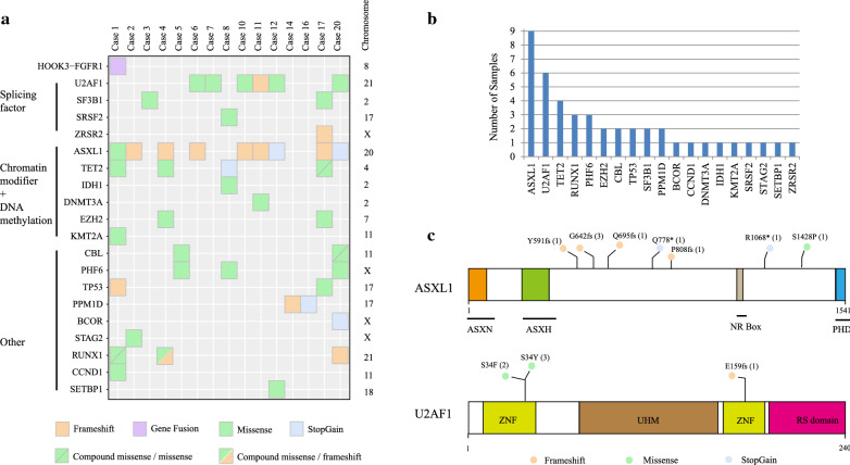
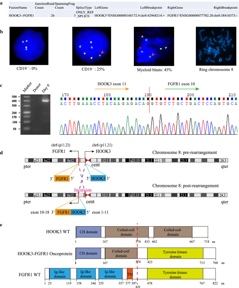
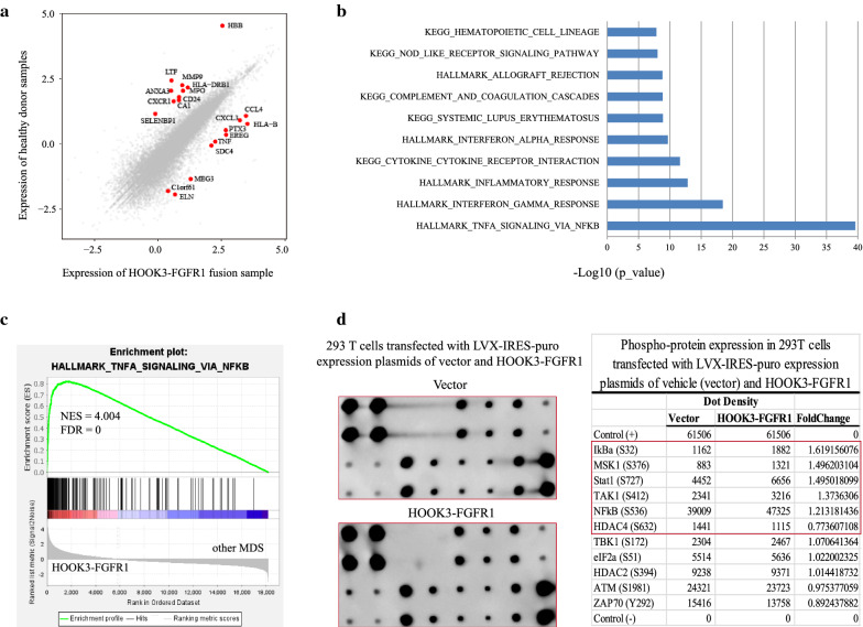
Results: We identified frequently recurrent mutations of ASXL1 and U2AF1 in the MDS cohort, which is consistent with previous reports. We also identified a novel in-frame HOOK3-FGFR1 fusion gene in one MDS case with abnormal monoclonal B-cell lymphocytosis and ring chromosome 8. FISH analysis detected the FGFR1 break-apart signal in myeloid blasts only. qRT-PCR and Sanger sequencing confirmed the HOOK3-FGFR1 fusion transcript with breakpoints located at the 11th exon of HOOK3 and 10th exon of FGFR1, and Western blot detected the chimeric HOOK3-FGFR1 fusion protein that is presumed to retain the entire tyrosine kinase domain of FGFR1. The transcriptional feature of HOOK3-FGFR1 fusion was characterized by the significant enrichment of the NF-kappaB pathway by comparing the expression profiling of FGFR1 fusion positive MDS with 8 healthy donors and FGFR1 fusion negative MDS patients. Further validation by phosphorylation antibody array also showed NF-kappaB activation, as evidenced by increased phosphorylation of p65 (Ser 536) and of IKBalpha (Ser 32).

Conclusions: The HOOK3-FGFR1 fusion gene may contribute to the pathogenesis of MDS and activate the NF-kappaB pathway. These findings highlight a potential novel approach for combination therapy for FGFR1 rearrangement patients.

Keywords: 8p11 myeloproliferative syndrome; Expression signature; HOOK3-FGFR1; NF-kappaB signaling pathway; RNA sequencing.


Conflict of interest statement

All authors have approved this submission and declare no competing interests.

Figures

Fig. 1
Fig. 1
Specific sequencing data of 20 patients with MDS. a Summary of mutation data in the MDS cohort. b The bar plot shows the number of specific mutations in our MDS cohort. c Functional structures and mutations of ASXL1 and U2AF1 proteins. ASX: Asx homology, ASXN: Asx N-terminal, NR box: nuclear receptor co-regulator binding motif, PHD: plant homeodomain, ZNF: zinc finger, UHM: U2AF homology motif.
Fig. 2
Fig. 2
Clinical presentation of the patient with the HOOK3-FGFR1 fusion gene. a The entire treatment process of the HOOK3-FGFR1 positive patient. b Wright stain of bone marrow aspirate smear from Case 1, with blast cells clearly seen in the primary and NR specimens. c Karyotype analysis showed complex abnormalities and one additional ring chromosome (indicated by the red arrow). d Flow cytometry analysis of immunophenotypic markers for the HOOK3-FGFR1 positive patient. Total cells are gated on SSC/FSC plot where viable cells are selected for following analysis. The bone marrow cells of this patient were labeled with monoclonal antibody CD45. The diagnosis stage flow results showed the blast cells were positive for CD117 and CD34 (myeloblasts: 2.43%), and positive for CD19 and cLambda (monoclonal B-lymphoid cells: 11%)
Fig. 3
Fig. 3
Validation and characterization of a novel HOOK3-FGFR1 fusion. a RNA sequencing analysis result revealed the chromosome positions of breakpoints in HOOK3 and FGFR1. b Interphase FISH analysis with FGFR1/D8Z2 Breakapart/Amplification probe (LPS018, CytoCell, UK) revealed a split FGFR1 signal pattern in the CD19- population and Myeloid blasts. The 5′ and 3′ FGFR1 are labeled with red and green, respectively; the D8Z2 (8p11-q11) region is labeled with blue as the control signal. The red arrow indicates a break-apart signal in the FGFR1 gene, and the percentages of positive signal detected in the bone marrow cells are showed in Fig. 3b. Metaphase FISH analysis exhibited a fluorescence signal in ring chromosome 8. c Validation of the HOOK3-FGFR1 fusion gene with the structure of HOOK3 exons 1-11 joining to FGFR1 exons 10-18 using PCR and Sanger sequencing. d Graphical representation of the organization process of the formation of the HOOK3-FGFR1 fusion at the chromosome level. e Schematic diagrams of the HOOK3, FGFR1, and HOOK3-FGFR1 fusion proteins. The break point is indicated by the red dashed line. CH Calponin-homology domain, TM transmembrane domain
Fig. 4
Fig. 4
HOOK3-FGFR1 fusion gene involved in the activation of NF-kappaB signaling pathway. a Comparative analysis of the HOOK3-FGFR1 positive patient (Case 1) and healthy donors (n = 8). The texts in the scatterplot correspond to the top 10 up-regulated and down-regulated genes (fold change > 2 and p < 0.01). b Bar plot of the differentially expressed genes from the comparison of the HOOK3-FGFR1 positive patient and healthy donors (FC > 2 and Q < 0.05) enriched in MSigDB. c Representative GSEA plots of one HOOK3-FGFR1 positive patient compared with the 19 HOOK3-FGFR1 negative MDS patients. The normalized enrichment score (NES) and nominal p-values are shown in the graph. d RayBiotech NF-kappaB Pathway Phosphorylation Array including 11 proteins was used to analyze the phosphorylation status of the signaling proteins of 293 T cells transfected with LVX-IRES-puro expression plasmids of vehicle (vector) and FLAG-HOOK3-FGFR1, respectively. Visualization and quantification of the results were performed using a Typhoon 7000 phosphorimager (GE Healthcare) and NIH ImageJ software. Left panels: images of the original blots; right panel: quantitative results. The fold change in the phosphoproteins of FLAG-HOOK3-FGFR1 was calculated relative to the vector

References

    1. Jackson CC, Medeiros LJ, Miranda RN. 8p11 myeloproliferative syndrome: a review. Hum Pathol. 2010;41(4):461–76. - PubMed
    1. Arber DA, Orazi A, Hasserjian R, Thiele J, Borowitz MJ, Le Beau MM, Bloomfield CD, Cazzola M, Vardiman JW. The 2016 revision to the World Health Organization classification of myeloid neoplasms and acute leukemia. Blood. 2016;127(20):2391–405. - PubMed
    1. Umino K, Fujiwara SI, Ikeda T, Toda Y, Ito S, Mashima K, Minakata D, Nakano H, Yamasaki R, Kawasaki Y, et al. Clinical outcomes of myeloid/lymphoid neoplasms with fibroblast growth factor receptor-1 (FGFR1) rearrangement. Hematology. 2018;23(8):470–477. - PubMed
    1. Villafuerte-Gutierrez P, Lopez Rubio M, Herrera P, Arranz E. A case of myeloproliferative neoplasm with BCR-FGFR1 rearrangement: favorable outcome after haploidentical allogeneic transplantation. Case Rep Hematol. 2018;2018:5724960. - PMC - PubMed
    1. Popovici C, Adelaide J, Ollendorff V, Chaffanet M, Guasch G, Jacrot M, Leroux D, Birnbaum D, Pebusque MJ. Fibroblast growth factor receptor 1 is fused to FIM in stem-cell myeloproliferative disorder with t(8;13) Proc Natl Acad Sci USA. 1998;95(10):5712–7. - PMC - PubMed