Review
. 2022 Jan;103(1):001730.
doi: 10.1099/jgv.0.001730.

Visualising Viruses

Affiliations
Review

Visualising Viruses

Annabel Slater et al. J Gen Virol. 2022 Jan.

Abstract

Viruses pose a challenge to our imaginations. They exert a highly visible influence on the world in which we live, but operate at scales we cannot directly perceive and without a clear separation between their own biology and that of their hosts. Communication about viruses is therefore typically grounded in mental images of virus particles. Virus particles, as the infectious stage of the viral replication cycle, can be used to explain many directly observable properties of transmission, infection and immunity. In addition, their often striking beauty can stimulate further interest in virology. The structures of some virus particles have been determined experimentally in great detail, but for many important viruses a detailed description of the virus particle is lacking. This can be because they are challenging to describe with a single experimental method, or simply because of a lack of data. In these cases, methods from medical illustration can be applied to produce detailed visualisations of virus particles which integrate information from multiple sources. Here, we demonstrate how this approach was used to visualise the highly variable virus particles of influenza A viruses and, in the early months of the COVID-19 pandemic, the virus particles of the then newly characterised and poorly described SARS-CoV-2. We show how constructing integrative illustrations of virus particles can challenge our thinking about the biology of viruses, as well as providing tools for science communication, and we provide a set of science communication resources to help visualise two viruses whose effects are extremely apparent to all of us.

Keywords: SARS-CoV-2; influenza virus; visualisation.


Conflict of interest statement

The authors declare that there are no conflicts of interest.

Figures
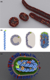
Fig. 1.
Fig. 1.
Modelling influenza A virus particles. (a) Variable morphologies of influenza A virus (IAV) particles, based on cryoelectron tomography of the strain A/Udorn/307/1972(H3N2). (b) A visual argument that the ribonucleoproteins (RNPs) encapsidating the longest IAV genome segments could only fit within a spherical virus particle of 120 nm outer diameter (of 80 nm internal diameter, shown in brown) if severely contorted, but that a bacilliform virus particle of equivalent surface area has the capacity to enclose the RNPs with minimal contortion, allowing them to form a parallel array. (c) A detailed model of a bacilliform IAV virus particle showing proteins encoded by both the virus and its host. Host proteins and membrane are in brown, reds and oranges; viral proteins are polymerase trimer (light green) and NP (light blue) in the RNPs; and HA (blue), NA (red), M2 (pink), M1 (dark green), NEP and NS1 (both purple).
Fig. 2.
Fig. 2.
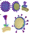
Modelling SARS-CoV-2 virus particles. (a) An initial model of a SARS-CoV-2 virion, produced in the early months of the COVID pandemic and partly based on studies of related coronaviruses. S proteins are purple, M proteins blue, E proteins green, N proteins yellow, and genomic RNA red. (b) A revised model incorporating information from experimental studies of SARS-CoV-2.
Fig. 3.
Fig. 3.
Using visualisations of virus particles in science communication. A selection of uses of the virus particle models described in this paper. (a) As branding for a virology reagent website (the MRC PPU and CVR Coronavirus Toolkit; image used with permission), (b) in educational colouring sheets, (c) as interactive 3D models on SketchFab (d) in the augmented reality app Visible Viruses, (e) as 3D prints and (f) as branding for the Journal of General Virology.

References

    1. Nee S, Smith JM. The evolutionary biology of molecular parasites. Parasitology. 1990;100 Suppl:S5–18. doi: 10.1017/s0031182000072978. - DOI - PubMed
    1. Koonin EV, Dolja VV, Krupovic M, Kuhn JH. Viruses defined by the position of the virosphere within the replicator sDefined by the Position of the Virosphere within the Replicator Space. Microbiol Mol Biol Rev. 2021;85:e0019320. doi: 10.1128/MMBR.00193-20. - DOI - PMC - PubMed
    1. Risco C, de Castro IF, Sanz-Sánchez L, Narayan K, Grandinetti G, et al. Three-dimensional imaging of viral infections. Annu Rev Virol. 2014;1:453–473. doi: 10.1146/annurev-virology-031413-085351. - DOI - PubMed
    1. Ho PT, Montiel-Garcia DJ, Wong J, Carrillo-Tripp M, Brooks CL, et al. VIPERdb: A tool for virus reseTool for Virus Research. Annu Rev Virol. 2018;5:477–488. doi: 10.1146/annurev-virology-092917-043405. - DOI - PubMed
    1. Twarock R, Luque A. Structural puzzles in virology solved with an overarching icosahedral design principle. Nat Commun. 2019;10:1–9. doi: 10.1038/s41467-019-12367-3. - DOI - PMC - PubMed

Publication types

MeSH terms