. 2022 May 4;20(5):699-711.
doi: 10.1158/1541-7786.MCR-21-0837.

Inactivation of p21-Activated Kinase 2 (Pak2) Inhibits the Development of Nf2-Deficient Tumors by Restricting Downstream Hedgehog and Wnt Signaling

Affiliations

Inactivation of p21-Activated Kinase 2 (Pak2) Inhibits the Development of Nf2-Deficient Tumors by Restricting Downstream Hedgehog and Wnt Signaling

Eleonora Sementino et al. Mol Cancer Res. .

Abstract

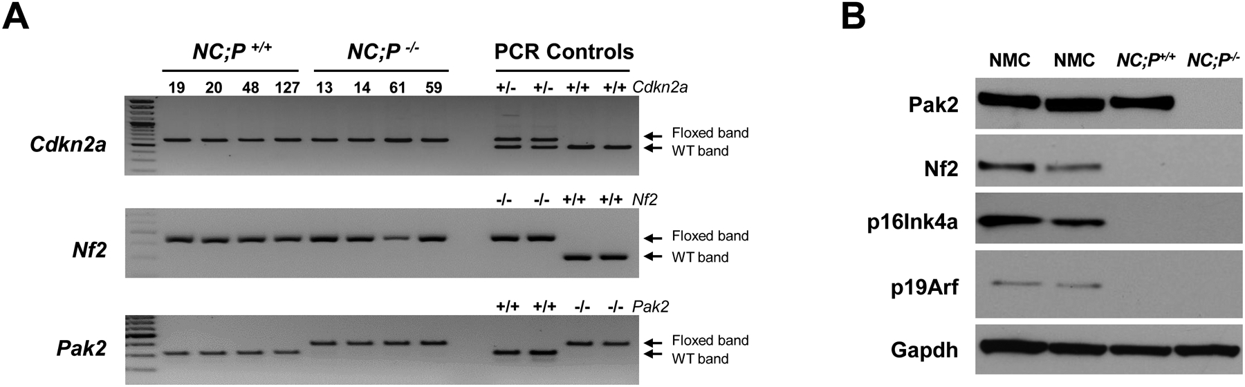
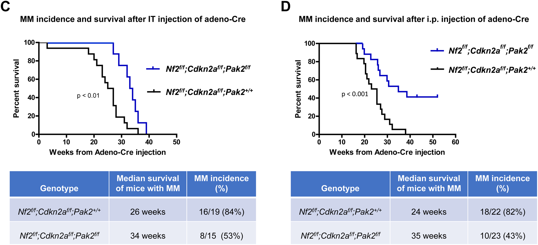
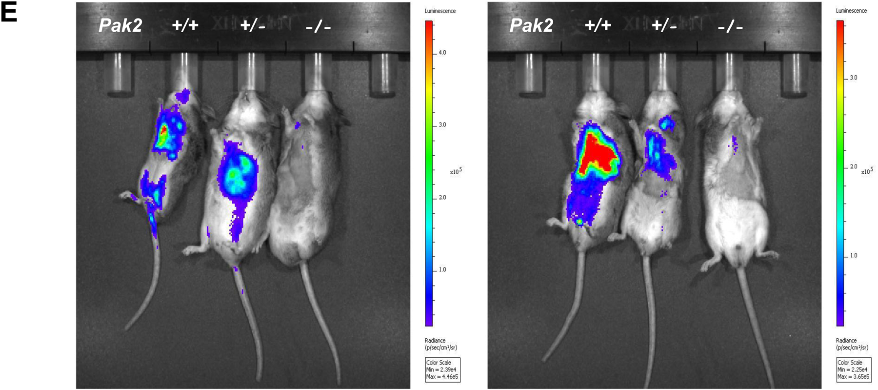
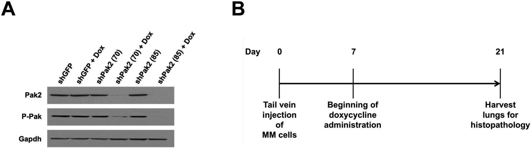
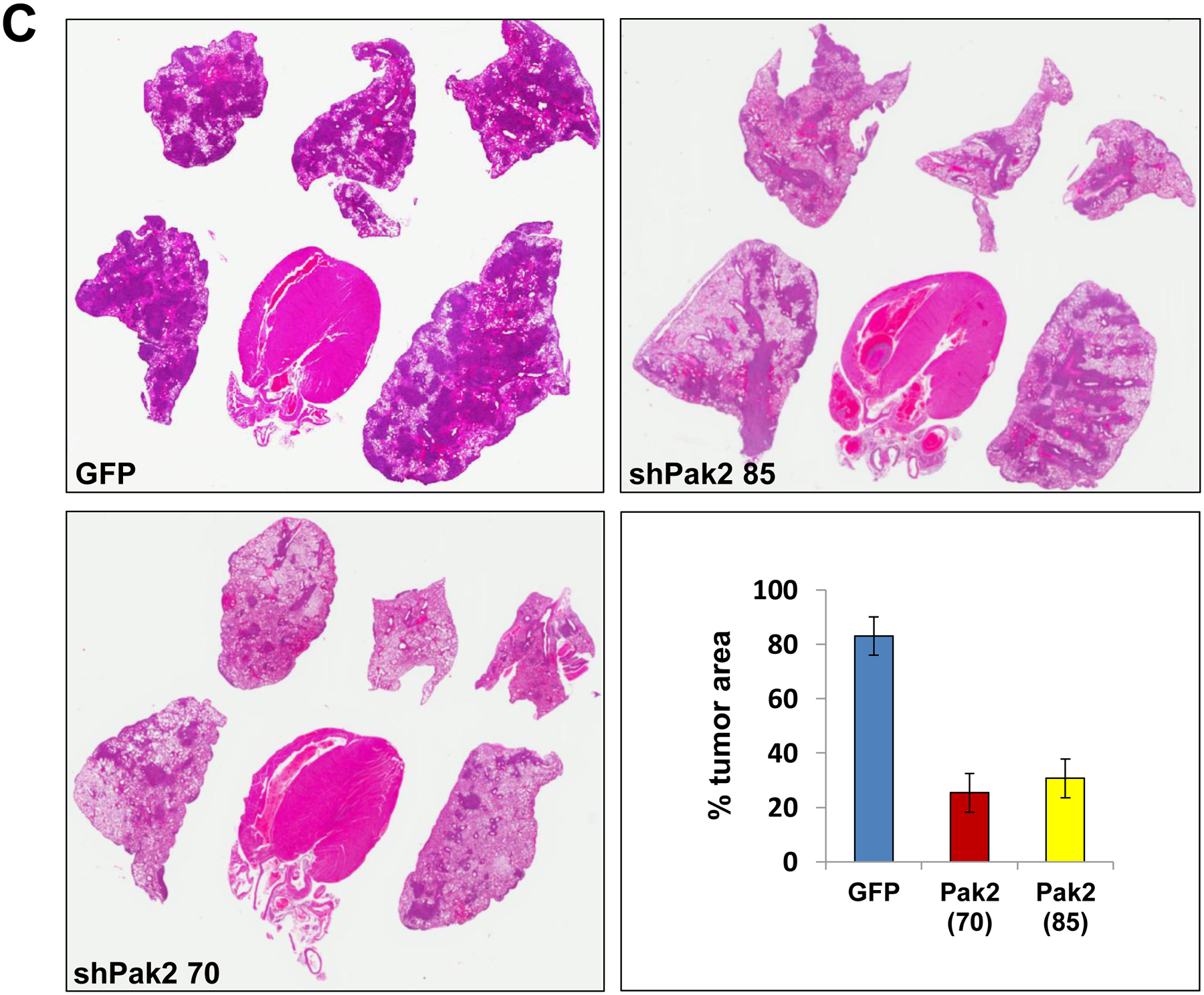
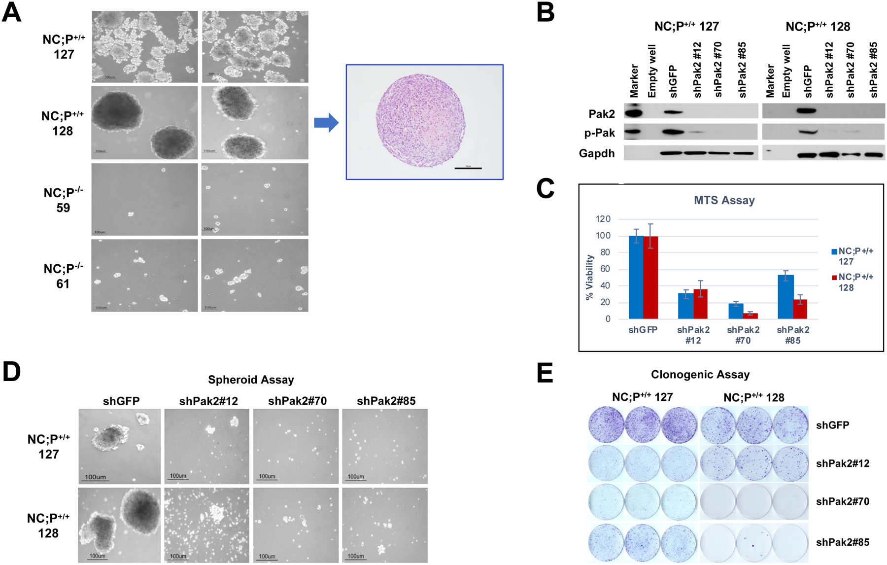
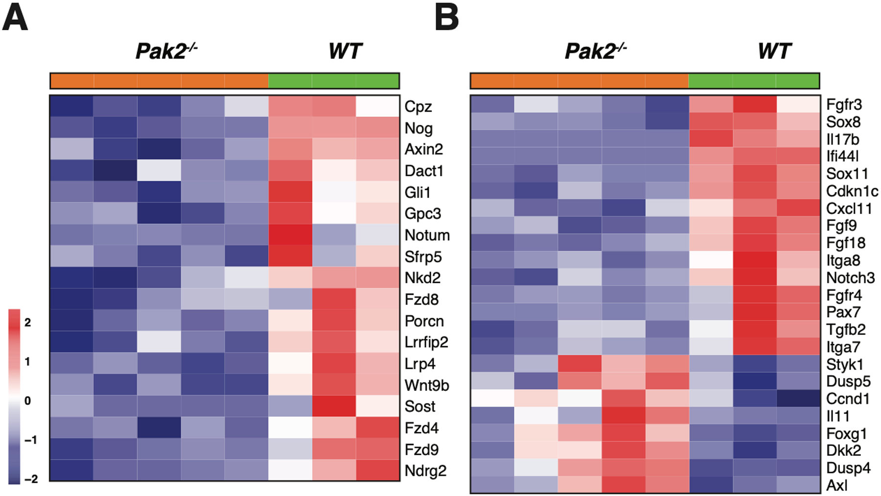
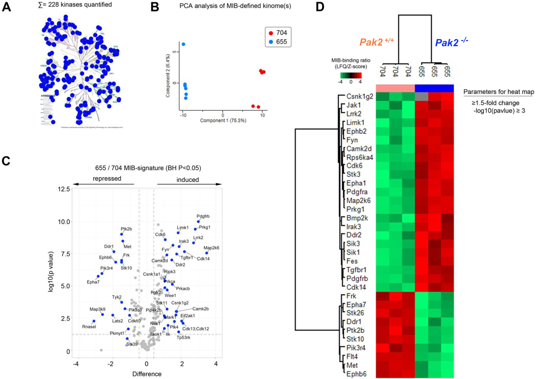
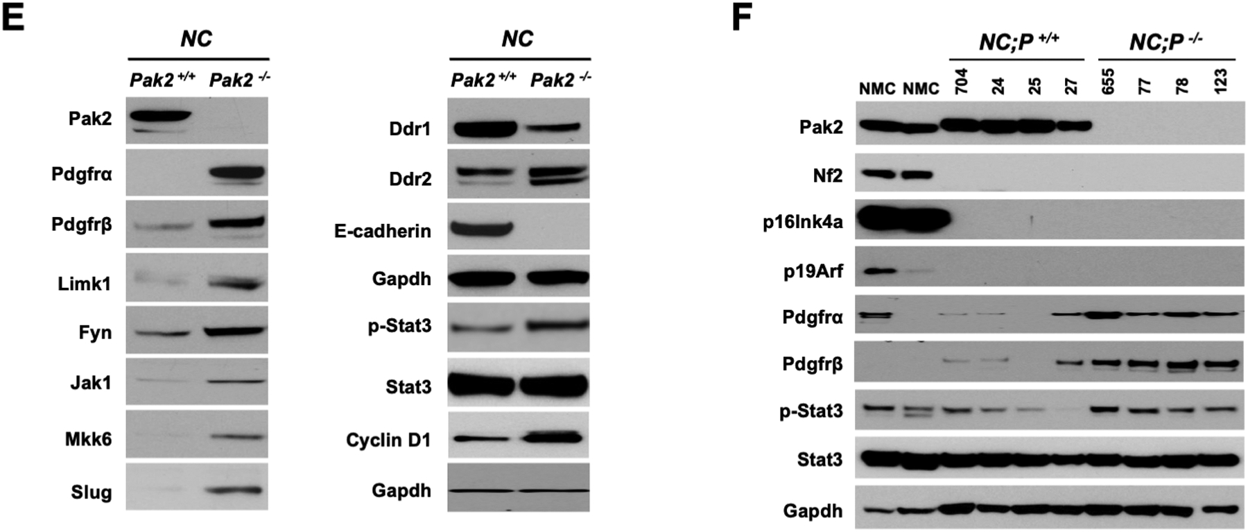
Because loss of the NF2 tumor suppressor gene results in p21-activated kinase (Pak) activation, PAK inhibitors hold promise for the treatment of NF2-deficient tumors. To test this possibility, we asked if loss of Pak2, a highly expressed group I PAK member, affects the development of malignant mesothelioma in Nf2;Cdkn2a-deficient (NC) mice and the growth properties of NC mesothelioma cells in culture. In vivo, deletion of Pak2 resulted in a markedly decreased incidence and delayed onset of both pleural and peritoneal malignant mesotheliomas in NC mice. In vitro, Pak2 deletion decreased malignant mesothelioma cell viability, migration, clonogenicity, and spheroid formation. RNA-sequencing analysis demonstrated downregulated expression of Hedgehog and Wnt pathway genes in NC;Pak2-/- mesothelioma cells versus NC;Pak2+/+ mesothelioma cells. Targeting of the Hedgehog signaling component Gli1 or its target gene Myc inhibited cell viability and spheroid formation in NC;P+/+ mesothelioma cells. Kinome profiling uncovered kinase changes indicative of EMT in NC;Pak2-/- mesothelioma cells, suggesting that Pak2-deficient malignant mesotheliomas can adapt by reprogramming their kinome in the absence of Pak activity. The identification of such compensatory pathways offers opportunities for rational combination therapies to circumvent resistance to anti-PAK drugs.

Implications: We provide evidence supporting a role for PAK inhibitors in treating NF2-deficient tumors. NF2-deficient tumors lacking Pak2 eventually adapt by kinome reprogramming, presenting opportunities for combination therapies to bypass anti-PAK drug resistance.


Conflict of interest statement

Conflict of Interest Statement

JRT has provided legal consultation regarding genetic aspects of mesothelioma. The remaining authors have no potential conflicts of interest with regard to the publication of this work.

Figures

Figure 1.
Figure 1.
Genotyping and characterization of mesothelioma development in, Nf2f/f;Cdknaf/f;Pak2+/+ and Nf2f/f;Cdknaf/f;Pak2f/f mice injected intrathoracically or intraperitoneally with adeno-Cre virus. (A) Genotyping of tail DNA from four representative Nf2f/f;Cdknaf/f;Pak2+/+ mice and four Nf2f/f;Cdknaf/f;Pak2f/f mice that developed peritoneal mesothelioma. PCR controls for floxed and wild type alleles of Cdkn2a, Nf2 and Pak2 were from tail DNA of heterozygous and homozygous conditional knockout mice. (B) Immunoblotting of two early passage cell lines derived from NC;P−/− and NC;P+/+ peritoneal mesotheliomas demonstrating loss of expression of conditionally knocked out genes in tumor cells. NMC, normal mesothelial cells. (C, D) Malignant mesothelioma progression, incidence and Kaplan-Meier survival curves of cohorts of Nf2f/f;Cdknaf/f;Pak2+/+ and Nf2f/f;Cdknaf/f;Pak2f/f mice injected intrathoracically (C) or intraperitoneally (D) with adeno-Cre virus and succumbing to mesothelioma. Abbreviations: IT, intrathoracic (injection); i.p., intraperitoneal (injection); MM, malignant mesothelioma. (E) Bioluminescent imaging reveals delayed mesothelioma progression in mice with Nf2-null mesothelial lining and excision of one or both alleles of Pak2. ffLucR;Nf2f/f;Cdkn2af/f mice were crossed to Pak2f/f mice to generate offspring having wild type (+/+) Pak2 or with one or both floxed (+/f or f/f, respectively) Pak2 alleles. Mice were injected IT with adeno-Cre virus to excise floxed alleles of thoracic mesothelial lining cells. Infection with adeno-Cre virus also removes a floxed polyadenylation sequence before the ORF of a luciferase reporter transgene (LucR). The latter permits luciferase expression to monitor tumor progression, using D-luciferin as a substrate and bioluminescent imaging with an IVIS Imaging System. Shown is bioluminescent imaging on two sets of ffLucR;NC littermates with three different Pak2 genotypes. Mice were injected with D-luciferin 6 months (left panel) or 7 months (right) after IT injection of adeno-Cre virus; mice with excision of Pak2 show delayed tumor progression as indicated by reduced intensity of luminescent signals. Experiment was repeated four times with similar results.
Figure 1.
Figure 1.
Genotyping and characterization of mesothelioma development in, Nf2f/f;Cdknaf/f;Pak2+/+ and Nf2f/f;Cdknaf/f;Pak2f/f mice injected intrathoracically or intraperitoneally with adeno-Cre virus. (A) Genotyping of tail DNA from four representative Nf2f/f;Cdknaf/f;Pak2+/+ mice and four Nf2f/f;Cdknaf/f;Pak2f/f mice that developed peritoneal mesothelioma. PCR controls for floxed and wild type alleles of Cdkn2a, Nf2 and Pak2 were from tail DNA of heterozygous and homozygous conditional knockout mice. (B) Immunoblotting of two early passage cell lines derived from NC;P−/− and NC;P+/+ peritoneal mesotheliomas demonstrating loss of expression of conditionally knocked out genes in tumor cells. NMC, normal mesothelial cells. (C, D) Malignant mesothelioma progression, incidence and Kaplan-Meier survival curves of cohorts of Nf2f/f;Cdknaf/f;Pak2+/+ and Nf2f/f;Cdknaf/f;Pak2f/f mice injected intrathoracically (C) or intraperitoneally (D) with adeno-Cre virus and succumbing to mesothelioma. Abbreviations: IT, intrathoracic (injection); i.p., intraperitoneal (injection); MM, malignant mesothelioma. (E) Bioluminescent imaging reveals delayed mesothelioma progression in mice with Nf2-null mesothelial lining and excision of one or both alleles of Pak2. ffLucR;Nf2f/f;Cdkn2af/f mice were crossed to Pak2f/f mice to generate offspring having wild type (+/+) Pak2 or with one or both floxed (+/f or f/f, respectively) Pak2 alleles. Mice were injected IT with adeno-Cre virus to excise floxed alleles of thoracic mesothelial lining cells. Infection with adeno-Cre virus also removes a floxed polyadenylation sequence before the ORF of a luciferase reporter transgene (LucR). The latter permits luciferase expression to monitor tumor progression, using D-luciferin as a substrate and bioluminescent imaging with an IVIS Imaging System. Shown is bioluminescent imaging on two sets of ffLucR;NC littermates with three different Pak2 genotypes. Mice were injected with D-luciferin 6 months (left panel) or 7 months (right) after IT injection of adeno-Cre virus; mice with excision of Pak2 show delayed tumor progression as indicated by reduced intensity of luminescent signals. Experiment was repeated four times with similar results.
Figure 2.
Figure 2.
Knockdown of Pak2 diminishes metastatic colonization of Nf2-deficient mesothelioma cells in the lung. Cell line MM87, which was derived from asbestos-induced mesothelioma from a Nf2+/−;Cdkn2a+/− mouse, was infected with tet-inducible lentiviruses against Pak2. (A) Immunoblot demonstrating expression of Pak2 after knockdown with shRNA. Two clones with robust knockdown of Pak2 (shPak2 #70 and shPak2 #85) and one clone infected with lentivirus against GFP, used as a control, were selected for tail vein injections into NSG mice. (B) Malignant mesothelioma clones were each injected into the tail vein of three different NGS mice, followed by injection with doxycycline 7 d later and every 2 d thereafter; all animals were sacrificed on d 21, and lungs were collected for histopathological assessment of tumor burden. (C) H&E staining illustrating representative tumor colonization (intensely stained areas) of lungs by MM87 cells expressing shGFP, shPak2 #70 or shPak2 #85. Tumor burden was quantified using an Aperio ScanScope CS2 scanner, as described in the Supplemental Materials and Methods.
Figure 2.
Figure 2.
Knockdown of Pak2 diminishes metastatic colonization of Nf2-deficient mesothelioma cells in the lung. Cell line MM87, which was derived from asbestos-induced mesothelioma from a Nf2+/−;Cdkn2a+/− mouse, was infected with tet-inducible lentiviruses against Pak2. (A) Immunoblot demonstrating expression of Pak2 after knockdown with shRNA. Two clones with robust knockdown of Pak2 (shPak2 #70 and shPak2 #85) and one clone infected with lentivirus against GFP, used as a control, were selected for tail vein injections into NSG mice. (B) Malignant mesothelioma clones were each injected into the tail vein of three different NGS mice, followed by injection with doxycycline 7 d later and every 2 d thereafter; all animals were sacrificed on d 21, and lungs were collected for histopathological assessment of tumor burden. (C) H&E staining illustrating representative tumor colonization (intensely stained areas) of lungs by MM87 cells expressing shGFP, shPak2 #70 or shPak2 #85. Tumor burden was quantified using an Aperio ScanScope CS2 scanner, as described in the Supplemental Materials and Methods.
Figure 3.
Figure 3.
Deletion of Pak2 results in decreased peritoneal malignant mesothelioma cell viability, clonogenicity, and spheroid formation. (A) NC;P−/− mesothelioma cell lines exhibit greatly deceased spheroid formation. Mesothelioma cells were seeded in ultra-low attachment 6-well plates (5,000 cells/well) in stem cell medium, and colonies were photographed after 10 d. H&E staining of a section of typical spheroid from mesothelioma cell line NC;P+/+ 128 is also shown (A). In other experiments shown, two different NC;P+/+ mesothelioma cell lines (127, 128) were infected with lentivirus expressing shGFP (control) and three separate shRNA (#12, #70, #85) against Pak2. After 4 days, the selection with puromycin was completed, and the cells were collected for immunoblotting and seeded for the experiments described below. (B) Immunoblot analysis demonstrating knockdown of Pak2 in the two NC;P+/+ mesothelioma cell lines 4 d after selection with puromycin. Cells with stable knockdown of Pak2 inhibited mesothelioma cell viability (C), spheroid formation (D), and clonogenicity (E). The MTS assay revealed a significant reduction in cell viability 96 h after seeding with each of the three shPak2 lentiviruses compared to the shGFP lentivirus. The clonogenic assay demonstrated a marked reduction in colony formation 2 weeks after seeding with each of the three shPak2, and knock down of Pak2 similarly caused a striking decrease in spheroid formation after 10 d of growth in vitro.
Figure 4.
Figure 4.
Heatmaps showing expression patterns of genes in NC;P+/+ and NC;P−/− primary malignant mesotheliomas. Heatmaps depict differentially expressed genes observed in peritoneal malignant mesotheliomas from 3 NC;P+/+ mice versus 5 peritoneal malignant mesotheliomas from NC;P−/− mice. (A) Genes involved in Wnt and Hedgehog signaling. (B) Differentially expressed genes involved in multiple pathways/functions, including Akt and Notch signaling, cardiac EMT, cell cycling, cancer stem cell (CSC) markers, and integrins. (C) Validation of a number of differentially expressed genes by semi-quantitative RT-PCR analysis. Left, Down-regulated genes in NC;P−/− primary malignant mesotheliomas include genes involved in Hedgehog signaling (Gli1, Gli2), CSC properties and metastasis (Sox8, Sox11), Wnt signaling (Fzd4, Fzd9, Axin2), and Akt signaling (Igf2, Fgfr4). Right, Validation of genes involved in Notch signaling (Notch3, Hey1) and genes involved in tumor reprogramming in NC;Pak2−/− mesothelioma cells. Note that NC;Pak2−/− cells show downregulation of IfI44L, a tumor suppressor gene that has been implicated in metastasis and drug resistance by regulating met/Src signaling (49), and upregulation of Dkk2 and Styk1, genes involved in cell proliferation and metastasis. Controls include malignant mesothelioma marker genes Msln (mesothelin), Wt1, and cytokeratin 18 (Krt18). Note that in addition to the mutant deleted Pak2 allele seen in NC;P−/− malignant mesotheliomas, there is also a weak wild type allele due to contaminating stroma in these tumor samples. NMC, normal mesothelial cells; *, wild type Pak2 allele; **, mutant Pak2 allele created after adeno-Cre excision of floxed allele. (D) Immunoblot analysis confirming the loss of expression of Pak2 (as well as of Nf2, p16Ink4a and p19Arf) in three NC;P−/− primary malignant mesotheliomas, whereas Pak2 expression is retained in the three NC;P+/+ tumors. NMC, normal mesothelial cells.
Figure 4.
Figure 4.
Heatmaps showing expression patterns of genes in NC;P+/+ and NC;P−/− primary malignant mesotheliomas. Heatmaps depict differentially expressed genes observed in peritoneal malignant mesotheliomas from 3 NC;P+/+ mice versus 5 peritoneal malignant mesotheliomas from NC;P−/− mice. (A) Genes involved in Wnt and Hedgehog signaling. (B) Differentially expressed genes involved in multiple pathways/functions, including Akt and Notch signaling, cardiac EMT, cell cycling, cancer stem cell (CSC) markers, and integrins. (C) Validation of a number of differentially expressed genes by semi-quantitative RT-PCR analysis. Left, Down-regulated genes in NC;P−/− primary malignant mesotheliomas include genes involved in Hedgehog signaling (Gli1, Gli2), CSC properties and metastasis (Sox8, Sox11), Wnt signaling (Fzd4, Fzd9, Axin2), and Akt signaling (Igf2, Fgfr4). Right, Validation of genes involved in Notch signaling (Notch3, Hey1) and genes involved in tumor reprogramming in NC;Pak2−/− mesothelioma cells. Note that NC;Pak2−/− cells show downregulation of IfI44L, a tumor suppressor gene that has been implicated in metastasis and drug resistance by regulating met/Src signaling (49), and upregulation of Dkk2 and Styk1, genes involved in cell proliferation and metastasis. Controls include malignant mesothelioma marker genes Msln (mesothelin), Wt1, and cytokeratin 18 (Krt18). Note that in addition to the mutant deleted Pak2 allele seen in NC;P−/− malignant mesotheliomas, there is also a weak wild type allele due to contaminating stroma in these tumor samples. NMC, normal mesothelial cells; *, wild type Pak2 allele; **, mutant Pak2 allele created after adeno-Cre excision of floxed allele. (D) Immunoblot analysis confirming the loss of expression of Pak2 (as well as of Nf2, p16Ink4a and p19Arf) in three NC;P−/− primary malignant mesotheliomas, whereas Pak2 expression is retained in the three NC;P+/+ tumors. NMC, normal mesothelial cells.
Figure 5.
Figure 5.
Loss of Pak2 in peritoneal mesothelioma cell lines results in down regulation of Hedgehog and Wnt signaling as well as CSC and metastasis markers. (A) Semi-quantitative PCR analysis of early passage (p. ≤8) peritoneal NC;P−/− and NC;P+/+ mesothelioma cell lines demonstrating down-regulation of several genes involved in Hedgehog signaling (Gli1), CSCs (Nanog, Nes, Sox2), migration/invasion (Adam8, Mmp9), Wnt signaling (Lrp4, Lrg6, Wnt10b, Stmn2). (B) Immunoblot analysis of NC;P−/− and NC;P+/+ mesothelioma cell lines demonstrating loss of expression of Pak2, Nf2, p16Ink4a, and p19Arf in all cell lines and loss of expression of Pak2 and the oncoprotein Myc specifically in NC;P−/− mesothelioma cells. (C) Cell fractionation analysis of peritoneal NC;P−/− and NC;P+/+ mesothelioma cells; immunoblotting demonstrates decreased expression of nuclear localized Gli1 and Myc proteins in NC;P−/− cell lines 59 and 61. C, cytoplasmic lysate; N, nuclear lysate.
Figure 6.
Figure 6.
Kinome profiling of NC;P−/− pleural mesothelioma cells and NC;P+/+ mesothelioma cells. Early passage pleural mesothelioma cell cultures were derived from tumors observed in Nf2f/f;Cdkn2af/f and Nf2f/f;Cdkn2af/f;Pak2f/f mice injected IT with adeno-Cre virus. (A) Kinome activity as measure by MIBs. (B) PCA analysis, showing differential kinome profiles. (C) Volcano plot depicts kinases exhibiting induced or repressed MIB binding in cells with knockout of Pak2. (D) Heat-map depicts statistical changes in kinase levels between one NC;P−/− mesothelioma cell line and one NC;P+/+ mesothelioma cell line, with experiment performed in triplicate. (E) Immunoblot confirmation of expression changes in selected kinases in these two cell lines. (F) Immunoblot analysis confirming consistently up-regulated expression of Pdgfrα, Pdgfrβ, and p-Stat3 in NC;P−/− versus NC;P+/+ mesothelioma cell lines.
Figure 6.
Figure 6.
Kinome profiling of NC;P−/− pleural mesothelioma cells and NC;P+/+ mesothelioma cells. Early passage pleural mesothelioma cell cultures were derived from tumors observed in Nf2f/f;Cdkn2af/f and Nf2f/f;Cdkn2af/f;Pak2f/f mice injected IT with adeno-Cre virus. (A) Kinome activity as measure by MIBs. (B) PCA analysis, showing differential kinome profiles. (C) Volcano plot depicts kinases exhibiting induced or repressed MIB binding in cells with knockout of Pak2. (D) Heat-map depicts statistical changes in kinase levels between one NC;P−/− mesothelioma cell line and one NC;P+/+ mesothelioma cell line, with experiment performed in triplicate. (E) Immunoblot confirmation of expression changes in selected kinases in these two cell lines. (F) Immunoblot analysis confirming consistently up-regulated expression of Pdgfrα, Pdgfrβ, and p-Stat3 in NC;P−/− versus NC;P+/+ mesothelioma cell lines.
Figure 7.
Figure 7.
Targeting of Gli1 or Myc each inhibits cell viability and spheroid formation in NC;P+/+ mesothelioma cells. (A) Early passage NC;P−/− and NC;P+/+ peritoneal mesothelioma cell lines were seeded in 96-well plates (2,500 cells/well) and treated with indicated concentrations of GANT61 or JQ1 for 72 h in medium supplemented with 2.5% FBS for GANT61 treatment and 10% FBS for JQ1. MTS assays show significant decreases in cell viability in NC;P+/+ cells compared to NC;P−/− cells at increasing concentrations of GANT61 and JQ1. IC50 values for the two NC;P+/+ cell lines were determined using GraphPad Prism. IC50’s were not determined for the NC;P−/− cell lines, as these cells were very insensitive to GANT61 and JQ1 due to their low expression of Gli1 and Myc, respectively. (B) Morphological appearance of spheroids in presence or absence of GANT61 and JQ1. NC;P−/− and NC;P+/+ cells were seeded in 6-well plates (5,000 cells/well) in stem cell medium and treated with GANT61 (2.5 and 5 μM) or JQ1 (39 and 78 nM), and cells were photographed after 10 d. Both GANT61 (at 5 μM) and JQ1 (both at 39 and 78 nM) markedly inhibited spheroid formation in NC;P+/+ cells compared to cells cultured in medium containing vehicle (DMSO). In contrast, neither drug had much effect on NC;P−/− cells. (C) Semi-quantitative PCR analysis was performed on NC;P+/+ cell lines treated as above, and after treatment with drugs for 72 h. RNA was extracted with TRIzol and semi-quantitative PCR was performed. (D) Immunoblot analysis of NC;P+/+ cells treated with different concentrations of JQ1. Cells were seeded in 6-well plates, treated with JQ1 for 72 h, lysed, and subjected to immunoblot analysis, which demonstrates inhibition of Myc expression by the drug treatment.

References

    1. Kindler HL. Peritoneal mesothelioma: the site of origin matters. Am Soc Clin Oncol Educ Book 2013;33:182–8. - PubMed
    1. Britton M The epidemiology of mesothelioma. Semin Oncol 2002;29:18–25. - PubMed
    1. Cheng JQ, Jhanwar SC, Klein WM, Bell DW, Lee W-C, Altomare DA, et al. p16 alterations and deletion mapping of 9p21-p22 in malignant mesothelioma. Cancer Res 1994;54:5547–51. - PubMed
    1. Sekido Y, Pass HI, Bader S, Mew DJY, Christman MF, Gazdar AF, et al. Neurofibromatosis type 2 (NF2) gene is somatically mutated in mesothelioma but not in lung cancer. Cancer Res 1995;55:1227–31. - PubMed
    1. Bianchi AB, Mitsunaga S-I, Cheng JQ, Klein WM, Jhanwar SC, Seizinger B, et al. High frequency of inactivating mutations in the neurofibromatosis type 2 gene (NF2) in primary malignant mesotheliomas. Proc Natl Acad Sci U S A 1995;92:10854–8. - PMC - PubMed

Publication types