

7T ultra-high-field neuroimaging for mental health: an emerging tool for precision psychiatry?
- PMID: 35082273
- PMCID: PMC8791951
- DOI: 10.1038/s41398-022-01787-3
7T ultra-high-field neuroimaging for mental health: an emerging tool for precision psychiatry?
Abstract
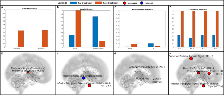
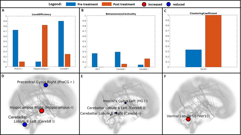
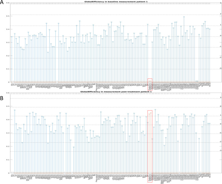
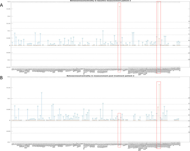
Given the huge symptom diversity and complexity of mental disorders, an individual approach is the most promising avenue for clinical transfer and the establishment of personalized psychiatry. However, due to technical limitations, knowledge about the neurobiological basis of mental illnesses has, to date, mainly been based on findings resulting from evaluations of average data from certain diagnostic groups. We postulate that this could change substantially through the use of the emerging ultra-high-field MRI (UHF-MRI) technology. The main advantages of UHF-MRI include high signal-to-noise ratio, resulting in higher spatial resolution and contrast and enabling individual examinations of single subjects. Thus, we used this technology to assess changes in the properties of resting-state networks over the course of therapy in a naturalistic study of two depressed patients. Significant changes in several network property measures were found in regions corresponding to prior knowledge from group-level studies. Moreover, relevant parameters were already significantly divergent in both patients at baseline. In summary, we demonstrate the feasibility of UHF-MRI for capturing individual neurobiological correlates of mental diseases. These could serve as a tool for therapy monitoring and pave the way for a truly individualized and predictive clinical approach in psychiatric care.
© 2022. The Author(s).
Conflict of interest statement
None of the authors who contributed to this paper have any disclosures/conflicts of interest to declare. All authors declare the absence of any competing interests or personal financial interests related to the work reported in the manuscript.
Figures
References
-
- James SL, Abate D, Abate KH, Abay SM, Abbafati C, Abbasi N, et al. Global, regional, and national incidence, prevalence, and years lived with disability for 354 diseases and injuries for 195 countries and territories, 1990–2017: a systematic analysis for the Global Burden of Disease Study 2017. Lancet. 2018;392:1789–858. - PMC - PubMed
-
- Insel TR, Cuthbert BN. Medicine. Brain disorders? Precisely. Science. 2015;348:499–500. - PubMed
-
- Silbersweig DA, Rauch SL. Neuroimaging in psychiatry: a quarter century of progress. Harv Rev Psychiatry 2017;25. https://journals.lww.com/hrpjournal/Fulltext/2017/09000/Neuroimaging_in_.... - PubMed
-
- Theysohn JM, Maderwald S, Kraff O, Moenninghoff C, Ladd ME, Ladd SC. Subjective acceptance of 7 Tesla MRI for human imaging. Magn Reson Mater Phys, Biol Med. 2007;21:63. - PubMed
Publication types
MeSH terms
LinkOut - more resources
Full Text Sources
Medical
