. 2022 Jan 26;12(1):1410.
doi: 10.1038/s41598-022-05271-2.

Overexpression of the dystrophins Dp40 and Dp40L170P modifies neurite outgrowth and the protein expression profile of PC12 cells

Affiliations

Overexpression of the dystrophins Dp40 and Dp40L170P modifies neurite outgrowth and the protein expression profile of PC12 cells

César García-Cruz et al. Sci Rep. .

Abstract

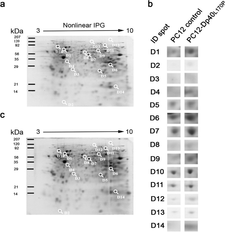
Dp40 is ubiquitously expressed including the central nervous system. In addition to being present in the nucleus, membrane, and cytoplasm, Dp40 is detected in neurites and postsynaptic spines in hippocampal neurons. Although Dp40 is expressed from the same promoter as Dp71, its role in the cognitive impairment present in Duchenne muscular dystrophy patients is still unknown. Here, we studied the effects of overexpression of Dp40 and Dp40L170P during the neuronal differentiation of PC12 Tet-On cells. We found that Dp40 overexpression increased the percentage of PC12 cells with neurites and neurite length, while Dp40L170P overexpression decreased them compared to Dp40 overexpression. Two-dimensional gel electrophoresis analysis showed that the protein expression profile was modified in nerve growth factor-differentiated PC12-Dp40L170P cells compared to that of the control cells (PC12 Tet-On). The proteins α-internexin and S100a6, involved in cytoskeletal structure, were upregulated. The expression of vesicle-associated membrane proteins increased in differentiated PC12-Dp40 cells, in contrast to PC12-Dp40L170P cells, while neurofilament light-chain was decreased in both differentiated cells. These results suggest that Dp40 has an important role in the neuronal differentiation of PC12 cells through the regulation of proteins involved in neurofilaments and exocytosis of synaptic vesicles, functions that might be affected in PC12-Dp40L170P.


Conflict of interest statement

The authors declare no competing interests.

Figures

Figure 1
Figure 1
Morphometric analysis of undifferentiated and NGF-differentiated PC12-Dp40 and PC12-Dp40L170P cells. PC12 control, PC12-Dp40 and PC12-Dp40L170P cells were differentiated with NGF for 9 days. (a) Morphometric analysis was performed using clear-field micrographs of the undifferentiated (top panels) and differentiated PC12 cells (bottom panels). (b) Quantification of the differentiation ratio of the NGF-differentiated PC12 control, PC12-Dp40 and PC12-Dp40L170P cells. Top panel: percentage of cells with neurites. Bottom panel: average of neurite lengths. *P ˂ 0.05, **P ˂ 0.01, ***P ˂ 0.0005 compared to the PC12 control or PC12-Dp40 versus PC12-Dp40L170P cells. The scale bar represents 10 µm.
Figure 2
Figure 2
Two-dimensional gel electrophoresis images of proteins from the NGF-differentiated PC12 control and PC12-Dp40L170P cells. Total protein extracts of the NGF-differentiated PC12 control and PC12-Dp40L170P cells from three independent experiments were collected after differentiation for 9 days and separated by isoelectric focusing using immobilized pH nonlinear strips (7 cm), followed by SDS-PAGE. Gels were stained with coomassie colloidal blue. (a) Protein profile of the PC12 control cells and (b) the PC12-Dp40L170P cells. Numbers 1–14 indicate the IDs of the differentially expressed protein spots. (c) Magnified views of the differentially expressed protein spots. Arrows represent the nonlinear immobilized pH gradient used for IEF. The positions of standard markers for the second dimension are shown on the left side of each image.
Figure 3
Figure 3
Expression of the proteins α-internexin, VAMP, HspB1, NF-L, Myc-Dp40 and Myc-Dp40L170P in the undifferentiated PC12-Dp40 and PC12-Dp40L170P cells. Protein extracts were obtained from the undifferentiated PC12 control, PC12-Dp40 and Dp40L170P cells and analyzed by WB. (a) Expression of α-internexin, VAMP, HspB1, NF-L, HspB1, Myc-Dp40 and Myc-Dp40L170P. (b) Relative expression of α-internexin, VAMP, and NF-L was determined as indicated in the materials and methods. The graph represents the mean ± SD from three independent experiments. *P ˂ 0.034 and 0.016 for α-internexin and NF-L comparing the PC12 control cells and the PC12-Dp40L170P cells. ***P ˂ 0.0006 and **P ˂ 0.0018 for Myc-Dp40 and Myc-Dp40L170P comparing the PC12-Dp40 and PC12-Dp40L170P with PC12 control cells. *P ˂ 0.0124 for Myc-Dp40 comparing the PC12-Dp40 and PC12-Dp40L170P cells. β-Actin was used as a loading control. Molecular weight of each protein is indicated in kDa. The blots of each protein are available in Supplementary Fig. 3.
Figure 4
Figure 4
Differential expression of α-internexin, VAMP, HspB1, NF-L, Myc-Dp40 and Myc-Dp40L170P in the NGF-differentiated PC12-Dp40 and PC12-Dp40L170P cells. Protein extracts were obtained from the NGF-differentiated PC12 control, PC12-Dp40 and PC12-Dp40L170P cells and analyzed by WB. (a) Expression of α-internexin, VAMP, HspB1, NF-L, Myc-Dp40 and Myc-Dp40L170P. (b) Relative expression of α-internexin, VAMP, HspB1, NF-L, Myc-Dp40 and Myc-Dp40L170P was determined as indicated in the materials and methods. The graph presents the mean ± SD from three independent experiments. **P ˂ 0.002 and *P ˂ 0.021 for α-internexin comparing the PC12-Dp40L170P with PC12 control and PC12-Dp40 cells, respectively. **P ˂ 0.008 and **P ˂ 0.007 for VAMP comparing the PC12 control with PC12-Dp40 and PC12-Dp40L170P cells, respectively, and *P ˂ 0.015 comparing PC12-Dp40 and PC12-Dp40L170P. *P ˂ 0.040 for NF-L comparing the PC12 control and PC12-Dp40L170P cells. *P ˂ 0.010 and *P ˂ 0.040 for Myc-Dp40 and Myc-Dp40L170P comparing the PC12-Dp40 and PC12-Dp40L170P with PC12 control cells. No statistical difference was observed for HspB1 (#P > 0.1). β-Actin was used as a loading control. Molecular weight of each protein is indicated in kDa. The blots of each protein are available in Supplementary Fig. 4.
Figure 5
Figure 5
Cellular distribution of α-internexin in undifferentiated and NGF-differentiated PC12-Dp40 and PC12-Dp40L170P cells. ImmF was performed on the PC12-Dp40, PC12-Dp40L170P and PC12 control cells after the induction of differentiation. ImmF staining for the Myc-Dp40 and Myc-Dp40L170P proteins was performed using the anti-c-Myc antibody (green) and α-internexin (red). Nuclei were stained with DAPI (blue). Images represent a single equatorial Z-section from confocal images to show the localization of each protein. (a) Undifferentiated and (b) differentiated PC12 control, PC12-Dp40 and PC12-Dp40L170P cells. Merged images correspond to the overlap of Myc-Dp40 and Myc-Dp40L170P with the α-internexin protein. The figure is a representative result of three independent experiments. The scale bar represents 20 µm.

References

    1. Tinsley JM, Blake DJ, Davies KE. Apo-dystrophin-3: A 2.2kb transcript from the DMD locus encoding the dystrophin glycoprotein binding site. Hum. Mol. Genet. 1993;2:521–524. - PubMed
    1. Fujimoto T, Itoh K, Yaoi T, Fushiki S. Somatodendritic and excitatory postsynaptic distribution of neuron-type dystrophin isoform, Dp40, in hippocampal neurons. Biochem. Biophys. Res. Commun. 2014;452:79–84. - PubMed
    1. Aragón J, et al. EF-hand domains are involved in the differential cellular distribution of dystrophin Dp40. Neurosci. Lett. 2015;600:115–120. - PubMed
    1. Blake DJ, Weir A, Newey SE, Davies KE. Function and genetics of dystrophin and dystrophin-related proteins in muscle. Physiol. Rev. 2002;82:291–329. - PubMed
    1. Suárez-Sánchez R, et al. Nucleocytoplasmic shuttling of the Duchenne muscular dystrophy gene product dystrophin Dp71d is dependent on the importin α/β and CRM1 nuclear transporters and microtubule motor dynein. Biochim. Biophys. Acta. 2014;1843:985–1001. - PubMed

Publication types

MeSH terms

LinkOut - more resources