Case Reports
. 2022 Jan 10:12:760164.
doi: 10.3389/fneur.2021.760164. eCollection 2021.

Case Report: Progressive Asymmetric Parkinsonism Secondary to CADASIL Without Dementia

Affiliations
Case Reports

Case Report: Progressive Asymmetric Parkinsonism Secondary to CADASIL Without Dementia

Weihang Guo et al. Front Neurol. .

Abstract

Parkinsonism is a rare phenotype of cerebral autosomal dominant arteriopathy with subcortical infarction and leukoencephalopathy (CADASIL), all of which involve cognitive decline. Normal cognition has not been reported in previous disease studies. Here we report the case of a 60-year-old female patient with a 2-year history of progressive asymmetric parkinsonism. On examination, she showed severe parkinsonism featuring bradykinesia and axial and limb rigidity with preserved cognition. Magnetic resonance imaging (MRI) revealed white matter hyperintensity in the external capsule and periventricular region. Dopaminergic response was limited. A missense mutation c.1630C>T (p.R544C) on the NOTCH3 gene was identified on whole-exome sequencing, which confirmed the diagnosis of vascular parkinsonism secondary to CADASIL. A diagnosis of CADASIL should be considered in asymmetric parkinsonism without dementia. Characteristic MRI findings support the diagnosis.

Keywords: case report; cerebral autosomal dominant arteriopathy with subcortical infarction and leukoencephalopathy; cognition; parkinsonism; rapid eye movement sleep behavior disorder (RBD).


Conflict of interest statement

The authors declare that the research was conducted in the absence of any commercial or financial relationships that could be construed as a potential conflict of interest.

Figures
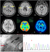
Figure 1
Figure 1
MRI, FDG-PET, VMAT2-PET, skin biopsy, and Sanger sequencing. (A1) FLAIR image shows white matter hyperintensity of the external capsule (yellow arrow). (A2) FLAIR image shows periventricular white matter hyperintensity (yellow arrow). (A3) Bilateral temporal poles show normal signal. (B) Flair image revealed no clearly cerebellar atrophy. (C) FDG-PET reveals hypometabolism in the right putamen. (D) VMAT2 indicates low-intensity signals in right putamen. (E) Skin biopsy shows granular osmiophilic material near the basement membrane of arteriolar smooth muscle cells (red arrow). (F) Sanger sequencing confirmed the mutation c. 1630C>T (p.R544C). MRI, magnetic resonance imaging; FDG-PET, fluorodeoxyglucose-positron emission tomography; FLAIR, fluid-attenuated inversion recovery; VMAT2, vesicular monoamine transporter type 2.

References

    1. Erro R, Lees AJ, Moccia M, Picillo M, Penco S, Mosca L, et al. . Progressive parkinsonism, balance difficulties, and supranuclear gaze palsy. JAMA Neurol. (2014) 71:104–7. 10.1001/jamaneurol.2013.5149 - DOI - PubMed
    1. Ragno M, Sanguigni S, Manca A, Pianese L, Paci C, Berbellini A, et al. . Parkinsonism in a pair of monozygotic CADASIL twins sharing the R1006C mutation: a transcranial sonography study. Neurol Sci. (2016) 37:875–81. 10.1007/s10072-016-2497-x - DOI - PubMed
    1. Wegner F, Strecker K, Schwarz J, Wagner A, Heinritz W, Sommerer F, et al. . Vascular parkinsonism in a CADASIL case with an intact nigrostriatal dopaminergic system. J Neurol. (2007) 254:1743–5. 10.1007/s00415-007-0529-4 - DOI - PubMed
    1. Fanciulli A, Wenning GK. Multiple-system atrophy. N Engl J Med. (2015) 372:249–63. 10.1056/NEJMra1311488 - DOI - PubMed
    1. Moreton FC, Razvi SS, Davidson R, Muir K. W. Changing clinical patterns and increasing prevalence in CADASIL. Acta Neurol Scand. (2014) 130:197–203. 10.1111/ane.12266 - DOI - PubMed

Publication types