. 2022 Jan 10:12:746021.
doi: 10.3389/fimmu.2021.746021. eCollection 2021.

Persistence of SARS CoV-2 S1 Protein in CD16+ Monocytes in Post-Acute Sequelae of COVID-19 (PASC) up to 15 Months Post-Infection

Affiliations

Persistence of SARS CoV-2 S1 Protein in CD16+ Monocytes in Post-Acute Sequelae of COVID-19 (PASC) up to 15 Months Post-Infection

Bruce K Patterson et al. Front Immunol. .

Abstract

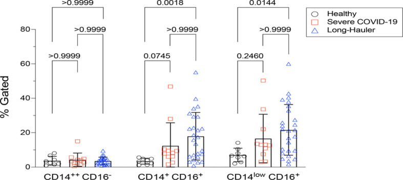
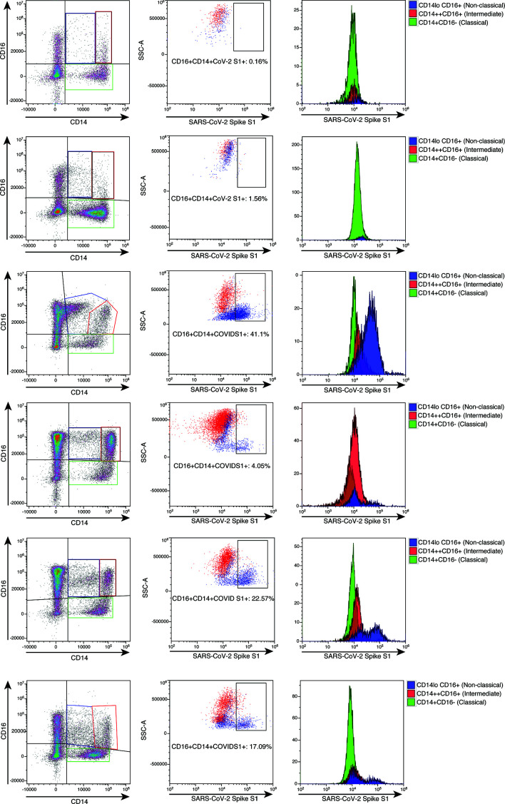
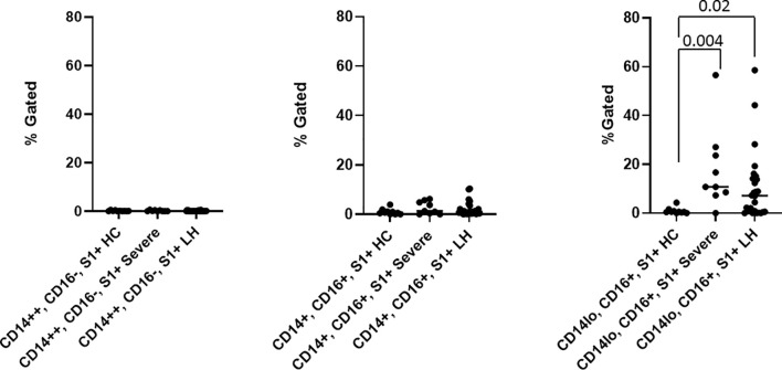
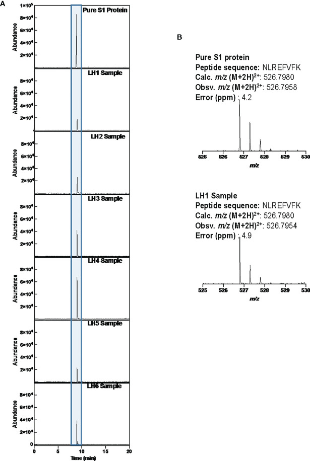
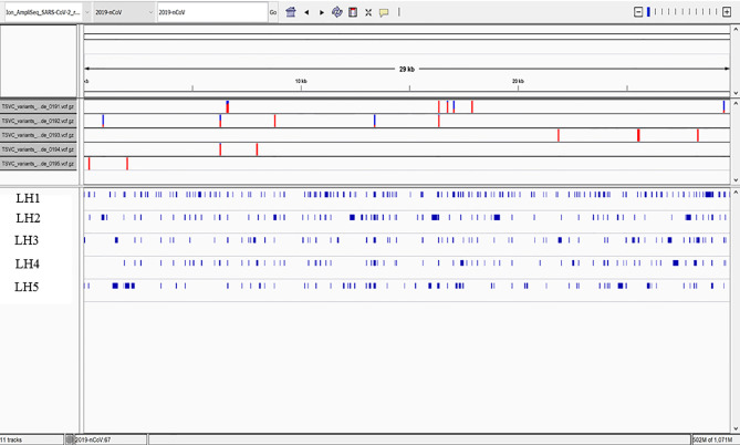
The recent COVID-19 pandemic is a treatment challenge in the acute infection stage but the recognition of chronic COVID-19 symptoms termed post-acute sequelae SARS-CoV-2 infection (PASC) may affect up to 30% of all infected individuals. The underlying mechanism and source of this distinct immunologic condition three months or more after initial infection remains elusive. Here, we investigated the presence of SARS-CoV-2 S1 protein in 46 individuals. We analyzed T-cell, B-cell, and monocytic subsets in both severe COVID-19 patients and in patients with post-acute sequelae of COVID-19 (PASC). The levels of both intermediate (CD14+, CD16+) and non-classical monocyte (CD14Lo, CD16+) were significantly elevated in PASC patients up to 15 months post-acute infection compared to healthy controls (P=0.002 and P=0.01, respectively). A statistically significant number of non-classical monocytes contained SARS-CoV-2 S1 protein in both severe (P=0.004) and PASC patients (P=0.02) out to 15 months post-infection. Non-classical monocytes were sorted from PASC patients using flow cytometric sorting and the SARS-CoV-2 S1 protein was confirmed by mass spectrometry. Cells from 4 out of 11 severe COVID-19 patients and 1 out of 26 PASC patients contained ddPCR+ peripheral blood mononuclear cells, however, only fragmented SARS-CoV-2 RNA was found in PASC patients. No full length sequences were identified, and no sequences that could account for the observed S1 protein were identified in any patient. That non-classical monocytes may be a source of inflammation in PASC warrants further study.

Keywords: CCR5; COVID-19; PASC; SARS CoV-2 S1 protein; fractalkine; non-classical monocytes.


Conflict of interest statement

BP, AP, HR, EL, and EF are employees of IncellDx, Inc. TT, PS, SH, and DM are employees of Avrok Laboratories, Inc. The remaining authors declare that the research was conducted in the absence of any commercial or financial relationships that could be construed as a potential conflict of interest.

Figures

Figure 1
Figure 1
Quantification of classical, intermediate and non-classical monocytes in PASC (LH). Non-classical monocytes were significantly elevated in severe COVID-19 and in PASC. Values for each category were based on the parental gating in Supplementary Figure 1 . Statistical analysis was performed using the Kluskal-Wallis test. P values <0.05 are considered significant.
Figure 2
Figure 2
High parameter flow cytometric quantification of SARS-CoV-2 S1 protein in monocytic subsets. Cells were gated as demonstrated in Supplementary Figure 1 . Each horizontal row represents an individual patient. A representative healthy control is demonstrated in the top row. All subsequent rows represent the spectrum of S1 containing monocytes. Classical monocytes are green, intermediate monocytes are red and non-classical monocytes are blue.
Figure 3
Figure 3
Quantification of SARS-CoV-2 S1 protein in monocyte subsets isolated from healthy 63 controls (HC), severe COVID-19 (severe), and PASC patients (LH). SARS-CoV-2 S1 protein was expressed in non-classical monocytes in both severe and PASC individuals. Values (median value-horizontal line) for each category were based on the parental gating in Supplementary Figure 1 . Statistical analysis was performed using the Kluskal-Wallis test. P values <0.05 are considered significant.
Figure 4
Figure 4
LCMS confirmation of the presence of S1 subunit in samples LH1-6. (A) Extracted ion chromatogram (EIC) displaying the NLREFVFK peptide. The retention time matches that of the NLREFVFK peptide in the commercial S1 standard. (B) Mass Spectra of the NLREFVFK from both the commercial standard and patient LH1. The Spectra show the same mass and isotope distribution.
Figure 5
Figure 5
High Quality Mutations in the Callable Regions. Only fragmented viral RNA was identified in the five patients, but multiple mutations throughout the viral genome were identified, the vast majority of which were unique to each patient. Overall coverage was less than 20%, and no complete sequence in any portion of the viral genome was detected, including in the spike gene encoding the S1 subunit identified by protein analysis in these patients.

References

    1. Rubin R. As Their Numbers Grow, COVID-19 "Long Haulers” Stump Experts. JAMA (2020) 324:1381–3. doi: 10.1001/jama.2020.17709 - DOI - PubMed
    1. (2021). Available at: https://www.cdc.gov/coronavirus/2019-ncov/hcp/clinical-care/post-covid-c....
    1. Yao XH, He ZC, Li TY, Zhang HR, Wang Y, Mou H, et al. . Pathological Evidence for Residual SARS-CoV-2 in Pulmonary Tissues of a Ready-for-Discharge Patient. Cell Res (2020) 30:541–3. doi: 10.1038/s41422-020-0318-5 - DOI - PMC - PubMed
    1. Nienhold R, Ciani Y, Koelzer VH, Tzankov A, Haslbauer JD, Menter T, et al. . Two Distinct Immunopathological Profiles in Autopsy Lungs of COVID-19. Nat Commun (2020) 11:5086. doi: 10.1038/s41467-020-18854-2 - DOI - PMC - PubMed
    1. Patterson BK, Guevarra-Coto J, Yogendra R, Francisco EB, Long E, Pise A, et al. . Immune-Based Prediction of COVID-19 Severity and Chronicity Decoded Using Machine Learning. Front Immunol (2021) 12:700782. doi: 10.3389/fimmu.2021.700782 - DOI - PMC - PubMed