. 2022 Jan 10:11:733088.
doi: 10.3389/fonc.2021.733088. eCollection 2021.

Intraoperative Magnetic Resonance Imaging Assisted Endoscopic Endonasal Resection of Clival Chordomas

Affiliations

Intraoperative Magnetic Resonance Imaging Assisted Endoscopic Endonasal Resection of Clival Chordomas

Beste Gulsuna et al. Front Oncol. .

Abstract

Background: Cranial base chordomas are typically indolent and usually appear as encapsulated tumors. They slowly grow by infiltrating the bone, along with the lines of least resistance. Due to its relationship with important neurovascular structures, skull base chordoma surgery is challenging.

Objective: The usefulness of intraoperative magnetic resonance imaging (IO-MRI) in achieving the goal of surgery, is evaluated in this study.

Methods: Between March 2018 and March 2020, 42 patients were operated on for resection of skull base chordomas in our institution. All of them were operated on under IO-MRI. Patients were analyzed retrospectively for identifying common residue locations, complications and early post-operative outcomes.

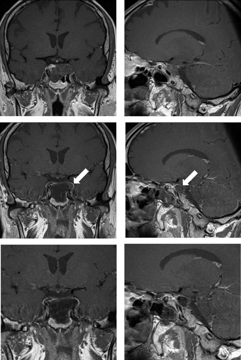
Results: In 22 patients (52,4%) gross total resection was achieved according to the final IO-MRI. In 20 patients (47,6%) complete tumor removal was not possible because of extension to the petrous bone (8 patients), pontocerebellar angle (6 patients), prepontine cistern (4 patients), temporobasal (1 patient), cervical axis (1 patient). In 13 patients, the surgery was continued after the first IO-MRI control was performed, which showed a resectable residual tumor. 7 of these patients achieved total resection according to the second IO-MRI, in the other 6 patients all efforts were made to ensure maximal resection of the tumor as much as possible without morbidity. Repeated IO-MRI helped achieve gross total resection in 7 patients (53.8%).

Conclusions: Our study proves that the use of IO-MRI is a safe method that provides the opportunity to show the degree of resection in skull base chordomas and to evaluate the volume and location of the residual tumor intraoperatively. Hence IO-MRI can improve the life expectancy of patients because it provides an opportunity for both gross total resection and maximal safe resection in cases where total resection is not possible.

Keywords: IO-MRI; chordoma; endoscopic; intraoperative magnetic resonance imaging; skull base.


Conflict of interest statement

The authors declare that the research was conducted in the absence of any commercial or financial relationships that could be construed as a potential conflict of interest.

Figures

Figure 1
Figure 1
Case sample 1, a mass is detected after the 6th cranial nerve paralysis on the left. Upper row: Preoperative coronal and sagittal contrast-enhanced T1 weighted MRI demonstrates a chordoma that invades the left cavernous sinus and passes lateral to the internal carotid artery. Middle row: Contrast-enhanced T1 weighted IO-MRI slices, the white arrow indicating residual tumor superior to the cavernous sinus. Bottom row: Final contrast-enhanced T1 weighted IO-MRI slices demonstrate gross total surgical resection.
Figure 2
Figure 2
Case sample 2, the patient presenting with headache, diplopia, and reduced sensation of the left side of the face. (A, B) Preoperative axial contrast-enhanced T1 weighted MRI shows a tumor invading the left cavernous sinus and compressing the brainstem. (C) Preoperative sagittal contrast-enhanced T1 weighted MRI shows a tumor invading the lower clivus and upper cervical spine. (D) Preoperative coronal contrast-enhanced T1 weighted MRI shows the tumor relation to the internal acoustic canal and brainstem. (E) Final contrast-enhanced T1 weighted IO-MRI demonstrates residual tumor located at the lower clivus and upper cervical spine. This IO-MRI slice also demonstrates the fat graft and nasal package closure. (F) Final contrast-enhanced T1 weighted IO-MRI demonstrates tumor resection.
Figure 3
Figure 3
Case sample 3, the patient presenting cranial nerve deficit because of the tumor which invades the right cavernous sinus. Upper row: Preoperative axial and coronal contrast-enhanced T1 weighted MRI shows a tumor invading the right cavernous sinus. Middle row: First contrast-enhanced T1 weighted IO-MRI demonstrates residual tumor located at the inferolateral cavernous sinus. Bottom row: Final contrast-enhanced T1 weighted IO-MRI slices demonstrate gross total surgical resection.
Figure 4
Figure 4
Case sample 4, the patient presenting with headache and impaired cognitive functions. (A, B) Preoperative axial T2 weighted MRI shows a huge tumor that extremely compresses the brainstem. (C, D) Preoperative sagittal and axial contrast-enhanced T1 weighted MRI shows tumor relation to neurovascular structures. (E–G) Final contrast-enhanced T1 weighted IO-MRI shows the gross total tumor resection and also our nasal closure procedure. (F, G) The patient developed hydrocephalus in the postoperative period and a ventriculoperitoneal shunt was placed, and these axial T2 weighted MRI images were taken before the patient was discharged after an intensive care period of approximately 3 months (H).

References

    1. Chambers KJ, Lin DT, Meier J, Remenschneider A, Herr M, Gray ST. Incidence and Survival Patterns of Cranial Chordoma in the United States. Laryngoscope (2014) 124(5):1097–102. doi: 10.1002/lary.24420 - DOI - PubMed
    1. Samii A, Gerganov VM, Herold C, Hayashi N, Naka T, Mirzayan MJ, et al. . Chordomas of the Skull Base: Surgical Management and Outcome. J Neurosurg (2007) 107(2):319–24. doi: 10.3171/JNS-07/08/0319 - DOI - PubMed
    1. Almefty K, Pravdenkova S, Colli BO, Al-Mefty O, Gokden M. Chordoma and Chondrosarcoma: Similar, But Quite Different, Skull Base Tumors. Cancer (2007) 110(11):2467–77. doi: 10.1002/cncr.23073 - DOI - PubMed
    1. Metwali H, Samii A, Gerganov V, Giordano M, Fahlbusch R, Samii M. The Significance of Intraoperative Magnetic Resonance Imaging in Resection of Skull Base Chordomas. World Neurosurg (2019) 128:e185–94. doi: 10.1016/j.wneu.2019.04.086 - DOI - PubMed
    1. di Maio S, Temkin N, Ramanathan D, Sekhar LN. Current Comprehensive Management of Cranial Base Chordomas: 10-Year Meta-Analysis of Observational Studies: Clinical Article. J Neurosurg (2011) 115(6):1094–105. doi: 10.3171/2011.7.JNS11355 - DOI - PubMed

LinkOut - more resources