. 2022 Jan 24;8(1):00375-2021.
doi: 10.1183/23120541.00375-2021. eCollection 2022 Jan.

Evidence for shared genetic risk factors between lymphangioleiomyomatosis and pulmonary function

Affiliations

Evidence for shared genetic risk factors between lymphangioleiomyomatosis and pulmonary function

Xavier Farré et al. ERJ Open Res. .

Abstract

Introduction: Lymphangioleiomyomatosis (LAM) is a rare low-grade metastasising disease characterised by cystic lung destruction. The genetic basis of LAM remains incompletely determined, and the disease cell-of-origin is uncertain. We analysed the possibility of a shared genetic basis between LAM and cancer, and LAM and pulmonary function.

Methods: The results of genome-wide association studies of LAM, 17 cancer types and spirometry measures (forced expiratory volume in 1 s (FEV1), forced vital capacity (FVC), FEV1/FVC ratio and peak expiratory flow (PEF)) were analysed for genetic correlations, shared genetic variants and causality. Genomic and transcriptomic data were examined, and immunodetection assays were performed to evaluate pleiotropic genes.

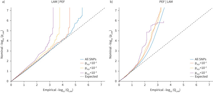
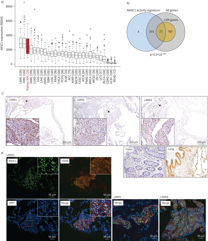
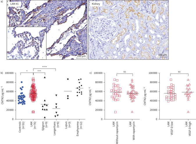
Results: There were no significant overall genetic correlations between LAM and cancer, but LAM correlated negatively with FVC and PEF, and a trend in the same direction was observed for FEV1. 22 shared genetic variants were uncovered between LAM and pulmonary function, while seven shared variants were identified between LAM and cancer. The LAM-pulmonary function shared genetics identified four pleiotropic genes previously recognised in LAM single-cell transcriptomes: ADAM12, BNC2, NR2F2 and SP5. We had previously associated NR2F2 variants with LAM, and we identified its functional partner NR3C1 as another pleotropic factor. NR3C1 expression was confirmed in LAM lung lesions. Another candidate pleiotropic factor, CNTN2, was found more abundant in plasma of LAM patients than that of healthy women.

Conclusions: This study suggests the existence of a common genetic aetiology between LAM and pulmonary function.


Conflict of interest statement

Conflict of interest: X. Farré has nothing to disclose. Conflict of interest: R. Espín has nothing to disclose. Conflict of interest: A. Baiges has nothing to disclose. Conflict of interest: E. Blommaert has nothing to disclose. Conflict of interest: W. Kim has nothing to disclose. Conflict of interest: K. Giannikou has nothing to disclose. Conflict of interest: C. Herranz has nothing to disclose. Conflict of interest: A. Román has nothing to disclose. Conflict of interest: B. Sáez has nothing to disclose. Conflict of interest: Á. Casanova has nothing to disclose. Conflict of interest: J. Ancochea has nothing to disclose. Conflict of interest: C. Valenzuela has nothing to disclose. Conflict of interest: P. Ussetti has nothing to disclose. Conflict of interest: R. Laporta has nothing to disclose. Conflict of interest: J.A. Rodríguez-Portal has nothing to disclose. Conflict of interest: C.H.M. van Moorsel has nothing to disclose. Conflict of interest: J.J. van der Vis has nothing to disclose. Conflict of interest: M.J.R. Quanjel has nothing to disclose. Conflict of interest: M. Tena-Garitaonaindia has nothing to disclose. Conflict of interest: F. Sánchez de Medina has nothing to disclose. Conflict of interest: F. Mateo has nothing to disclose. Conflict of interest: M. Molina-Molina has nothing to disclose. Conflict of interest: S. Won has nothing to disclose. Conflict of interest: D.J. Kwiatkowski has nothing to disclose. Conflict of interest: R. de Cid has nothing to disclose. Conflict of interest: M.A. Pujana has nothing to disclose. Support statement: This research was supported by Asociación Española de LAM; The LAM Foundation Seed Grant 2019; Carlos III Institute of Health grants PI18/01029, PI21/01306 and ICI19/00047 (co-funded by European Regional Development Fund (ERDF), “A way to build Europe”); Ministry of Economy and Competitivity grant SAF2017-88457-R; the Generalitat de Catalunya SGR 2017-449 and 2017-529; PERIS PFI-Salut SLT017-20-000076; and the CERCA Program to IDIBELL and Institut Germans Trias i Pujol. X. Farré is supported by the VEIS project (001-P-001647, ERDF Operational Programme of Catalonia 2014–2020; co-funded by ERDF, “A way to build Europe”). Funding information for this article has been deposited with the Crossref Funder Registry.

Figures

FIGURE 1
FIGURE 1
Stratified Q-Q plots including lymphangioleiomyomatosis (LAM) and peak expiratory flow (PEF) genome-wide association study (GWAS) results. a) Q-Q plot (nominal versus empirical −log10 p-values, corrected for inflation) conditioning LAM on PEF; and b) Q-Q plot conditioning PEF on LAM. Leftwards deflection from the null distribution of the observed p-value as the thresholds become more stringent indicates genetic overlap between the two traits. SNP: single nucleotide polymorphism.
FIGURE 2
FIGURE 2
NR3C1 gene expression in angiomyolipoma (AML) tumours and NR3C1 protein expression in lymphangioleiomyomatosis (LAM) lung lesions. a) Comparison of NR3C1 expression in kidney AMLs (red font) with other neoplasms (TCGA data: 2463 tumours of 27 histological types). Cancer abbreviations: LAML: acute myeloid leukaemia; KIRC: kidney renal clear cell carcinoma; SARC: sarcoma; LUAD: lung adenocarcinoma; LGG: low-grade glioma; LIHC: liver hepatocellular carcinoma; HNSC: head and neck squamous cell carcinoma; GBM: glioblastoma multiforme; LUSC: lung squamous cell carcinoma; PAAD: pancreatic adenocarcinoma; THCA: thyroid carcinoma; PCPG: phaeochromocytoma and paraganglioma; DLBC: lymphoid neoplasm diffuse large B-cell lymphoma; ACC: adrenocortical carcinoma; KIRP: kidney renal papillary cell carcinoma; SKCM: skin cutaneous melanoma; CESC: cervical squamous cell carcinoma and endocervical adenocarcinoma; KICH: kidney chromophobe; PRAD: prostate adenocarcinoma; BRCA: breast invasive carcinoma; MESO: mesothelioma; OV: ovarian serous cystadenocarcinoma; UCS: uterine carcinosarcoma; BLCA: bladder urothelial carcinoma; COAD: colon adenocarcinoma; UCEC: uterine corpus endometrial carcinoma; READ: rectum adenocarcinoma. The numbers in parentheses are the sample sizes of the indicated cancer types. The median, interquartile range and 95% range are shown for each setting, with outliers indicated by circles. Gene expression is shown as RSEM (RNA sequencing by expectation maximisation) values. b) Venn diagram showing the identity overlap (n=27) between genes identified in the NR3C1-activity signature and genes differentially expressed in LAM single cells. c) Representative images from immunohistochemistry assays for the detection of NR3C1 expression in LAM lung lesions of three patients (LAM #1–3). The arrows indicate the magnified lesion areas in the insets: in magnified lung nodules, epithelioid and spindle-like diseased cells are apparent from the observed nuclear shapes of positive NR3C1 staining. The positive control results from colon tissue are also shown, as well as those from normal lung tissue showing positivity in the alveolar epithelium, and luminal and basal layers of the bronchioles. d) Representative images of immunohistofluorescence detection and colocalisation of NR3C1 and αSMA in LAM lung lesions; nuclei stained blue with DAPI (merged). Lung nodules of three LAM patients are shown.
FIGURE 3
FIGURE 3
Evaluation of additional pleiotropic factors. a) Representative images from immunohistochemistry assays for detecting NTN4 expression in lymphangioleiomyomatosis (LAM) lung lesions. Lung nodules appear negative, while the alveolar layer is positive. The positive control of kidney tissue is shown. b) Overabundance of CNTN2 in LAM plasma relative to healthy women and two related pulmonary diseases, as indicated. The number of samples analysed in each setting (n) is indicated. Asterisks indicate significant differences based on two-sided Mann–Whitney tests (*p<0.05, ***p<0.001 and ****p<0.0001). Mean values are indicated with horizontal black lines. c) Absence of significant differences (n.s.: not significant) of CNTN2 plasma levels between LAM patients receiving and not receiving rapamycin treatment (left panel), and between LAM patients with high and low vascular endothelial growth factor-D (VEGF-D) plasma levels (right panel).

References

    1. Johnson SR, Cordier JF, Lazor R, et al. . Review panel of the ERS LAM task force. European Respiratory Society guidelines for the diagnosis and management of lymphangioleiomyomatosis. Eur Respir J 2010; 35: 14–26. doi:10.1183/09031936.00076209 - DOI - PubMed
    1. McCormack FX, Gupta N, Finlay GR, et al. . Official American Thoracic Society/Japanese Respiratory Society clinical practice guidelines: lymphangioleiomyomatosis diagnosis and management. Am J Respir Crit Care Med 2016; 194: 748–761. doi:10.1164/rccm.201607-1384ST - DOI - PMC - PubMed
    1. Martignoni G, Pea M, Reghellin D, et al. . Molecular pathology of lymphangioleiomyomatosis and other perivascular epithelioid cell tumors. Arch Pathol Lab Med 2010; 134: 33–40. doi:10.5858/2008-0542-RAR1.1 - DOI - PubMed
    1. Harari S, Torre O, Cassandro R, et al. . The changing face of a rare disease: lymphangioleiomyomatosis. Eur Respir J 2015; 46: 1471–1485. doi:10.1183/13993003.00412-2015 - DOI - PubMed
    1. Dongre A, Clements D, Fisher AJ, et al. . Cathepsin K in lymphangioleiomyomatosis: LAM cell-fibroblast interactions enhance protease activity by extracellular acidification. Am J Pathol 2017; 187: 1750–1762. doi:10.1016/j.ajpath.2017.04.014 - DOI - PubMed