. 2022 Jan-Dec:18:17448069221074991.
doi: 10.1177/17448069221074991.

Offset analgesia is associated with opposing modulation of medial versus dorsolateral prefrontal cortex activations: A functional near-infrared spectroscopy study

Affiliations

Offset analgesia is associated with opposing modulation of medial versus dorsolateral prefrontal cortex activations: A functional near-infrared spectroscopy study

Benedict J Alter et al. Mol Pain. 2022 Jan-Dec.

Abstract

Offset analgesia is defined by a dramatic drop in perceived pain intensity with a relatively small decrease in noxious input. Although functional magnetic resonance imaging studies implicate subcortical descending inhibitory circuits during offset analgesia, the role of cortical areas remains unclear. The current study identifies cortical correlates of offset analgesia using functional near infrared spectroscopy (fNIRS). Twenty-four healthy volunteers underwent fNIRS scanning during offset (OS) and control (Con) heat stimuli applied to the forearm. After controlling for non-neural hemodynamic responses in superficial tissues, widespread increases in cortical oxygenated hemoglobin concentration were observed, reflecting cortical activation during heat pain. OS-Con contrasts revealed deactivations in bilateral medial prefrontal cortex (mPFC) and bilateral somatosensory cortex (SSC) associated with offset analgesia. Right dorsolateral prefrontal cortex (dlPFC) showed activation only during OS. These data demonstrate opposing cortical activation patterns during offset analgesia and support a model in which right dlPFC underlies ongoing evaluation of pain intensity change. With predictions of decreasing pain intensity, right dlPFC activation likely inhibits ascending noxious input via subcortical pathways resulting in SSC and mPFC deactivation. This study identifies cortical circuitry underlying offset analgesia and introduces the use of fNIRS to study pain modulation in an outpatient clinical environment.

Keywords: Offset analgesia; descending inhibition; endogenous analgesia; functional near-infrared spectroscopy; heat pain; human; pain.


Conflict of interest statement

Declaration of conflicting interests: The author(s) declared no potential conflicts of interest with respect to the research, authorship, and/or publication of this article.

Figures

Figure 1.
Figure 1.
Experimental design. A. Participant setup, depicting thermode placement on the left forearm, the computerized visual analogue scale for real-time pain intensity rating controlled with the right hand, and placement of the NIRS cap. B. The two different suprathreshold heat stimuli used to measure offset analgesia. T1 is the individually tailored temperature that evokes a moderate pain (50/100 mm on the COVAS). T2 is 1 C° hotter than T1. C. NIRS head probe with sources and detectors positioned across the frontal and sensorimotor cortices. For region-of-interest (ROI) analysis, Brodmann areas (BA) are shown with the corresponding color intensity reflecting channel weights for a given ROI.
Figure 2.
Figure 2.
Cortical activation during prolonged noxious heat stimulation of the forearm. Whole-brain T-score heatmaps are displayed for each channel during constant control (A) and offset (B) stimuli. Forty-five seconds during noxious stimulation were contrasted with an immediately preceding baseline time period. In C., [OS-Con] contrast during the full 45-second stimulus time period is shown. Solid lines reflect statistically significant contrasts surviving correction for multiple comparisons (q < 0.05). Dashed lines are not statistically significant.
Figure 3.
Figure 3.
Temperature offset is associated with divergent patterns of cortical activation. Whole-brain T-score heatmaps reflecting constant control (Con)–baseline (A), offset stimulus (OS)–baseline (B), and OS-Con (C) contrasts during the 20 seconds following the step down from T2 to T1 during the OS. This 20–40 second epoch is commonly examined to measure offset analgesia. Solid lines reflect statistically significant contrasts surviving correction for multiple comparisons (q < 0.05). Dashed lines are not statistically significant. In C., for the oxyhemoglobin montage, regional activations and deactivations are labeled in red and blue, respectively.
Figure 4.
Figure 4.
Behavioral measures of offset analgesia. A. An example of pain intensity continuously rated over time during offset (OS) and constant control stimuli (Con) from a single subject. Cutaneous thermode temperatures for different time periods are noted above the graph. In this participant, T1 = 47°C and T2 = 48°C. The black arrow represents a standard measure of offset analgesia, derived by subtracting the minimum pain intensity in that time period during the OS with Con pain intensity at the same time point. B. Group-level data demonstrate offset analgesia, as measured by comparing pain intensity at the time point of the minima following OS temperature offset during both OS and Con stimuli. Group mean values are plotted with error bars representing 95% CI, ** paired t-test, p < .01. Pain intensity difference (OS–Con) at each time point for a single participant (C.) and across the group at the OS pain intensity minima (D.). E. Pain difference curves, similar to C., are plotted for all participants (thin lines, colored by median split subgroup: “robust offset analgesia” are participants with pain intensity differences in D. more negative than the median and “no offset analgesia” less negative than the median). Thick lines represent the median of the subgroups at each time point.
Figure 5.
Figure 5.
Offset analgesia is associated with decreases in mPFC and SSC activation and increases dlPFC activation. Whole-brain T-score heatmaps reflecting [OS-Con] contrasts during maximal offset analgesia, 26–36 seconds after the start of the OS. Solid lines reflect statistically significant contrasts surviving correction for multiple comparisons (q < 0.05). Dashed lines are not statistically significant. For the oxyhemoglobin montage, regional activations and deactivations are labeled in red and blue, respectively.
Figure 6.
Figure 6.
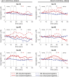
Dynamic changes in oxygenated and deoxygenated hemoglobin during offset and control stimuli. Group median HbO (red) and HbR (blue) from right and left medial prefrontal cortex (BA-10), dorsolateral prefrontal cortex (BA-45), and somatosensory cortex (BA-40) are plotted at a sampling rate of 1 Hz during both offset stimuli (OS) and constant control stimuli (Con). Stars indicate significant differences (q < 0.05) from the ROI analysis between OS and Con during the 20–40 second interval examined for offset analgesia. Red stars represent oxyhemoglobin OS-Con, and blue stars represent deoxyhemoglobin OS-Con. Borderline significance is noted with the q-value.
Figure 7.
Figure 7.
Subgroup sensitivity analysis. HbO and HbR activation maps reflecting OS-Con contrast T-scores in participants with robust offset analgesia (A., N = 12) and without offset analgesia (B., N = 12). Solid lines reflect statistically significant contrasts surviving correction for multiple comparisons (q < 0.05). Dashed lines are not statistically significant.

References

    1. Grill JD, Coghill RC. Transient analgesia evoked by noxious stimulus offset. J Neurophysiol 2002; 87: 2205–2208. DOI: 10.1152/jn.00730.2001. - DOI - PubMed
    1. Yelle MD, Rogers JM, Coghill RC. Offset analgesia: a temporal contrast mechanism for nociceptive information. Pain 2008; 134: 174–186. DOI: 10.1016/j.pain.2007.04.014. - DOI - PMC - PubMed
    1. Szikszay TM, Adamczyk WM, Luedtke K. The magnitude of offset analgesia as a measure of endogenous pain modulation in healthy subjects and patients with chronic pain - a systematic review and meta-analysis. Clin J Pain 2018; 35, 189, 204. DOI: 10.1097/ajp.0000000000000657. - DOI - PubMed
    1. Lockwood S, Dickenson AH. What goes up must come down: insights from studies on descending controls acting on spinal pain processing. J Neural Transm (Vienna) 2020; 127: 541–549. DOI: 10.1007/s00702-019-02077-x - DOI - PMC - PubMed
    1. Georgopoulos V, Akin-Akinyosoye K, Zhang W, McWilliams DF, Hendrick P, Walsh DA. Quantitative sensory testing and predicting outcomes for musculoskeletal pain, disability, and negative affect: a systematic review and meta-analysis. Pain 2019; 160: 1920–1932. DOI: 10.1097/j.pain.0000000000001590. - DOI - PMC - PubMed

Publication types

LinkOut - more resources