Comparative Study
. 2022 Jan;8(1):000749.
doi: 10.1099/mgen.0.000749.

Genome diversity of domesticated Acinetobacter baumannii ATCC 19606T strains

Affiliations
Comparative Study

Genome diversity of domesticated Acinetobacter baumannii ATCC 19606T strains

Irene Artuso et al. Microb Genom. 2022 Jan.

Abstract

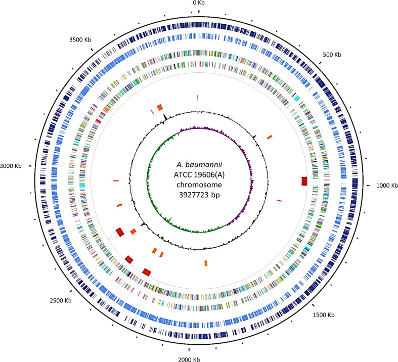
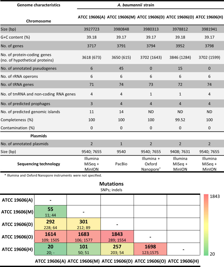
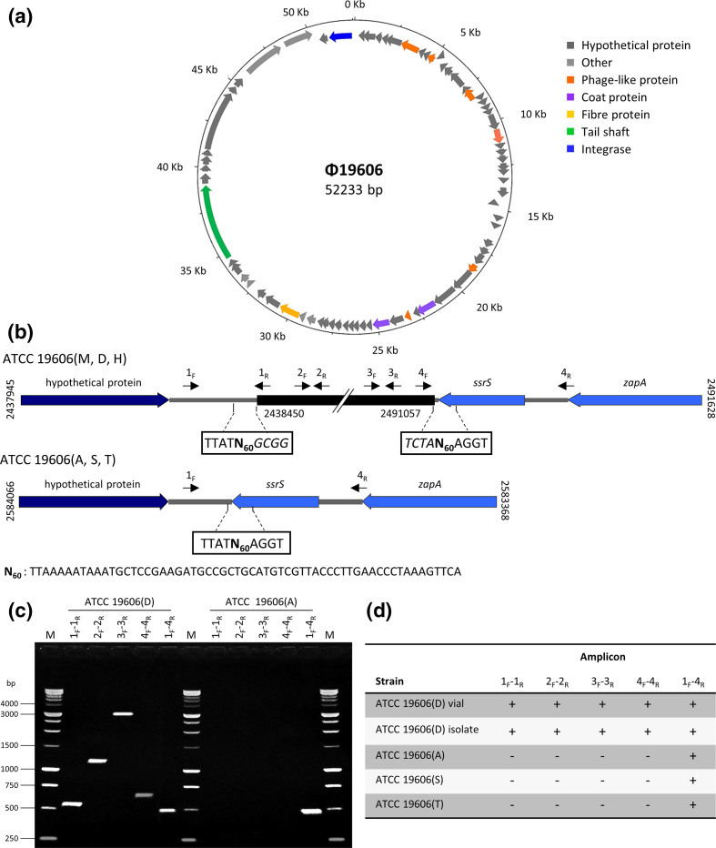
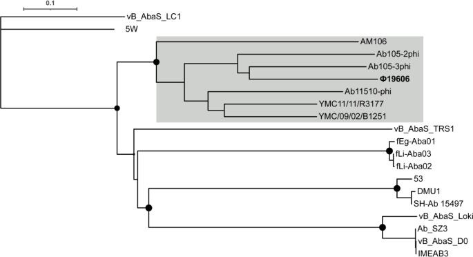
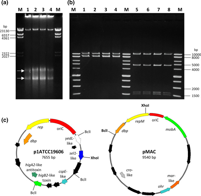
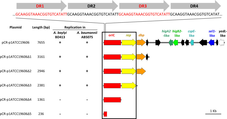
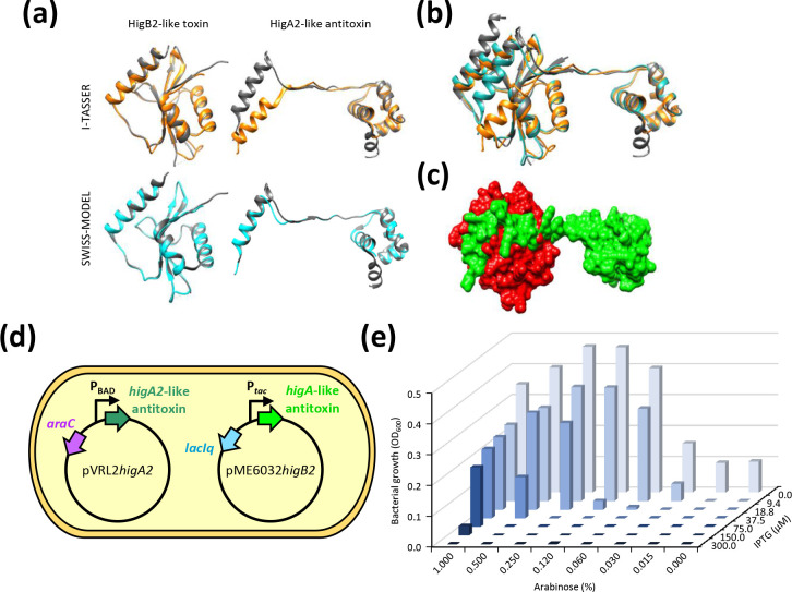
Acinetobacter baumannii has emerged as an important opportunistic pathogen worldwide, being responsible for large outbreaks for nosocomial infections, primarily in intensive care units. A. baumannii ATCC 19606T is the species type strain, and a reference organism in many laboratories due to its low virulence, amenability to genetic manipulation and extensive antibiotic susceptibility. We wondered if frequent propagation of A. baumannii ATCC 19606T in different laboratories may have driven micro- and macro-evolutionary events that could determine inter-laboratory differences of genome-based data. By combining Illumina MiSeq, MinION and Sanger technologies, we generated a high-quality whole-genome sequence of A. baumannii ATCC 19606T, then performed a comparative genome analysis between A. baumannii ATCC 19606T strains from several research laboratories and a reference collection. Differences between publicly available ATCC 19606T genome sequences were observed, including SNPs, macro- and micro-deletions, and the uneven presence of a 52 kb prophage belonging to genus Vieuvirus. Two plasmids, pMAC and p1ATCC19606, were invariably detected in all tested strains. The presence of a putative replicase, a replication origin containing four 22-mer direct repeats, and a toxin-antitoxin system implicated in plasmid stability were predicted by in silico analysis of p1ATCC19606, and experimentally confirmed. This work refines the sequence, structure and functional annotation of the A. baumannii ATCC 19606T genome, and highlights some remarkable differences between domesticated strains, likely resulting from genetic drift.

Keywords: Acinetobacter baumannii ATCC 19606T; genome refinement; native plasmids; strain domestication; Φ19606 phage.


Conflict of interest statement

The authors declare that there are no conflicts of interest.

Figures

Fig. 1.
Fig. 1.
Chromosome map of A. baumannii ATCC 19606(A). Circular map created by the CGView server. From the outermost to innermost, the tracks show the genes on positive (dark blue) and negative (light blue) strands, ORFs on positive and negative strands (with colours indicating COG classifications; [119]), prophages (red) with dotted lines indicating the excision site of the missing prophage, GIs (orange), GC content (green) and GC skew (purple and light green for positive and negative, respectively). Position 1 in ATCC 19606(A) corresponds to position 3772737 in ATCC 19696(H) and position 1094161 in ATCC 19606(M). Both genomes are in reverse orientation relative to ATCC 19606(A).
Fig. 2.
Fig. 2.
Relevant features of genome sequences of different A. baumannii ATCC 19606T strains.
Fig. 3.
Fig. 3.
A. baumannii Φ19606 phage. (a) Circular map of the Φ19606 genome drawn with DNAPlotter. The genome map illustrates putative ORFs along with the direction of transcription indicated with arrows. Functional proteins predicted by PHASTER are depicted in different colours. (b) Integration site of Φ19606 (black) into the ATCC 19606(M, D, H) chromosomes (top). The double slash denotes a phage region that is not shown. Positions refer to the ATCC 19606(M) genome sequence. Structure of ATCC 19606(A, S, T) after phage loss (bottom). Positions refer to ATCC 19606(A) genome sequence. Sequences flanking the insertion site are boxed, with predicted phage nucleotides italicized. Primer positions are indicated with black arrows. N60 stands for the 60-nucleotide sequence generated by phage insertion/excision. (c) Agarose gel electrophoresis of the PCR products obtained by using different primer pairs indicated in (b). (d) Presence (+) or absence (-) of amplicons detected in the different A. baumannii ATCC 19606T strains.
Fig. 4.
Fig. 4.
Phylogenetic tree of Acinetobacter phages belonging to the Shiphoviridae family. The tree was generated by VICTOR using the complete genome sequences of the Shiphoviridae family members. Filled circles at the nodes are GBDP pseudo-bootstrap support values >70 % from 100 replications. The scale bar indicates the number of substitutions per variable site. Phages belonging to the genus Vieuvirus are grey-shaded, Ф19606 is in bold. The tree was rooted at the midpoint.
Fig. 5.
Fig. 5.
Plasmids p1ATCC19606 and pMAC harboured by A. baumannii ATCC 19606T strains. (a) Agarose gel electrophoresis of clear lysates of A. baumannii ATCC 19606(A) (lane 1), ATCC 19606(D) (lane 2), ATCC 19606(S) (lane 3) and ATCC 19606(T) (lane 4). M, Lambda DNA/HindIII marker (ThermoFisher). White arrows indicate the closed circular forms of pMAC (upper band) and p1ATCC19606 (lower band). (b) p1ATCC19606 and pMAC were copurified from A. baumannii strains ATCC 19606(A) (lanes 1 and 5), ATCC 19606(D) (lanes 2 and 6), ATCC 19606(S) (lanes 3 and 7) and ATCC 19606(T) (lanes 4 and 8), and digested with XhoI (lanes 1–4) and BclI (lanes 5–8). M, BenchTop 1 kb DNA Ladder (Promega). (c) Physical and functional maps of the p1ATCC19606 and pMAC plasmids. Restriction sites for the enzymes used to generate the electropherogram in (b) are shown. Unique cutter restriction enzymes are indicated in bold. Nomenclature of p1ATCC19606: rep, putative replicase; dbp, gene encoding a predicted DNA-binding protein; cspE-like, putative cold-shock protein gene; sel1-like, putative gene coding for a Sel1-repeat family protein; yedL-like, gene coding for the putative YedL N-acetyltransferase; oriC, predicted origin of replication. Nomenclature of pMAC: repM, replication protein M; dbp, gene encoding a predicted DNA-binding protein; ohr, gene encoding an organic hydroperoxide resistance protein, mobA, plasmid mobilization protein; oriC, origin of replication. ORFs shown in black are predicted to encode for hypothetical proteins. All genes are reported in scale over the total length of each plasmid. Images were obtained by the use of the SnapGene software (GSL Biotech).
Fig. 6.
Fig. 6.
Deletion analysis of p1ATCC19606 to determine the minimal region required for autonomous plasmid replication in Acinetobacter spp. Deletion fragments of p1ATCC19606 were generated by PCR amplification with primers listed in Table S1 and cloned into pCR. The resulting p1ATCC19606 deletion derivatives were introduced in A. baylyi BD413 and A. baumannii AB5075 to map the minimal self-replicating region (black box). Relevant coding regions are indicated with colours: red, predicted minimal origin of replication (oriC); yellow, putative replicase (rep); orange, gene encoding a predicted DNA-binding protein (dbp); dark green, putative higA2-like antitoxin gene; light green, putative higB2-like toxin gene; cyan, putative cold-shock protein gene (cspE); blue, gene coding for putative a Sel1-repeat family protein (sel1); white, gene coding for the putative YedL N-acetyltransferase (yedL). Four copies of the 22-mer direct repeat (DR1–DR4) in the predicted origin of replication are shown on top. ORFs in black are predicted to encode for hypothetical proteins. All genes are reported in scale over the total length of the plasmid. Images were obtained by the use of the SnapGene software (GSL Biotech).
Fig. 7.
Fig. 7.
HigB2-like and HigA2-like components the TA system of p1ATCC19606. (a) Superimposition of the HigBA2-like TA complex on the Vibrio cholerae HigBA2 TA crystal structure (5JAA). The query structure is shown in grey, while the structural analogue is displayed in orange or cyan for I-TASSER- and SWISS-MODEL-based models, respectively. Only the first-ranked model predicted by I-TASSER and SWISS-MODEL for each query is shown. Torsion angles of amminoacid residues 26–30 of the I-TASSER-based model of the predicted HigA2-like antitoxin were modified to orient the α-helix involved in the interaction with HigB2-like toxin. (b) Superimposition of the predicted p1ATCC19606 TA complex models (I-TASSER, orange; SWISS-MODEL, cyan) over the crystal structure of HigB2-HigA2 (grey; 5JAA). (c) GRASP surface representation of the HigB2-like toxin (red)-HigA2-like antitoxin (green) complex based on the SWISS-MODEL predictions, displaying the interaction between the putative toxin and antitoxin proteins. The images shown in (a–c) were obtained using UCSF Chimaera. (d) Schematic illustration of HigB2-like toxin neutralization by the HigA2-like antitoxin. The arabinose-inducible expression of the higA2-like antitoxin gene provided in trans from pVRL2 allows the growth of E. coli DH5α expressing the IPTG-inducible higB2-like toxin gene from plasmid pME6032higB2. (e) Bacterial growth assessed after 24 h incubation at 37 °C in LB supplemented with the appropriate antibiotic concentration. To induce the expression of the higA2-like antitoxin gene from the arabinose-inducible PBAD promoter and of the higB2-like toxin gene from the IPTG-inducible P tac promoter, the medium was supplemented with the indicated arabinose and IPTG concentrations, respectively. OD600 values are representative of three independent experiments giving similar results.

References

    1. Peleg AY, Seifert H, Paterson DL. Acinetobacter baumannii: emergence of a successful pathogen. Clin Microbiol Rev. 2008;21:538–582. doi: 10.1128/CMR.00058-07. - DOI - PMC - PubMed
    1. Visca P, Seifert H, Towner KJ. Acinetobacter infection--an emerging threat to human health. IUBMB Life. 2011;63:1048–1054. doi: 10.1002/iub.534. - DOI - PubMed
    1. Antunes LCS, Visca P, Towner KJ. Acinetobacter baumannii: evolution of a global pathogen. Pathog Dis. 2014;71:292–301. doi: 10.1111/2049-632X.12125. - DOI - PubMed
    1. Harding CM, Hennon SW, Feldman MF. Uncovering the mechanisms of Acinetobacter baumannii virulence. Nat Rev Microbiol. 2018;16:91–102. doi: 10.1038/nrmicro.2017.148. - DOI - PMC - PubMed
    1. Lee C-R, Lee JH, Park M, Park KS, Bae IK, et al. Biology of Acinetobacter baumannii: pathogenesis, antibiotic resistance mechanisms, and prospective treatment options. Front Cell Infect Microbiol. 2017;7:55. doi: 10.3389/fcimb.2017.00055. - DOI - PMC - PubMed

Publication types

MeSH terms

LinkOut - more resources