Review
. 2022 May 25;122(10):9571-9642.
doi: 10.1021/acs.chemrev.1c00708. Epub 2022 Jan 27.

Paramagnetic Chemical Probes for Studying Biological Macromolecules

Affiliations
Review

Paramagnetic Chemical Probes for Studying Biological Macromolecules

Qing Miao et al. Chem Rev. .

Abstract

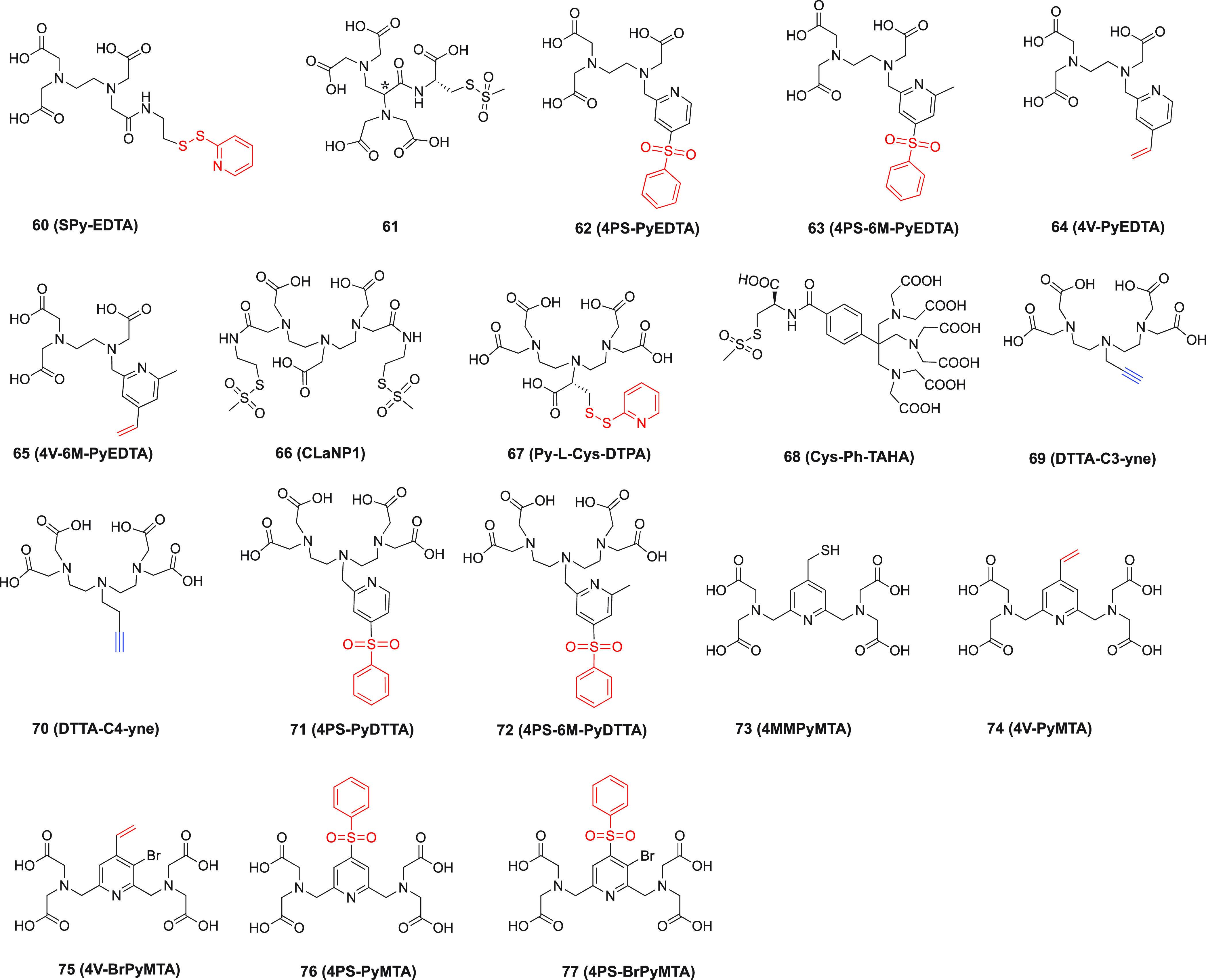
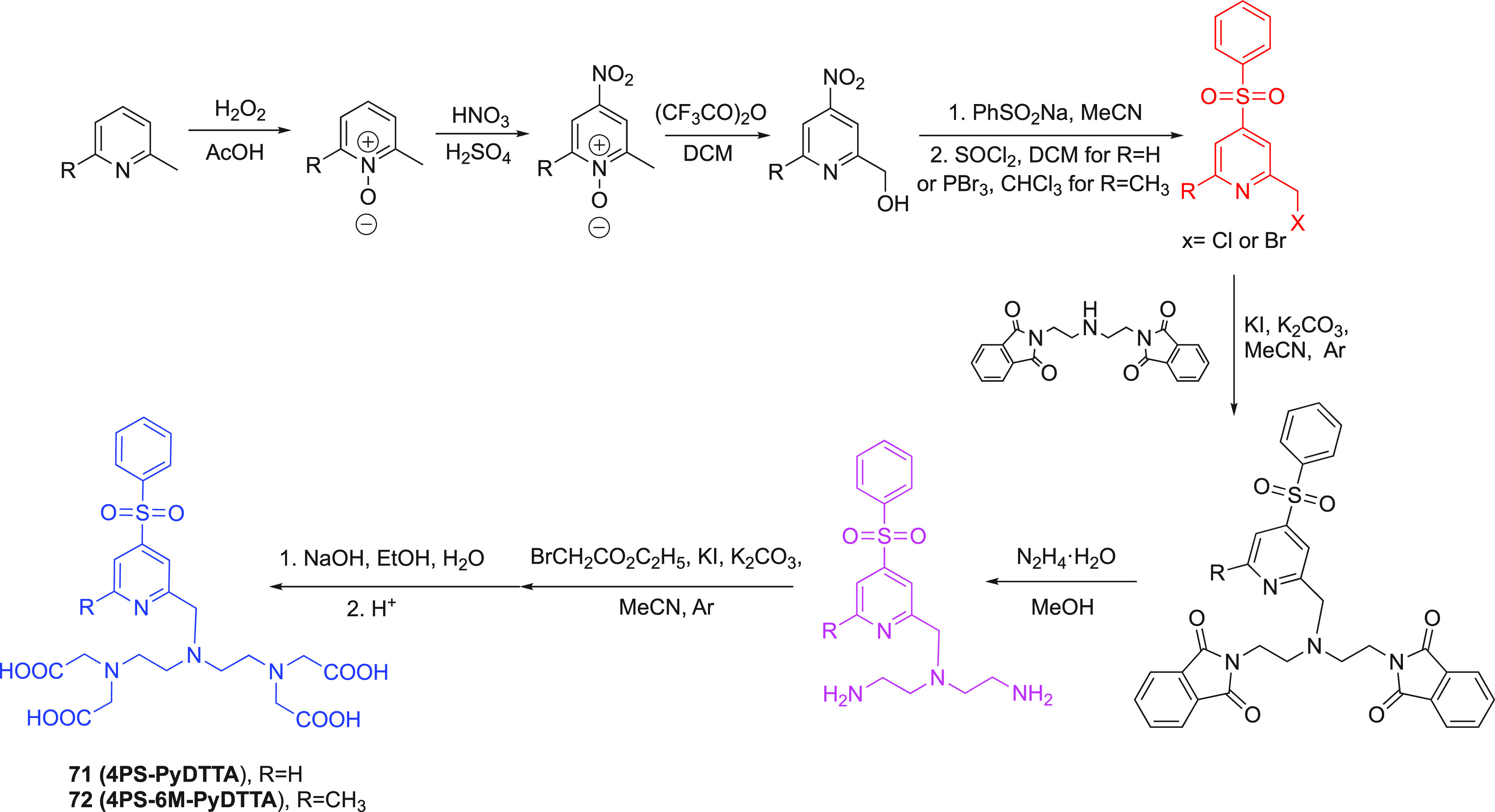
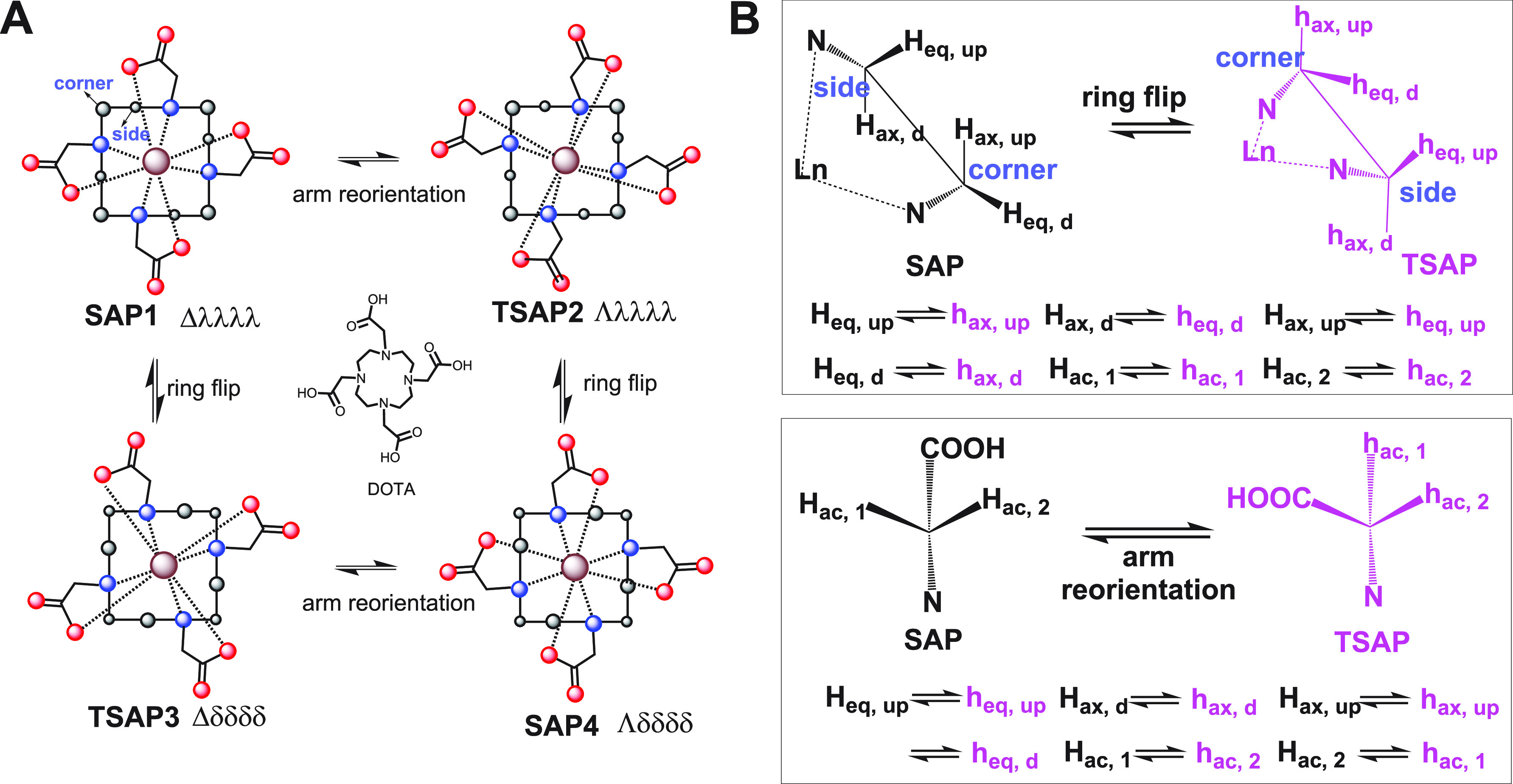
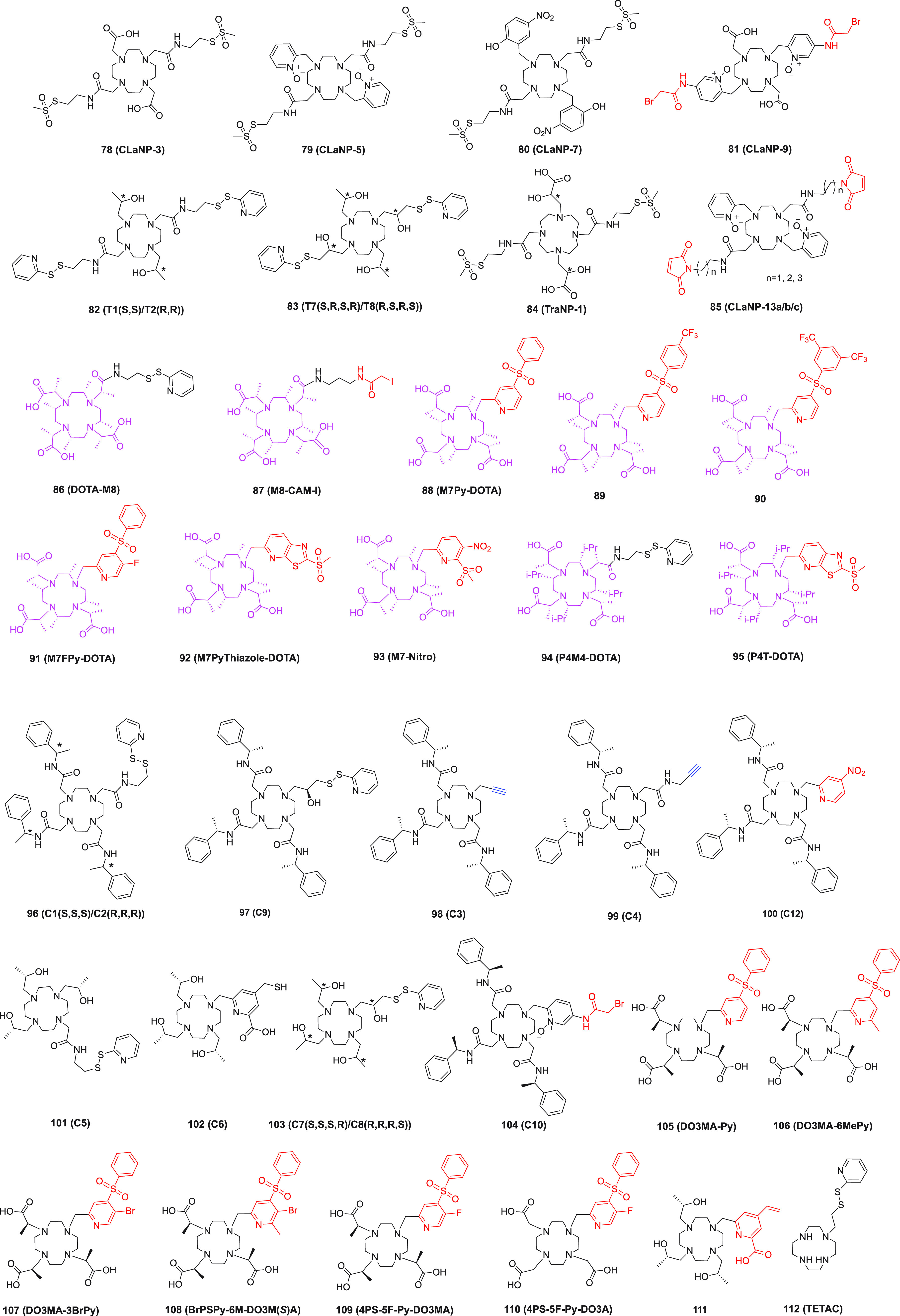
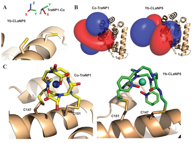
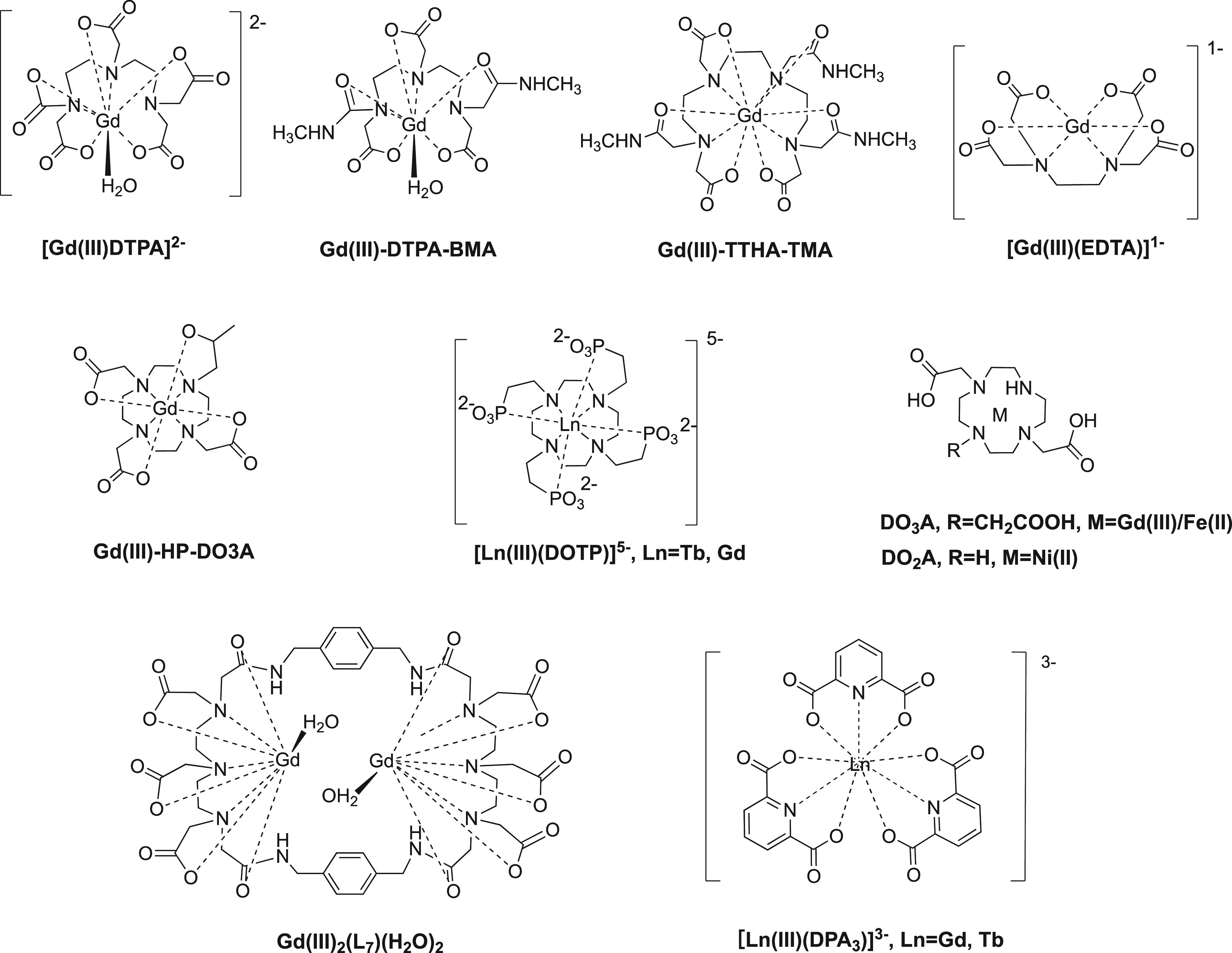
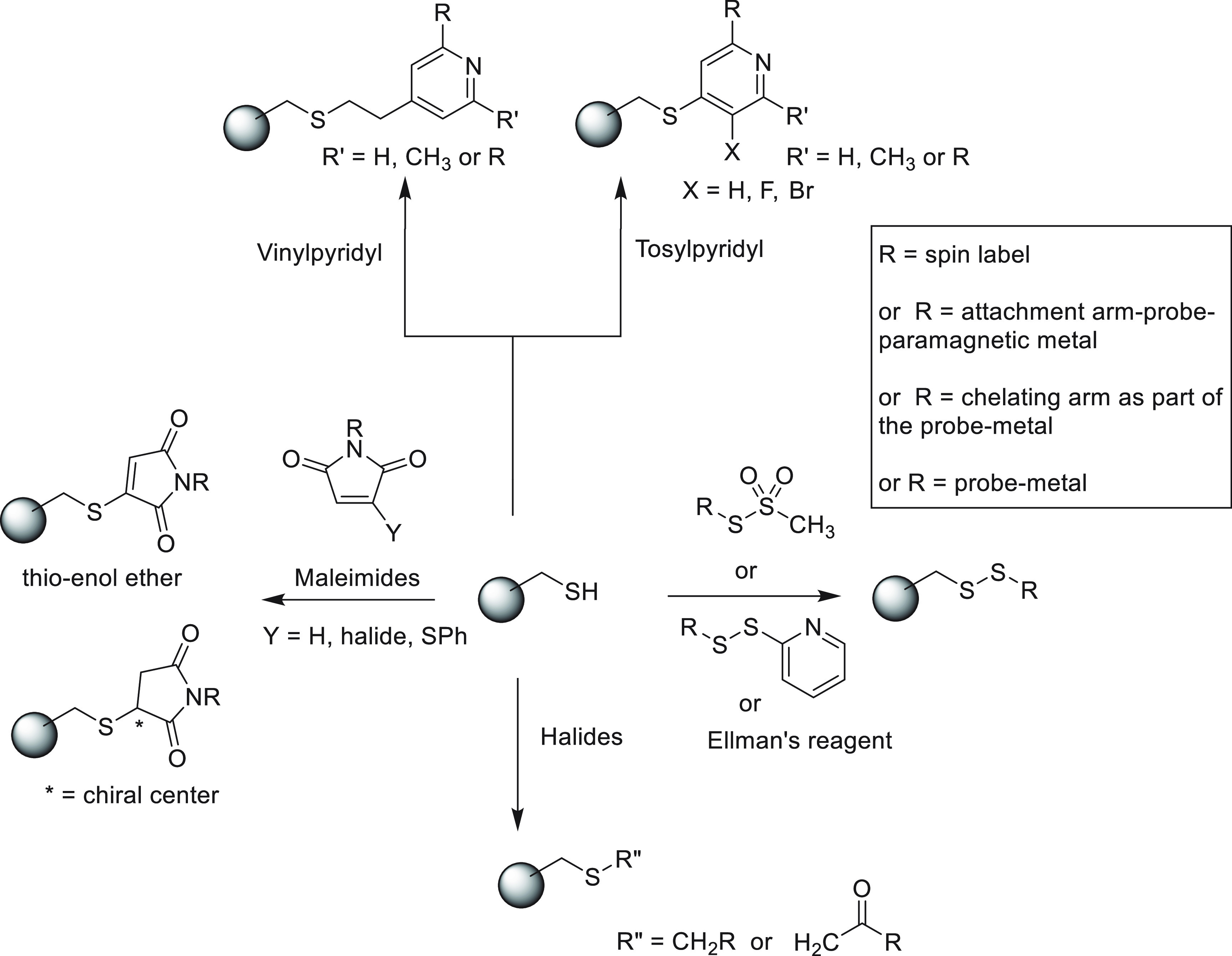
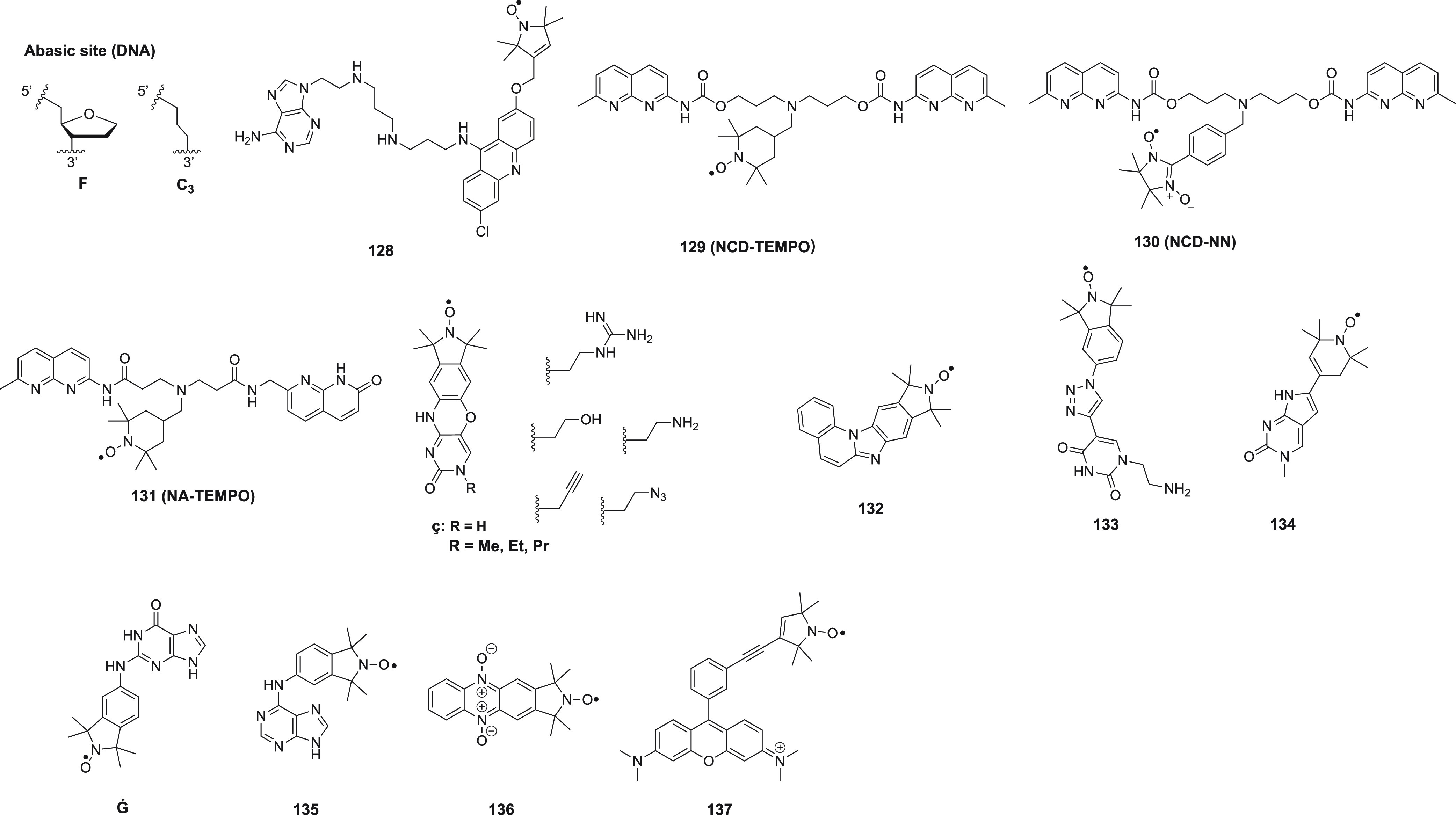
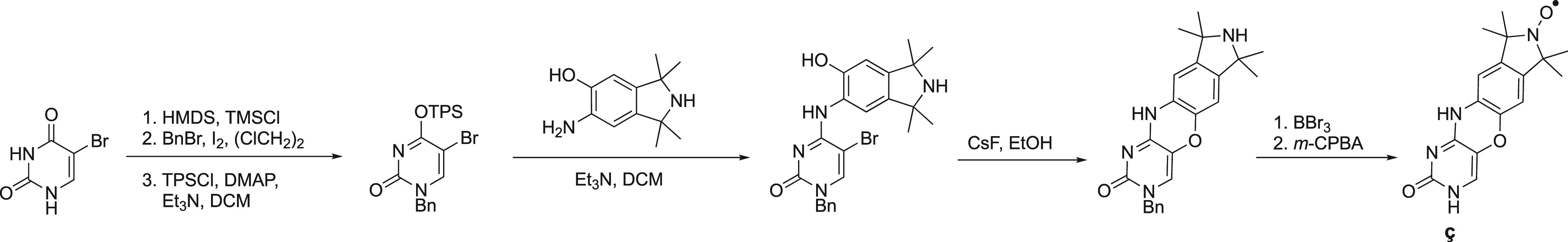
Paramagnetic chemical probes have been used in electron paramagnetic resonance (EPR) and nuclear magnetic resonance (NMR) spectroscopy for more than four decades. Recent years witnessed a great increase in the variety of probes for the study of biological macromolecules (proteins, nucleic acids, and oligosaccharides). This Review aims to provide a comprehensive overview of the existing paramagnetic chemical probes, including chemical synthetic approaches, functional properties, and selected applications. Recent developments have seen, in particular, a rapid expansion of the range of lanthanoid probes with anisotropic magnetic susceptibilities for the generation of structural restraints based on residual dipolar couplings and pseudocontact shifts in solution and solid state NMR spectroscopy, mostly for protein studies. Also many new isotropic paramagnetic probes, suitable for NMR measurements of paramagnetic relaxation enhancements, as well as EPR spectroscopic studies (in particular double resonance techniques) have been developed and employed to investigate biological macromolecules. Notwithstanding the large number of reported probes, only few have found broad application and further development of probes for dedicated applications is foreseen.


Conflict of interest statement

The authors declare no competing financial interest.

Figures

Figure 1
Figure 1
Schematic representation of the paramagnetic effects of PCS, RDC, PRE, and CCR, respectively, where CCR refers to cross-correlation between DSA and dipole–dipole relaxation. The top panel depicts the geometric dependencies relative to the frame of a χ tensor. The bottom panel illustrates the effects in the NMR spectra of paramagnetic (red) versus diamagnetic (black) samples.
Figure 2
Figure 2
Schematic diagrams of 3d block ions coordination geometries.
Figure 3
Figure 3
General structure of nitroxides. R represents an alkyl group.
Figure 4
Figure 4
Chemical structures of some natural (1), noncanonical (29, 11) amino acids, and nitroxide radicals (10, 12).
Scheme 1
Scheme 1. Synthetic Strategies for Modification of Amino Acids
Scheme 2
Scheme 2. Synthetic Routes of p-Azido-l-phenylalanine (A), Compound 4 (B),BpyAla (C), and HQA (D),
The functional groups of the ncAAs are colored in blue.
Scheme 3
Scheme 3. Two Synthetic Approaches toward Nitroxide Radicals
(a) Oxidation with m-CPBA or H2O2/cat. Na2WO2. (b) Oxidation with MnO2.
Figure 5
Figure 5
Structures of nitroxide probes. The functional groups for covalent attachment to cysteine via a thioether are highlighted in red and the functional groups for covalent attachment to a ncAA are highlighted in blue. Probe 13(207) is derived from MTSL; 14(211) and 15(212) are maleimide functionalized nitroxides; probes 16,17, and 18(391) are iodide functionalized nitroxides; probes 20,21,, and 22(224) are double-anchored nitroxides; probes 23,24, and 25(225) are azide functionalized nitroxides; probes 26,27,28,29, and 30(229) are alkyne functionalized nitroxides; probes 31(230) and 32(231) are iso(thio)cyanide functionalized nitroxides; probe 33(231) is a carbodiimide functionalized nitroxide; probe 34(232) is a hydroxylamine ether functionalized nitroxide; probe 35(207) is a nitroxide functionalized with a hydroxysuccinimide ester.
Figure 6
Figure 6
(A) Reaction of MTSL with a cysteine residue of a protein generates a flexible linkage with five rotatable bonds (red arrows). (B) Schematic representation of two different enantiomeric EDTA complexes produced by different coordination of the metal ion. M denotes the metal ion. O denotes the carboxyl groups. Curved lines represent the ethylene groups and dashed lines trace the octahedral coordination.
Scheme 4
Scheme 4. Preparation of Various Nitroxides from Tetramethylpiperidone
(A) i. NaBH4, H2O2/cat. Na2WO2 (to give 37a); ii. reductive amination with NaBH3CN (to give 37b); iii. active amine with methanesulfonyl to substitute with NaN3 (to give 37c); iv. carboxylation with tosylmethylisocyanide (to give 37d). (B) Cyclohexanone, NH4Cl, H2O2/cat. Na2WO2 (to give 38ac). (C) Raney–nickel (to give 40). (D) i. Bromination (to give 42, 43); ii. Favorskii rearrangement (to give 44, 45)., Blue colored arrows indicate the synthetic route to five-membered ring nitroxides.
Scheme 5
Scheme 5. Synthetic Route to α-Tetrasubstituted Piperidone from a Bisphosphonate Precursor
(a) i. LDA (lithium diisopropylamide) at 0 °C. ii. BuLi (butyllithium) at −35 °C. iii. excess 3-pentanone. (b) i. LDA, THF, ii. acetone. (c) i. NH4OH at 105 °C. ii. m-CPBA. (d) Na2WO4, H2O2. (e) i. TMSCHN2 ((diazomethyl)trimethylsilane), BF3·OEt2; ii. Na2WO4, H2O2..
Scheme 6
Scheme 6. Synthetic Routes Towards Isoindolinyl Nitroxide
(a) RMgBr. (b) Excess RMgBr. (c) i. Pd–C/H2; ii. m-CPBA.
Figure 7
Figure 7
Structures of aminopoly(carboxylic acid) based probes. Functional groups for covalent attachment to a cysteine residue in the target protein via thioether formation are highlighted in red. Alkyne groups for attachment to an azide group in a ncAA via “click” reaction are highlighted in blue. Probes 6065 are EDTA based. Probe 61 has two chiral centers. Probe 66(255) is double-anchored. Probes 7377 are PyMTA based. The asterisk identifies one of the chiral carbon atoms.
Scheme 7
Scheme 7. Synthetic Route to Probes 71 (4PS-PyDTTA) and 72 (4PS-6M-PyDTTA).
Figure 8
Figure 8
(A) Schematic representation of four different stereoisomers populated by DOTA lanthanoid complexes and their conformational exchange. The cyclen ring is drawn as a square with solid lines; nitrogen, oxygen, and carbon atoms are shown as blue, red, and black spheres, respectively; the metal ion is represented by a brown sphere. (B) Newman projections showing the effect of the ring flip on the positions of the hydrogen atoms of two neighboring carbon atoms. Adapted from Q. Miao (2019) Design, synthesis and application of paramagnetic NMR probes for protein structure studies. PhD Thesis, Leiden University.
Figure 9
Figure 9
Chemical structures of cyclen-based paramagnetic probes. The functional groups for attachment to cysteine via thioether formation are highlighted in red. The functional groups for attachment to an azide group in ncAA via “click” reaction are highlighted in blue. The cyclen rings with substituents are shown in magenta. 78,79,,80,81,82,83, and 84(280) are double-armed probes. Asterisks identify chiral carbon atoms.
Figure 10
Figure 10
Comparison of Co(II)-TraNP-1-SS and Yb(III)-CLaNP-5 attached to T4Lys K147C/T151C. (A) Metal positions and tensor orientations. (B) PCS iso-surfaces (±0.4 ppm for Yb(III)-CLaNP-5, ±0.2 ppm for Co(II)-TraNP-1-SS). (C) Models of the protein–probe structures. The protein is shown in ribbon representation; the probes and cysteine residues are shown as sticks and the metal ions as spheres. Reproduced without changes from ref (280). Copyright 2019 the authors, under CCA license (https://creativecommons.org/licenses/by/4.0/).
Scheme 8
Scheme 8. Three Strategies for Cyclen Ring Modulation
The magenta colored structures show the final cyclized ring with different substituents. The compounds in magenta are applied for paramagnetic probe design.
Figure 11
Figure 11
Chemical structures of small molecule probes. Functional groups designed for attachment via thioether formation are highlighted in red. 116,117,118,121,122, and 124(265) are DPA-based probes. 119 and 120 are TDA based probes. 123(176) is based on 8-hydroxyquinoline. 125(327) is an NTA based probe. 126(328) and 127(329) are IDA based probes.
Figure 12
Figure 12
Chemical structures of nitroxide based cosolute probes.
Figure 13
Figure 13
Chemical structures of metal ion based cosolute probes.
Figure 14
Figure 14
Most relevant chemical methods to connect NMR or EPR probes to one or more cysteine residues in proteins. The aim of the figure is to provide an overview of the currently used chemistry for the modification of cysteine residues, suitable for the attachment of a chemically sensitive paramagnetic center. The use of maleimide as reactive group results in the generation of a chiral center, and therefore, this type of linkage is not recommended in NMR studies.
Figure 15
Figure 15
Site-directed noncovalent spin labels for DNA and RNA.
Scheme 9
Scheme 9. Synthesis of the Noncovalent DNA Spin Label ç from 5-Bromouracil
5-Bromouracil is regioselective benzylated, subsequently converted into its O4-sulfonylated derivative (TPS = 2,4,6-triisopropylbenzenesulfonyl) and coupled to an isoindol aminophenol derivative, followed by ring-closure. The benzyl group is removed and N-oxidation gives the nitroxide.
Scheme 10
Scheme 10. Synthesis of the Noncovalent RNA Spin label Ǵ from 2-Bromohypoxanthine via a Nucleophilic Aromatic Substitution
Figure 16
Figure 16
Strategies for the attachment of probes to the phosphodiester backbone or terminal phosphate groups of oligonucleotides.
Figure 17
Figure 17
Probe attachment to (A) a 2′ amino group of internal nucleotides (blue arrows indicate the labeling reactions) and (B) alternative 5′ modifications.
Figure 18
Figure 18
Probe attachment via “click” chemistry to the 2′ position.
Figure 19
Figure 19
X-ray crystal structure (PDB code 6QJS) of a 12 base pair DNA duplex containing the spin label 24 (highlighted in pink) attached in the 2′ position via “click” chemistry. The phosphate backbone is shown as a ribbon. The distance between both nitroxide radicals determined by DEER is 30 Å.
Figure 20
Figure 20
Attachment of the triarylmethyl (TAM)-based spin label 151 to the 5′ end of DNA. The immobilized oligonucleotide is treated with CDI and piperazine and cleaved from the support using standard conditions. The resulting oligonucleotide is treated with cetyltrimethylammonium bromide (CTAB), taken up in DMSO and treated with 151 as the chloride in the presence of base. The two remaining acid chloride functions were hydrolyzed. Two alternative TAM attachments strategies are shown.
Figure 21
Figure 21
Postsynthetic probe attachment to thiopyrimidines and thiopurines.
Figure 22
Figure 22
Convertible nucleotides for postsynthetic transformation into spin labels. The 2-fluoro-hypoxanthine derivative has also been used as O-NPE protected version.
Figure 23
Figure 23
Various strategies for postsynthetic probe attachment to nucleotide bases.
Figure 24
Figure 24
Paramagnetic nucleotides for incorporation during oligonucleotide synthesis.
Figure 25
Figure 25
X-ray crystal structure (PDB code 3OT0) of a 10 base pair DNA duplex containing the Ç spin label (highlighted in pink). Two uridine residues are 2′-methylated. The phosphate backbone is shown as a ribbon. The distance between both nitroxide radicals (18 Å) is indicated. Hydrogen bonds between guanine and Ç are indicated by dashed lines.
Figure 26
Figure 26
Carbohydrate probes and their synthesis.
Figure 27
Figure 27
Strategy for using PREs, PCSs, and RDCs for structure determination of protein–protein complexes using differently labeled samples. PREs are measured using a tag with slow electronic relaxation (e.g., Gd(III)) or a paramagnetic center with little χ tensor anisotropy (e.g., Er(III)). PCSs and RDCs are obtained using the tag with an anisotropic paramagnetic center (e.g., Tm(III)). The data are measured with respect to a diamagnetic reference produced with the tag containing, for example, a Lu(III) ion. To simplify the NMR spectra and distinguish between intermolecular and intramolecular effects, the complex can be prepared with isotope labeling (symbolized by green dots) of one of the proteins only. The intramolecular restraints are used to fit the position of the paramagnetic center and orient the Δχ tensor. 2D NMR spectra (typically [15N,1H]-HSQC spectra) can be sufficient to obtain a complete set of inter- and intramolecular PREs, RDCs, and PCSs, which allow determining the relative position and orientation of the two proteins in a rigid body docking approach. Reproduced with permission from ref (30). Copyright 2014 Elsevier.
Figure 28
Figure 28
Transferred PCSs identify the binding site and binding orientation of ligand molecules that are in fast exchange with excess free ligand. (A) tPCSs (the difference between the resonance positions for the paramagnetic (para) and diamagnetic (dia) samples) observed in the 1H 1D NMR spectra of excess ligand in complex with the protein, which is tagged with either paramagnetic or diamagnetic ions (one site at a time). The dashed and solid lines identify the ligand resonances in the diamagnetic and paramagnetic samples, respectively. (B) Superposition of the averaged NOE structure (in green) of the FKBP12–1 complex and the best five structures (in orange) calculated using tPCSs. The protein backbone is represented as a gray ribbon except for the residues Asp37 and Tyr82 (yellow). The average RMSD of the ligand from PCS calculations relative to the NOE calculation is 2.8 ± 0.4 Å. Reproduced with permission from ref (620). Copyright 2013. American Chemical Society.

References

    1. Overhauser A. W. Polarization of Nuclei in Metals. Phys. Rev. 1953, 92, 411–415. 10.1103/PhysRev.92.411. - DOI
    1. Brüschweiler R.; Case D. A. Charaterization of Biomolecular Structure and Dynamics by NMR Cross Relaxation. Prog. Nucl. Magn. Reson. Spectrosc. 1994, 26, 27–58. 10.1016/0079-6565(94)80003-0. - DOI
    1. Barry C. D.; North A. C. T.; Glasel J. A.; Williams R. J. P.; Xavier A. V. Quantitative Determination of Mononucleotide Conformations in Solution Using Lanthanide Ion Shift and Broadening NMR Probes. Nature 1971, 232, 236–245. 10.1038/232236a0. - DOI - PubMed
    1. Campbell I. D.; Dobson C. M.; Williams R. J. P.; Xavier A. V. The Determination of the Structure of Proteins in Solution: Lysozyme. Ann. N. Y. Acad. Sci. 1973, 222, 163–174. 10.1111/j.1749-6632.1973.tb15259.x. - DOI - PubMed
    1. Sculimbrene B. R.; Imperiali B. Lanthanide-Binding Tags as Luminescent Probes for Studying Protein Interactions. J. Am. Chem. Soc. 2006, 128, 7346–7352. 10.1021/ja061188a. - DOI - PubMed

Publication types

MeSH terms