. 2022 Jan 27;17(1):e0262532.
doi: 10.1371/journal.pone.0262532. eCollection 2022.

Sterilized human skin graft with a dose of 25 kGy provides a privileged immune and collagen microenvironment in the adhesion of Nude mice wounds

Affiliations

Sterilized human skin graft with a dose of 25 kGy provides a privileged immune and collagen microenvironment in the adhesion of Nude mice wounds

Jurandir Tomaz de Miranda et al. PLoS One. .

Abstract

This study aimed to report the effects of different doses of ionizing radiation on inflammatory and repair stage of human skin graft adherence in Nude mice wounds. Animals were divided into transplanted with irradiated human skin grafts (IHSG) at 25 and 50 kGy (IHSG 25 kGy; IHSG 50 kGy) and non-IHSG and euthanized on the 3rd, 7th and 21st days after the surgery, by gross and microscopic changes, immunostaining for human type I collagen (Col I) and mouse Col I and Col III and inflammatory cells. We found an effectiveness of human split-thickness graft adherence in mice transplanted with IHSG 25 kGy, as well decrease in dermo-epidermal necrosis and neutrophils, lower loss of skin thickness, epithelization and neo-vascularization. Day 21 post-transplantation with IHSG 25 kGy was observed a well-preserved human skin in the border of the graft, a prominent granulation tissue in an organization by proliferated fibroblasts, Col III deposition and increased B-cells and macrophages. A complete adherence of human skin graft occurred with IHSG 25 kGy. We suggest that the ionizing radiation at 25 kGy mediates inflammation and the repair stage of human skin graft adherence in murine model, thus emerging as a potential tool in healing cutaneous wounds.


Conflict of interest statement

The authors have declared that no competing interests exist.

Figures

Fig 1
Fig 1. Macroscopic evolution of the human skin graft in Nude mouse over time.
Human skin graft control (non-irradiation), group 3 (A), 7 (B), 21 (C) and 90 (D) days after surgery, (B) tissue was occupied by an extensive area of necrosis and haemorrhage. Human skin graft with dose 25 kGy group 3 (E), 7 (F), 21 (G) and 90 (H), in (G) only small central crusted island observed, (H) greater tissue contraction. Human skin graft with dose 50 kGy group 3 (I), 7 (J), 21 (K) and 90 (L), the graft was retracted and pale (J) the border was occupied by an extensive area of necrosis and haemorrhage, (K) central crusted island remains. All grafts show complete regeneration in 90 days (D), (H), (L).
Fig 2
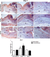
Fig 2. Histological analysis of irradiated and non-irradiated human skin grafts, 3 days after grafting.
(A-C) non-irradiated group. Human skin graft (Hu), mouse skin (Mo). Dermoepidermal necrosis, neutrophil exudate (Ne, A). Total graft thickness (dotted arrow, B). Human skin collagen (**, C). (D-F) group irradiated 25 kGy. Dermoepidermal necrosis, neutrophil exudate (Ne, D, E), reepithelialization, macrophage infiltration (Ma, E, F), collagen, fibroblasts, blood vessels (Col, FI, Ve, E-F), of the animal. (G-I) group irradiated 50 kGy. Dermoepidermal necrosis (Ne, G), macrophage infiltration (Ma, H, I), collagen degradation (Col I). (J) statistical differences between inflammation and repair process in mice grafted with IHSG 25 kGy. Dermoepidermal necrosis, neutrophils, graft thickness decreased compared with non-irradiated group. Epithelialization, vascularization at 25 kGy increased compared to other groups. (***p<0.0001). Original magnification: 100X (A, D, G); 200X (B, E, H); 400X (C, F, I).
Fig 3
Fig 3. Histological analysis of irradiated and non-irradiated human skin grafts, 7 days after grafting.
(A-C) non-irradiated group. Human skin graft (Hu), mouse skin (Mo), granulation tissue filling (GrT), macrophage infiltration (Ma), collagen (Col). (D-F) group irradiated 25 kGy. Macrophage infiltration (Ma), filling of granulation tissue (GrT) that progresses from the mouse dermis towards the 25 kGy IHSG graft compared to other groups. Reepithelization and collagen deposition (Col). (G-I) group irradiated 50 kGy. filling of granulation tissue (GrT), infiltration of macrophages (Ma), collagen (Col). (J) statistical differences between the process of epithelial repair in the matrix with increased macrophages and revascularization in skin wounds of mice transplanted with IHSG 25 kGy (***p<0.0001). Original magnification: 100X (A, D, G); 200X (B, E, H); 400X (C, F, I).
Fig 4
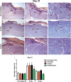
Fig 4. Histological analysis of irradiated and non-irradiated human skin grafts, 21 days after grafting.
(A-C) non-irradiated group. Human skin graft (Hu), mouse skin (Mo), granulation tissue filling (GrT), blood vessels (Ve). (D-F) group irradiated 25 kGy. Human skin (Hu, D) detached fragment covering the center of the healing area, large blood vessels (Ve, E-F), granulation tissue (GrT, D-F) organized by proliferated fibroblasts and deposition of fine collagen fibers (Col, E-F). (G-I) group irradiated 50 kGy. Human skin graft (Hu), mouse skin (Mo) still in the healing process. (J) Statistical differences between the reepithelialization process and granulation tissue, fibroblasts, collagen in the epithelial and matrix repair process in mice grafted with IHSG 25 kGy (***p<0.0001). Original magnification: 100X (A, D, G); 200X (B, E, H); 400X (C, F, I).
Fig 5
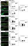
Fig 5. Characterization by immunofluorescence of the distribution of collagen fibers in dermis, 21 days after grafting.
Human type I collagen (A) greater statistical significance in the center and border of the non-IHSG group compared to irradiated groups, this difference being more pronounced 25 kGy IHSG tissues. Mouse type III collagen (B) greater statistical significance in the center and border of the IHSG 25 kGy group compared to non-IHSG and IHSG 50kGy. Mouse type I collagen (C), similar distribution in the three groups. Graphs showing significant differences between the repair process of the collagen matrix in skin wounds of grafted mice (A, B; *p<0.01; ***p<0.0001). Original magnification: 100X (A, B, C).
Fig 6
Fig 6. Distribution of B cells (CD20 +) and macrophages (CD68 +), 21 days after grafting.
Tissue non-IHSG and IHSG 25 kGy and 50 kGy. Increased density of B-cell lymphocytes (CD20 +, arrow, A) and macrophages (CD68 +, arrow, B) did not show statistical significance, but a trend in mice from the IHSG 25 kGy group compared to the other groups. Original magnification: 400X (A, B); Lower figures: Zoom of the dashed region in A and B.

References

    1. Souto EB, Ribeiro AF, Ferreira MI, Teixeira MC, Shimojo AAM, Soriano JL, et al.. New Nanotechnologies for the Treatment and Repair of Skin Burns Infections. Int J Mol Sci. 2020;21(2): 393. doi: 10.3390/ijms21020393 - DOI - PMC - PubMed
    1. Tuğcu H, Zor F, Toygar M, Balandız H. Comparison of antemortem clinical diagnosis and postmortem findings in burn-related deaths. Ulus Travma Acil Cerrahi Derg. 2015. 21(6): 491–5. doi: 10.5505/tjtes.2015.36604 - DOI - PubMed
    1. Finnerty CC, Jeschke MG, Branski LK, Barret JP, Dziewulski P, Herndon DN. Hypertrophic scarring: the greatest unmet challenge after burn injury. Lancet. 2016;388(10052): 1427–36. doi: 10.1016/S0140-6736(16)31406-4 - DOI - PMC - PubMed
    1. Lobmann R, Ambrosch A, Schultz G, Waldmann K, Schiweck S, Lehnert H. Expression of matrix-metalloproteinases and their inhibitors in the wounds of diabetic and non-diabetic patients. Diabetologia. 2002;45(7): 1011–6. doi: 10.1007/s00125-002-0868-8 - DOI - PubMed
    1. Kellar RS, Diller RB, Tabor AJ, Dominguez DD, Audet RG, Bardsley TA, et al.. Improved wound closure rates and mechanical properties resembling native skin in murine diabetic wounds treated with a tropoelastin and collagen wound healing device. J diabetes Clin Res. 2020;2(3): 86–99. doi: 10.33696/diabetes.1.024 - DOI - PMC - PubMed

Publication types

Substances