Review
. 2022 Feb 23;14(2):10.1088/1758-5090/ac4fb5.
doi: 10.1088/1758-5090/ac4fb5.

Bioprinted microvasculature: progressing from structure to function

Affiliations
Review

Bioprinted microvasculature: progressing from structure to function

Alexis J Seymour et al. Biofabrication. .

Abstract

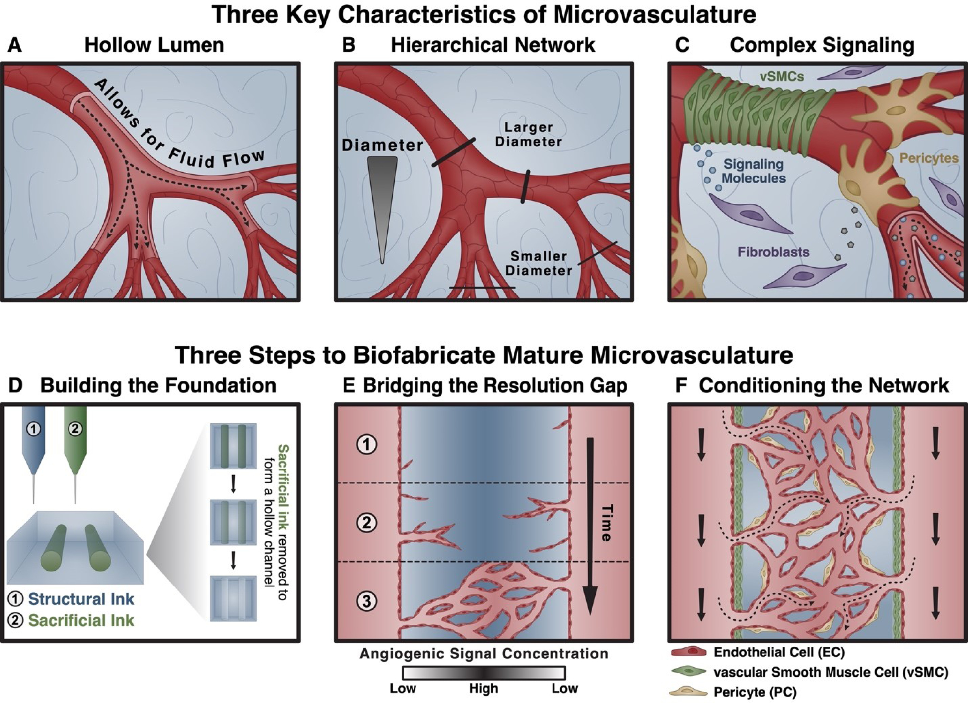
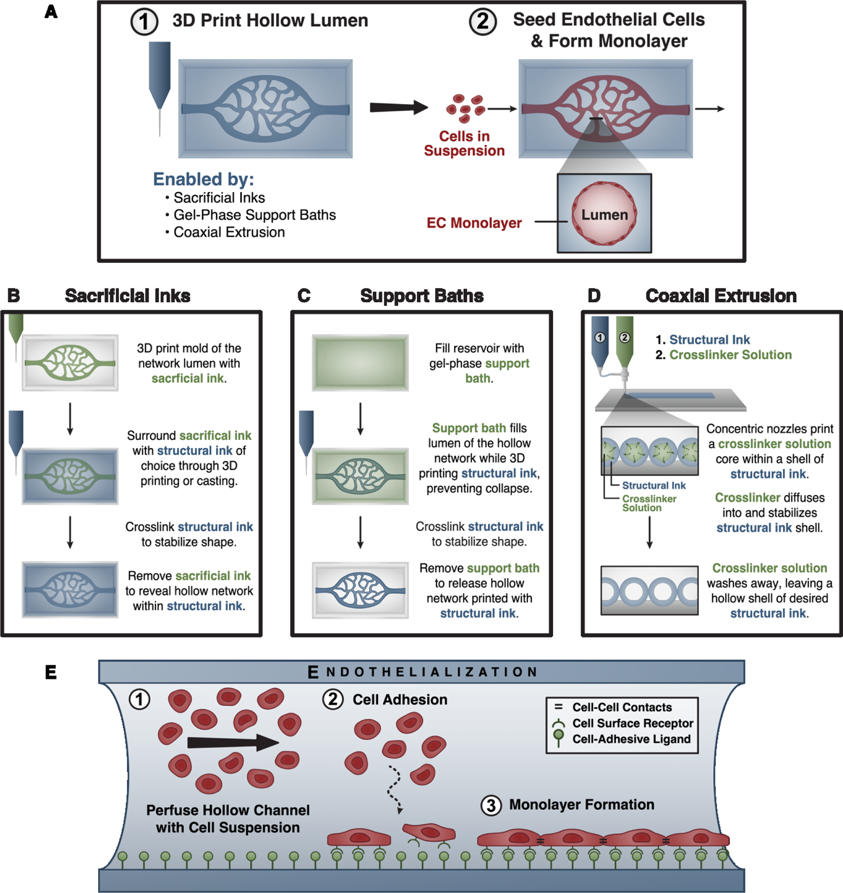
Three-dimensional (3D) bioprinting seeks to unlock the rapid generation of complex tissue constructs, but long-standing challenges with efficientin vitromicrovascularization must be solved before this can become a reality. Microvasculature is particularly challenging to biofabricate due to the presence of a hollow lumen, a hierarchically branched network topology, and a complex signaling milieu. All of these characteristics are required for proper microvascular-and, thus, tissue-function. While several techniques have been developed to address distinct portions of this microvascularization challenge, no single approach is capable of simultaneously recreating all three microvascular characteristics. In this review, we present a three-part framework that proposes integration of existing techniques to generate mature microvascular constructs. First, extrusion-based 3D bioprinting creates a mesoscale foundation of hollow, endothelialized channels. Second, biochemical and biophysical cues induce endothelial sprouting to create a capillary-mimetic network. Third, the construct is conditioned to enhance network maturity. Across all three of these stages, we highlight the potential for extrusion-based bioprinting to become a central technique for engineering hierarchical microvasculature. We envision that the successful biofabrication of functionally engineered microvasculature will address a critical need in tissue engineering, and propel further advances in regenerative medicine andex vivohuman tissue modeling.

Keywords: 3D printing; endothelial sprouting; extrusion-based bioprinting; microvasculature; vascular function; vascular structure.


Figures

Figure 1.
Figure 1.. Biofabricating functional microvasculature in three steps.
At a high level, three characteristics of microvasculature are both crucial for function and difficult to biofabricate using any one technique alone: (A) the presence of a hollow lumen that enables fluid (blood) flow; (B) a hierarchically branched network topology; and (C) a complex signaling environment composed of biophysical (fluid flow, extracellular matrix mechanics) and biochemical (diverse cell types, signaling molecules, matrix ligands) cues for tissue homeostasis. We propose overcoming this challenge using a three-step approach that leverages existing techniques. (D) First, a foundational, hollow network structure is fabricated using extrusion-based bioprinting. (E) Second, proangiogenic signaling is harnessed to induce endothelial cell sprouting and the formation of a capillary-scale network. (F) Third, the network is matured using signals known to condition and stabilize nascent vascular networks, including the introduction of supporting cell types (vascular smooth muscle cells, pericytes).
Figure 2.
Figure 2.. Building the foundation using extrusion-based bioprinting.
(A, panel 1) Several 3D bioprinting techniques enable the fabrication of hollow networks by using materials or crosslinker solutions that fill and/or physically support the hollow lumen being created. (A, panel 2) Because these techniques often preclude the direct deposition of endothelial cell-laden inks, hollow channels are endothelialized – or seeded with endothelial cells – after fabrication. (B) Sacrificial inks are used to create a mold of the desired hollow space, which physically supports the surrounding ink as it is printed or cast around it. After crosslinking the surrounding ink, the sacrificial ink is removed to form a hollow lumen. (C) Gel-phase support baths similarly fill and physically support a hollow structure during printing. These materials locally fluidize around the printing nozzle as it passes, and rapidly self-heal to confine the deposited ink. After crosslinking the biomaterial ink, the support bath is removed. (D) Coaxial extrusion systems allow for direct printing of hollow channels by extruding both a biomaterial ink and its crosslinker from concentric nozzles to crosslink the material in situ. (E) During endothelialization, the 3D printed lumen is perfused with a suspension of endothelial cells. The cells are allowed to adhere to the construct surface, which is crucially dependent upon factors such as the presentation of cell-adhesive ligands. After removing unattached cells, the construct is cultured to allow endothelial monolayer formation.
Figure 3.
Figure 3.. Biomaterials and biofabrication approaches to bridge the critical resolution gap through induced spouting.
Angiogenic sprouting progresses through well-orchestrated stages. (A) First, local endothelial cells become activated and initiate sprouting in response to an angiogenic signal. In this first step, endothelial basement membrane is degraded, endothelial junctions weaken, and a tip cell is formed. (B) Next, the liberated tip cell directionally migrates toward the angiogenic signal, followed by a leaky nascent vessel composed of proliferating stalk cells. (C) Finally, after sprout fusion, perfusion helps to restore a quiescent phalanx endothelial phenotype, basement membrane is deposited, and mural cells (such as pericytes, shown) mature and stabilize the vessel. (D) A host of biochemical and biophysical signals are known to direct angiogenic sprouting, including proangiogenic growth factor signaling, hypoxia, the presentation of cell-adhesive ligands, matrix stiffness, matrix density, and matrix degradation. Biomaterials and biofabrication-based approaches have been used to spatiotemporally control angioinductive cues (E-G). (E) Photochemistry allows the user to tether one or more bioactive molecules in complex spatial patterns within biomaterial constructs, with the ability to further control release. (F) Microfluidic channels are commonly used to create stable gradients of soluble signaling molecules. (G) Meanwhile, bioprinting may be used for the patterned deposition of multiple materials with different matrix stiffnesses or densities, tethered ligands or growth factors, or degradation profiles.
Figure 4.
Figure 4.. Conditioning microvascular constructs to promote stability and maturation.
Several methods exist for conditioning microvascular networks, including: (A) the use of co-culture systems that mirror the heterotypic cell-cell signaling inherent within the vascular niche, (B) heterogeneous substrate cues, and (C) the use of fluid flow to mechanically condition the network. Topographic patterns are one aspect of substrate heterogeneity that can be fabricated through a variety of methods (D-F). (D) Soft lithography can be used to make simple gratings, hierarchical gratings, convex microlenses, concave microlenses, pillars, and holes at the micro- and nanoscale. (E) Electrospinning can create nanofibers aligned parallel or perpendicular to the seeded cells, or not aligned at all. (F) Based on print resolution, the printed construct can contain grooves and surface features that influence cell alignment, migration, and adhesion similarly to the patterns formed in (D) and (E).

References

    1. Shin Y, Choi SH, Kim E, Bylykbashi E, Kim JA, Chung S, Kim DY, Kamm RD and Tanzi RE Blood–Brain Barrier Dysfunction in a 3D In Vitro Model of Alzheimer’s Disease. Adv. Sci. 6, 1900962 (2019). doi:10.1002/advs.201900962 - DOI - PMC - PubMed
    1. Gertler FB, Condeelis JS, Zervantonakis IK, Charest JL, Kamm RD and Hughes-Alford SK Three-dimensional microfluidic model for tumor cell intravasation and endothelial barrier function. Proc. Natl. Acad. Sci. 109, 13515–13520 (2012). doi:10.1073/pnas.1210182109 - DOI - PMC - PubMed
    1. Wang YI, Abaci HE and Shuler ML Microfluidic blood–brain barrier model provides in vivo-like barrier properties for drug permeability screening. Biotechnol. Bioeng. 114, 184–194 (2017). doi:10.1002/bit.26045 - DOI - PMC - PubMed
    1. Mirabella T, Macarthur JW, Cheng D, Ozaki CK, Woo YJ, Yang MT and Chen CS 3D-printed vascular networks direct therapeutic angiogenesis in ischaemia. Nat. Biomed. Eng. 1, (2017). doi:10.1038/s41551-017-0083 - DOI - PMC - PubMed
    1. Fitzsimmons RE, Aquilino MS, Quigley J, Chebotarev O, Tarlan F and Simmons CA Generating vascular channels within hydrogel constructs using an economical open-source 3D bioprinter and thermoreversible gels. Bioprinting 9, 7–18 (2018). doi:10.1016/j.bprint.2018.02.001 - DOI

Publication types

LinkOut - more resources