Review
. 2023 Aug;29(4):488-505.
doi: 10.1177/10738584211069071. Epub 2022 Jan 27.

Prefrontal Cortical Control of Anxiety: Recent Advances

Affiliations
Review

Prefrontal Cortical Control of Anxiety: Recent Advances

Nancy R Mack et al. Neuroscientist. 2023 Aug.

Abstract

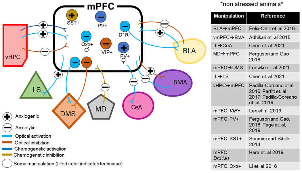
Dysfunction in the prefrontal cortex is commonly implicated in anxiety disorders, but the mechanisms remain unclear. Approach-avoidance conflict tasks have been extensively used in animal research to better understand how changes in neural activity within the prefrontal cortex contribute to avoidance behaviors, which are believed to play a major role in the maintenance of anxiety disorders. In this article, we first review studies utilizing in vivo electrophysiology to reveal the relationship between changes in neural activity and avoidance behavior in rodents. We then review recent studies that take advantage of optical and genetic techniques to test the unique contribution of specific prefrontal cortex circuits and cell types to the control of anxiety-related avoidance behaviors. This new body of work reveals that behavior during approach-avoidance conflict is dynamically modulated by individual cell types, distinct neural pathways, and specific oscillatory frequencies. The integration of these different pathways, particularly as mediated by interactions between excitatory and inhibitory neurons, represents an exciting opportunity for the future of understanding anxiety.

Keywords: anxiety; avoidance; interneurons; oscillations; prefrontal cortex; rodent.


Conflict of interest statement

Conflict of Interest

The authors declare no competing financial interests.

Figures

Figure 1.
Figure 1.
Organization of the rodent medial prefrontal cortex (mPFC). Schematic depicting a coronal section of the mouse brain and the four major subdivisions typically referred to when using the term ‘mPFC’ – secondary motor cortex (M2), anterior cingulate cortex (ACC), prelimbic cortex (PL) and infralimbic cortex (IL). In this review paper, a majority of the papers cited focused exclusively on the PL and IL subregions. The PL and IL are laminarly organized, with layers 1, 2/3, 5 and 6. Rodents lack a prominent layer 4 compared to human and non-human primates. Created with BioRender.com
Figure 2.
Figure 2.
Differential modulation of pyramidal cell (PC) activity by two types of interneurons. An AP burst in PC (grey) causes both synchronous and asynchronous release (AR) of glutamate at synapses onto SST+ Martinotti cell (MC, green). Synchronous release produces facilitating EPSCs/EPSPs and thus delayed AP firing in MC, while the occurrence of delayed AR desynchronizes and prolongs MC firing, resulting in imprecise and long-lasting inhibition in neighboring PCs (blue). This late-onset slow recurrent inhibition would decrease and desynchronize PC firing for a long period of time, presumably causing a reduction of LFP power (green box). In contrast, the PC burst only causes synchronous release at synapses onto PV+ fast-spiking cells (FS, orange). Because of the strong short-term depression, the initial EPSC/EPSP is normally large in size, resulting in a transient discharge in FS cell and thus early-onset inhibition in neighboring PCs. The sudden presence and withdrawal of this fast recurrent inhibition may enhance the synchronization of downstream PCs, thus increasing the LFP power (orange box). Grey shadows indicate the time window of AP burst of PC1. The figure was modified with permission from Deng and others 2020.
Figure 3.
Figure 3.
Membrane potential-dependent modulation of recurrent inhibition. A. AP burst (15 APs at 100 Hz) in PC1 induced low-threshold spiking (LTS) cell-mediated slow disynaptic inhibition in PC2. LTS cells are putative SST+ interneurons. * indicates individual IPSPs. Arrow indicates the onset of AP burst in PC1. B. Example PC-PC paired recording showing that the amplitude of slow disynaptic IPSPs was associated with presynaptic Vm levels (color-coded: red represents Vm near the AP threshold, blue represents Vm at the resting membrane potential). Group data show that the peak amplitude of IPSPs is positively correlated with the presynaptic membrane potential depolarization (bottom right). C. Bath application of a KV1 channel blocker α-dendrotoxin (α-DTX, 100 nM) mimicked the depolarization-induced increases of slow disynaptic IPSPs (left, blue) and occluded the increased IPSCs induced by PC depolarization (left, red). Group data indicate no significant additional increases in the amplitude of disynaptic IPSPs after α-DTX application (right). D. The amplitude of fast disynaptic IPSC mediated by FS also depended on presynaptic Vm levels. This figure was modified with permission from Zhu and others 2011.
Figure 4.
Figure 4.
In Vivo neural recordings reveal distinct changes in the mPFC during approach-avoidance conflict. A) vHPC-mPFC theta synchrony is significantly increased in the open field and elevated plus maze (EPM) relative to a familiar environment. B) The change in vHPC-mPFC theta synchrony during risk assessment depends on whether the animals’ future action is to approach (green) or avoid (red). C) Fold increase in mPFC theta power compared to a familiar environment is significantly increased in the more anxiogenic regions (periphery of open field and open arms of elevated plus maze) relative to the other zones. D) mPFC theta power during exposure to the open field or EPM is significantly greater in 5-HT1A KO mice relative to wild-type control mice. E) mPFC single units have task-related firing patterns in the EPM. Upper panel: The gray represents the animals’ track in the EPM, and the green represents the spatial distribution of single units that preferentially fired in the closed arms (left), open arms (middle), or center (right) of the EPM. The lower panel is the spatial firing map of the same single units. Part A, C, and D were modified with permission from Adhikari and others 2010, Elsevier. Part B was modified with permission from Jacinto and others, 2016. Part E was modified with permission from Adhikari and others 2011, Elsevier. Panels A-D were created with BioRender.com.
Figure 5.
Figure 5.
Circuits and cell types of the rodent medial prefrontal cortex play a causal role in behavior during approach-avoidance conflict. BLA – basal lateral amygdala; BMA – basomedial amygdala; CeA – central amygdala; D1R – dopamine receptor subtype 1; DMS – dorsal medial striatum LS – lateral septum; MD – mediodorsal thalamus; mPFC – medial prefrontal cortex; Oxtr – oxytocin receptor; PV – parvalbumin; SST – somatostatin; vHPC – ventral hippocampus; VIP – vasointestinal peptide. Anxiogenic, or producing anxiety, is used in reference to studies where manipulations increased avoidance behaviors whereas anxiolytic, or reducing anxiety, is used in reference to studies where manipulations decreased avoidance behaviors.
Figure 6.
Figure 6.
Hypothetical model supported by the recent literature, where strong theta frequency inputs from ventral hippocampal (vHPC) recruits VIP interneurons in the rodent medial prefrontal cortex (mPFC). The activation of VIP interneurons creates a disinhibitory effect within the mPFC microcircuit, leading to pyramidal cell (PC) activation. Synchronous PC firing leads to robust PV interneuron activation, and in return, PCs may receive feedback inhibition from PV+ interneurons to act as a rhythm generator in theta frequencies. The internal theta rhythm generator may reactivate SST+ interneurons, resulting in persistent and desynchronized lateral inhibition to non-task-related PCs.

Similar articles

Cited by

References

    1. Abbas AI, Sundiang MJM, Henoch B, Morton MP, Bolkan SS, Park AJ, Harris AZ, Kellendonk C, Gordon JA (2018) Somatostatin Interneurons Facilitate Hippocampal-Prefrontal Synchrony and Prefrontal Spatial Encoding. Neuron 100:926–939. - PMC - PubMed
    1. Adhikari A, Topiwala MA, Gordon JA (2010) Synchronized activity between the ventral hippocampus and the medial prefrontal cortex during anxiety. Neuron 65:257–269. - PMC - PubMed
    1. Adhikari A, Topiwala MA, Gordon JA (2011) Single units in the medial prefrontal cortex with anxiety-related firing patterns are preferentially influenced by ventral hippocampal activity. Neuron 71:898–910. - PMC - PubMed
    1. Adhikari A, Lerner TN, Finkelstein J, Pak S, Jennings JH, Davidson TJ, Ferenczi E, Gunaydin LA, Mirzabekov JJ, Ye L, Kim SY, Lei A, Deisseroth K (2015) Basomedial amygdala mediates top-down control of anxiety and fear. Nature 527:179–185. - PMC - PubMed
    1. Albert PR, Vahid-Ansari F, Luckhart C (2014) Serotonin-prefrontal cortical circuitry in anxiety and depression phenotypes: pivotal role of pre- and post-synaptic 5-HT1A receptor expression. Front Behav Neurosci 8:199. - PMC - PubMed

Publication types