. 2022 Sep 29;60(3):2003697.
doi: 10.1183/13993003.03697-2020. Print 2022 Sep.

The methyl-CpG-binding domain 2 facilitates pulmonary fibrosis by orchestrating fibroblast to myofibroblast differentiation

Affiliations

The methyl-CpG-binding domain 2 facilitates pulmonary fibrosis by orchestrating fibroblast to myofibroblast differentiation

Yi Wang et al. Eur Respir J. .

Abstract

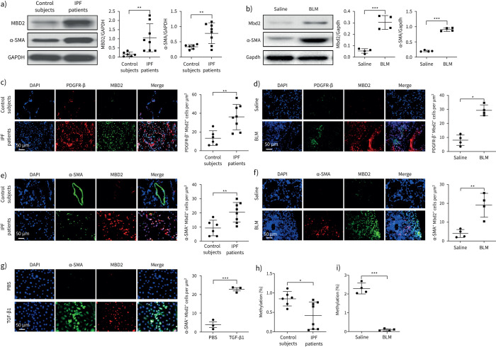
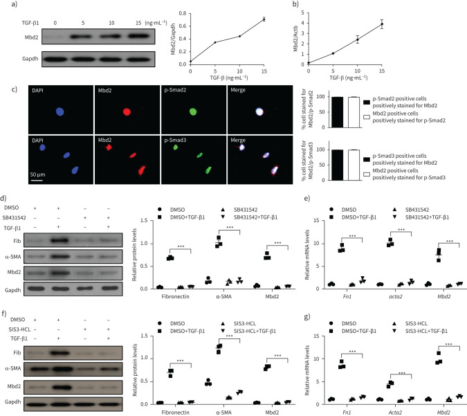
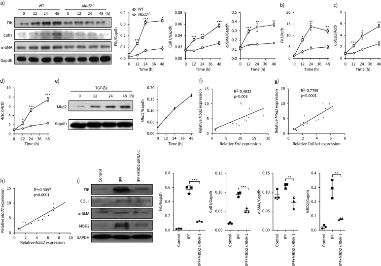
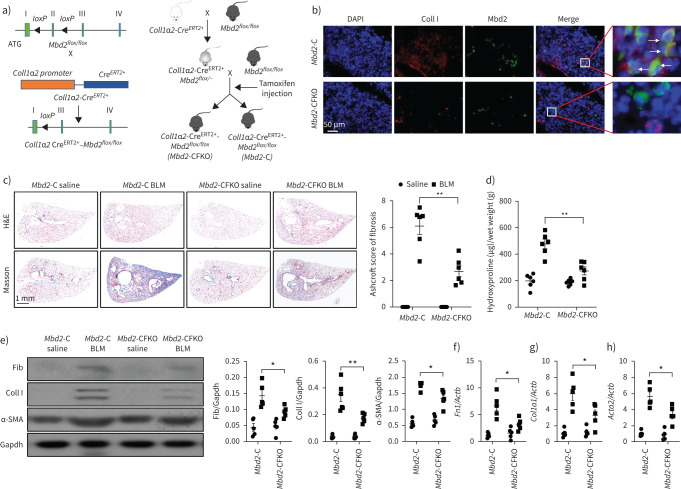
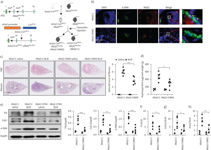
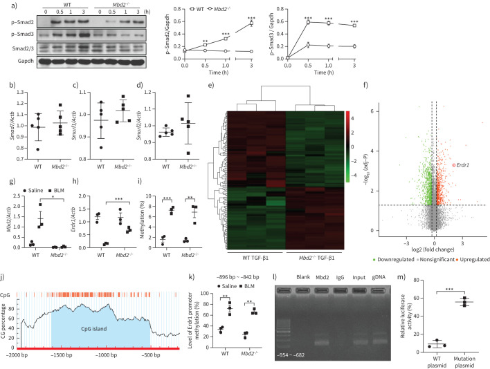
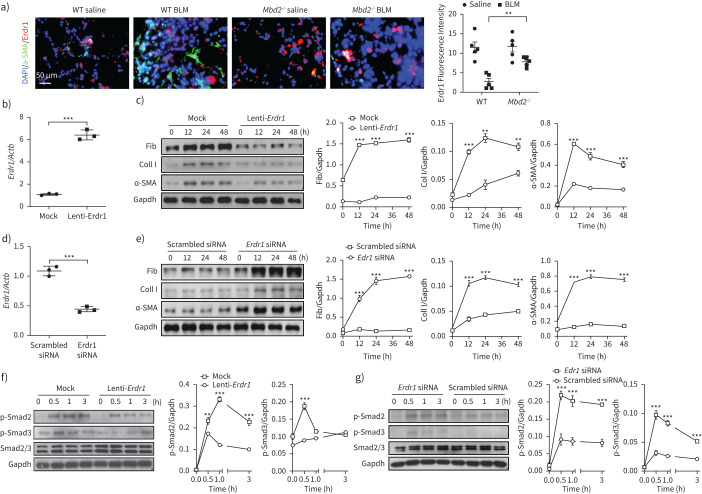
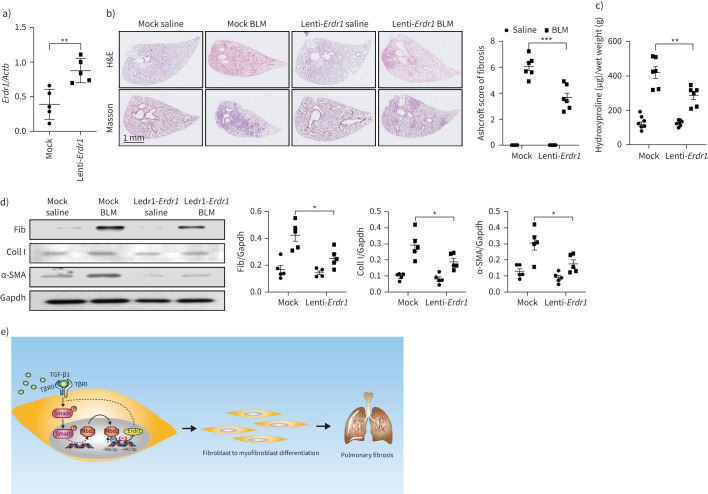
Although DNA methylation has been recognised in the pathogenesis of idiopathic pulmonary fibrosis (IPF), the exact mechanisms are yet to be fully addressed. Herein, we demonstrate that lungs originated from IPF patients and mice after bleomycin (BLM)-induced pulmonary fibrosis are characterised by altered DNA methylation along with overexpression in myofibroblasts of methyl-CpG-binding domain 2 (MBD2), a reader responsible for interpreting DNA methylome-encoded information. Specifically, depletion of Mbd2 in fibroblasts or myofibroblasts protected mice from BLM-induced pulmonary fibrosis coupled with a significant reduction of fibroblast differentiation. Mechanistically, transforming growth factor (TGF)-β1 induced a positive feedback regulatory loop between TGF-β receptor I (TβRI), Smad3 and Mbd2, and erythroid differentiation regulator 1 (Erdr1). TGF-β1 induced fibroblasts to undergo a global DNA hypermethylation along with Mbd2 overexpression in a TβRI/Smad3 dependent manner, and Mbd2 selectively bound to the methylated CpG DNA within the Erdr1 promoter to repress its expression, through which it enhanced TGF-β/Smad signalling to promote differentiation of fibroblast into myofibroblast and exacerbate pulmonary fibrosis. Therefore, enhancing Erdr1 expression strikingly reversed established pulmonary fibrosis. Collectively, our data support that strategies aimed at silencing Mbd2 or increasing Erdr1 could be viable therapeutic approaches for prevention and treatment of pulmonary fibrosis in clinical settings.


Conflict of interest statement

Conflict of interest: The authors declare no competing financial interests.

Figures

FIGURE 1
FIGURE 1
Analysis of methyl-CpG-binding domain 2 (MBD2) expression and DNA methylation in idiopathic pulmonary fibrosis (IPF) patients and mice with bleomycin (BLM) induction. a) Western blot analysis of MBD2 and α-smooth muscle actin (SMA) expression in the lungs of control subjects and IPF patients. b) Western blot analysis of Mbd2 and α-SMA expression in the lungs of mice following 21 days of BLM induction. c, d) Representative results for co-immunostaining of MBD2 and platelet-derived growth factor receptor (PDGFR)-β in lung sections from patients: c) control subjects and IPF patients and d) BLM-induced lung sections. e–g) Results for co-immunostaining of MBD2 and α-SMA, a myofibroblast marker, in e) lung sections from patients with IPF and control subjects, f) BLM-induced lung sections and g) transforming growth factor (TGF)-β1-induced fibroblasts. The nuclei were stained blue using 4′,6-diamidino-2-phenylindole (DAPI), and the images were taken under original magnification ×400. h) Global DNA methylation rate in the lungs of control subjects and IPF patients. i) Global DNA methylation rate in the lungs of mice following BLM induction. Eight patients with IPF and six control subjects were analysed. Five mice were analysed in each group. Data are presented as mean±sd. *: p<0.05, **: p<0.01, ***: p<0.001.
FIGURE 2
FIGURE 2
Transforming growth factor (TGF)-β1 induces methyl-CpG-binding domain 2 (Mbd2) in a TβRI and Smad3-dependent manner. a, b) Western blot and reverse transcriptase (RT)-PCR analysis of Mbd2 expression in the primary lung fibroblasts following different doses of TGF-β1 induction. c) Results for co-immunostaining of Mbd2 and p-Smad2 and Mbd2 and p-Smad3 in the primary lung fibroblasts following TGF-β1 induction for 1 h. The nuclei were stained blue using 4′,6-diamidino-2-phenylindole (DAPI), and the images were taken under original magnification ×400. d, e) Western blot and RT-PCR analysis of Mbd2 expression in the primary lung fibroblasts pre-treated with SB431542 following TGF-β1 induction. f, g) Western blot and RT-PCR analysis of Mbd2 expression in the primary lung fibroblasts pre-treated with SIS3-HCL following TGF-β1 induction. Data are presented as mean±sem of three independent experiments. Gapdh: glyceraldehyde 3-phosphate dehydrogenase; Fib: fibronectin; DMSO: dimethyl sulfoxide; SMA: smooth muscle actin. ***: p<0.001.
FIGURE 3
FIGURE 3
Loss of methyl-CpG-binding domain 2 (Mbd2) attenuated fibroblast differentiation into myofibroblast. a) Results for time-course Western blot analysis of fibronectin (Fib), collagen I (Coll I) and α-smooth muscle actin (SMA) expression in mouse lung fibroblasts following transforming growth factor (TGF)-β1 stimulation. Left: representative Western blot; right: bar graph showing the results with three replications. b–d) Results for time-course reverse transcriptase (RT)-PCR analysis of b) Fn1, c) Coll1a1 and d) Acta2 in primary mouse lung fibroblasts following TGF-β1 treatment. e) Results for time-course Western blot analysis of Mbd2 expression in mouse lung fibroblasts following TGF-β1 stimulation. f–h) RT-PCR analysis of the correlation between Mbd2 and f) Fn1, g) Coll1a1 and h) Acta2 expression after TGF-β1 induction. i) Western blot analysis of the expression of fibronectin, collagen I, α-SMA and MBD2 in control subject and idiopathic pulmonary fibrosis (IPF) patient lung-derived fibroblasts. Gapdh: glyceraldehyde 3-phosphate dehydrogenase; WT: wild type. Data are presented as mean±sem of three independent experiments. *: p<0.05, **: p<0.01, ***: p<0.001.
FIGURE 4
FIGURE 4
Comparison of the severity of lung fibrosis between methyl-CpG-binding domain 2 (Mbd2)-CFKO and Mbd2-C mice after bleomycin (BLM) induction. a) Mbd2flox/flox mice were generated by inserting two loxP sequences in the same direction into the introns flanked with the exon 2 of MBD2 based on the clustered regularly interspaced short palindromic repeats (CRISPR)–Cas9 system, which could produce a nonfunctional Mbd2 protein by generating a stop codon in exon 3 after Cre-mediated gene deletion. Mbd2flox/flox then crossed with the Coll1α2-CreERT2+ transgenic mice to get the fibroblast-specific Mbd2-knockout mice following intraperitoneal injection of tamoxifen for five consecutive days. b) Representative results for co-immunostaining of Coll I and Mbd2 in lung sections from Mbd2-C and Mbd2-CFKO mice. Nuclei were stained blue using 4′,6-diamidino-2-phenylindole (DAPI), and the images were taken at an original magnification of ×400. c) Histological analysis of the severity of lung fibrosis in mice after BLM induction. Left: representative images for haematoxylin and eosin (H&E) and Masson staining. Right: quantitative mean score of the severity of fibrosis. d) Quantification of hydroxyproline contents in Mbd2-CFKO and Mbd2-C mice after BLM challenge. e) Western blot analysis of levels of fibronectin (Fib), collagen (Coll) I and α-smooth muscle actin (SMA). f–h) Results for reverse transcriptase (RT)-PCR analysis of f) Fn1, g) Coll1a1 and h) Acta2. Five to six mice were included in each study group. Gapdh: glyceraldehyde 3-phosphate dehydrogenase. Data are presented as mean±sd. *: p<0.05, **: p<0.01.
FIGURE 5
FIGURE 5
Comparison of the severity of lung fibrosis between methyl-CpG-binding domain 2 (Mbd2)-CMKO and Mbd2-C mice after bleomycin (BLM) induction. a) Schematic illustration of the generation of myofibroblast specific Mbd2 knockout mice. b) Representative results for co-immunostaining of α-smooth muscle actin (SMA) and Mbd2 in lung sections derived from Mbd2-C and Mbd2-CMKO mice. Nuclei were stained blue by 4′,6-diamidino-2-phenylindole (DAPI), and the images were taken at an original magnification of ×400. c) Histological analysis of the severity of lung fibrosis in mice after BLM induction. Left: representative images for haematoxylin and eosin (H&E) and Masson staining; right: Ashcroft scores. d) Quantification of hydroxyproline contents in Mbd2-CMKO and Mbd2-C mice after BLM challenge. e) Western blot analysis of levels of fibronectin (Fib), collagen I (CollI) and α-SMA. f–h) Results for reverse transcriptase (RT)-PCR analysis of f) Fn1, g) Coll1a1 and h) Acta2. Five to six mice were included in each study group. Data are presented as mean±sd. Gapdh: glyceraldehyde 3-phosphate dehydrogenase. *: p<0.05, **: p<0.01, ***: p<0.001.
FIGURE 6
FIGURE 6
Methyl-CpG-binding domain 2 (Mbd2) represses the expression of erythroid differentiation regulator (Erdr)1 in fibroblasts following transforming growth factor (TGF)-β1 treatment. a) Results for time-course Western blot analysis of p-Smad2, p-Smad3 and Smad2/3 expression in fibroblasts following TGF-β1 stimulation. Left: representative Western blot result for p-Smad2, p-Smad3 and Smad2/3 expression at different time points after TGF-β1 stimulation. Right: bar graph showing the results for three replicates. b–d) Reverse transcriptase (RT)-PCR results for analysis of b) Smad7, c) Smurf1 and d) Smurf2 expression in lung fibroblasts originated from wild-type (WT) and Mbd2−/− mice following TGF-β1 stimulation. e) Heat map and f) volcano plot for the differentially expressed genes identified by RNA-sequencing analysis. The colour of the heat map represents the fold enrichment in each sample. g) and h) RT-PCR analysis of g) Mbd2 and h) Erdr1 expression in lung fibroblasts from WT and Mbd2−/− following TGF-β1 stimulation. i) Global DNA methylation rate in fibroblasts after TGF-β1 treatment. j) Predicted CpG island of Erdr1 promoter. k) Results for the bisulfite DNA sequencing analysis of the Erdr1 promoter. l) Chromatin immunoprecipitation results for analysis of Mbd2 binding activity to the Erdr1 promoter. m) Relative luciferase activity in fibroblasts following TGF-β1 induction. Smurf: SMAD-specific E3 ubiquitin protein ligase. Data are presented as mean±sem of three independent experiments. Gapdh: glyceraldehyde 3-phosphate dehydrogenase. *: p<0.05, **: p<0.01, ***: p<0.001.
FIGURE 7
FIGURE 7
Erdr1 is necessary and sufficient for inhibiting fibroblast differentiation. a) Results for co-immunostaining of erythroid differentiation regulator (Erdr)1 and α-smooth muscle actin (SMA) in lung sections originated from wild-type (WT) and methyl-CpG-binding domain 2 (MBD2) knockout (Mbd2−/−) mice. b) Reverse transcriptase (RT)-PCR analysis of Erdr1 expression in WT fibroblasts following Erdr1 lentiviral transduction. c) Results for time-course Western blot analysis of fibronectin (Fib), collagen (Coll) I and α-SMA expression in transforming growth factor (TGF)-β1-induced WT fibroblasts following mock or Erdr1 lentiviral transduction. Left: representative Western blot result; right: results with three replications. d) RT-PCR results for analysis of Erdr1 expression in TGF-β1-induced Mbd2−/− fibroblasts transfected with an Erdr1 small interfering (si)RNA. e) Results for a time-course Western blot analysis of fibronectin, collagen I and α-SMA expression in TGF-β1-induced Mbd2−/− fibroblasts transfected with a scrambled or an Erdr1 siRNA. Left: representative Western blot result; right: results with three replications. f) Results for time-course Western blot analysis of p-Smad2 and p-Smad3 levels in mock or Erdr1 lentiviral transduced WT fibroblasts following TGF-β1 stimulation. g) A time-course Western blot analysis of p-Smad2 and p-Smad3 levels in scrambled or Erdr1 siRNA transfected Mbd2−/− fibroblasts following TGF-β1 stimulation. BLM: bleomycin. Data are represented as the mean±sem of three independent experiments. Gapdh: glyceraldehyde 3-phosphate dehydrogenase. **: p<0.01, ***: p<0.001.
FIGURE 8
FIGURE 8
Ectopic erythroid differentiation regulator (Erdr)1 expression protected mice from bleomycin (BLM)-induced lung fibrosis. a) Reverse transcriptase (RT)-PCR analysis of Erdr1 expression in BLM-induced lungs following Erdr1 lentiviral transduction. b) Lentiviral-delivered Erdr1 expression protected mice from BLM-induced pulmonary fibrosis. Left: representative results for haematoxylin and eosin (H&E) and Masson staining; right: semiquantitative Ashcroft scores relevant to the severity of fibrosis. c) Quantification of hydroxyproline contents in mice transduced with lentiviruses after BLM challenge. d) Western blot for analysis of fibronectin (Fib), collagen (Coll)I and α-smooth muscle actin (SMA) expression. e) Mechanisms underlying methyl-CpG-binding domain 2 (MBD2) regulation of fibroblast differentiation, in which transforming growth factor (TGF)-β induces Mbd2 overexpression in a TβRI/Smad3-dependent manner. Then, Mbd2 selectively binds to the regions of Erdr1 promoter to repress its expression, through which it enhances TGF-β/Smads signalling to promote fibroblast differentiating into myofibroblast. Five to six mice were included in each study group. Data are presented as mean±sd. Gapdh: glyceraldehyde 3-phosphate dehydrogenase. *: p<0.05, **: p<0.01; ***: p<0.001.

Comment in

References

    1. Richeldi L, Collard HR, Jones MG. Idiopathic pulmonary fibrosis. Lancet 2017; 389: 1941–1952. doi:10.1016/S0140-6736(17)30866-8 - DOI - PubMed
    1. Yao Y, Wang Y, Zhang Z, et al. . Chop deficiency protects mice against bleomycin-induced pulmonary fibrosis by attenuating M2 macrophage production. Mol Ther 2016; 24: 915–925. doi:10.1038/mt.2016.36 - DOI - PMC - PubMed
    1. Kolb M, Raghu G, Wells AU, et al. . Nintedanib plus sildenafil in patients with idiopathic pulmonary fibrosis. N Engl J Med 2018; 379: 1722–1731. doi:10.1056/NEJMoa1811737 - DOI - PubMed
    1. Sack C, Raghu G. Idiopathic pulmonary fibrosis: unmasking cryptogenic environmental factors. Eur Respir J 2019; 53: 1801699. doi:10.1183/13993003.01699-2018 - DOI - PubMed
    1. Boutanquoi PM, Burgy O, Beltramo G, et al. . TRIM33 prevents pulmonary fibrosis by impairing TGF-β1 signalling. Eur Respir J 2020; 55: 1901346. doi:10.1183/13993003.01346-2019 - DOI - PubMed

Publication types

MeSH terms

LinkOut - more resources