Review
. 2022 Jan 27;7(1):26.
doi: 10.1038/s41392-022-00884-5.

Structural biology of SARS-CoV-2: open the door for novel therapies

Affiliations
Review

Structural biology of SARS-CoV-2: open the door for novel therapies

Weizhu Yan et al. Signal Transduct Target Ther. .

Abstract

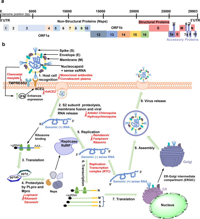
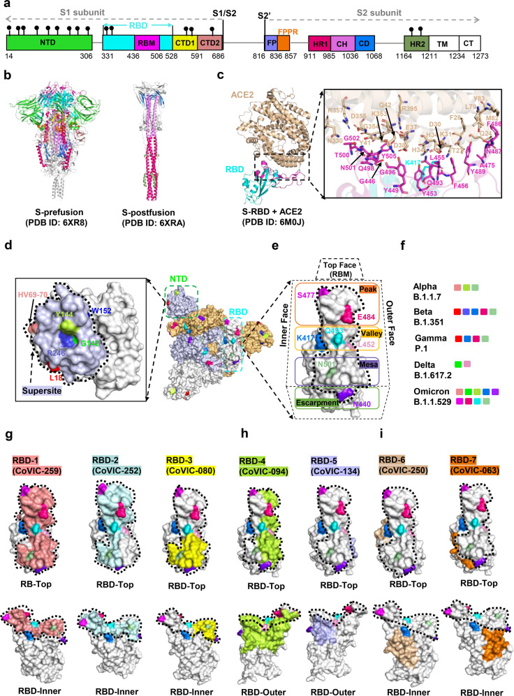
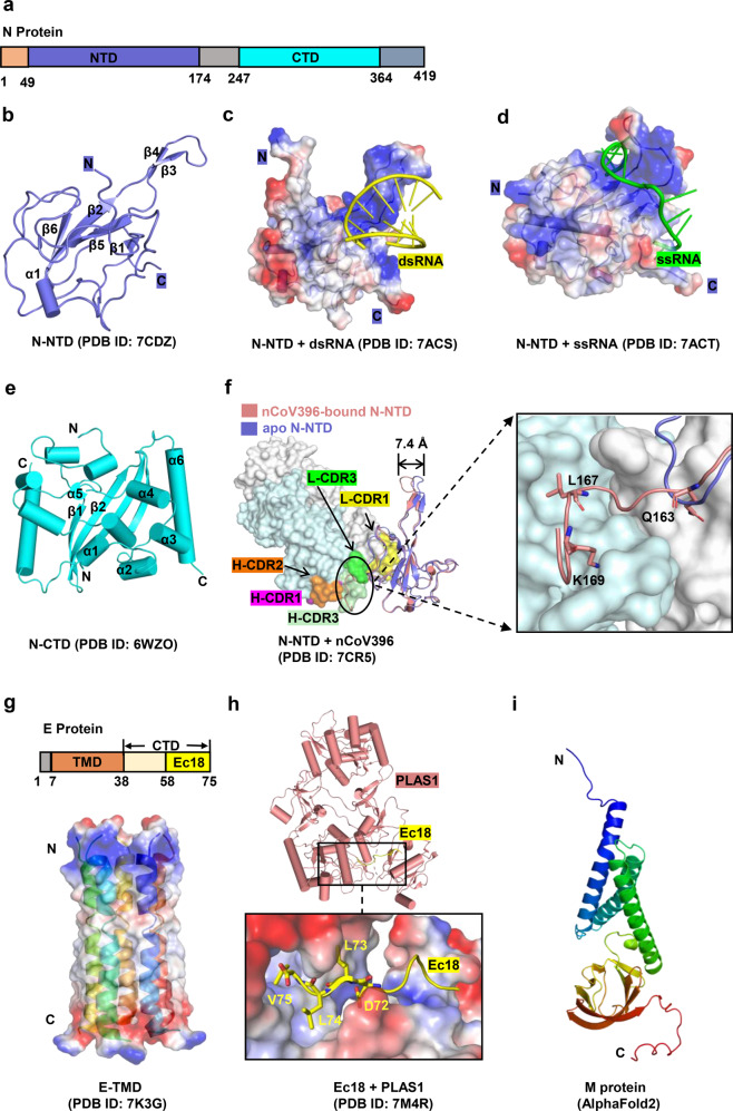
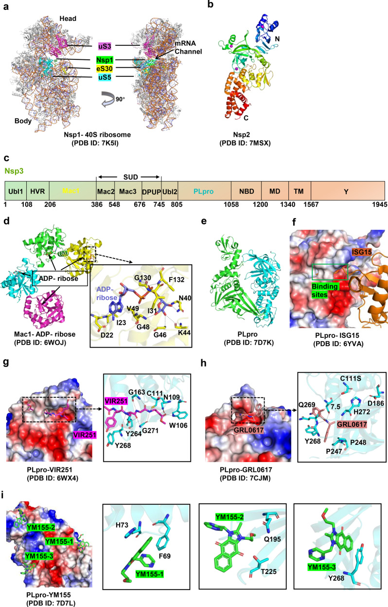
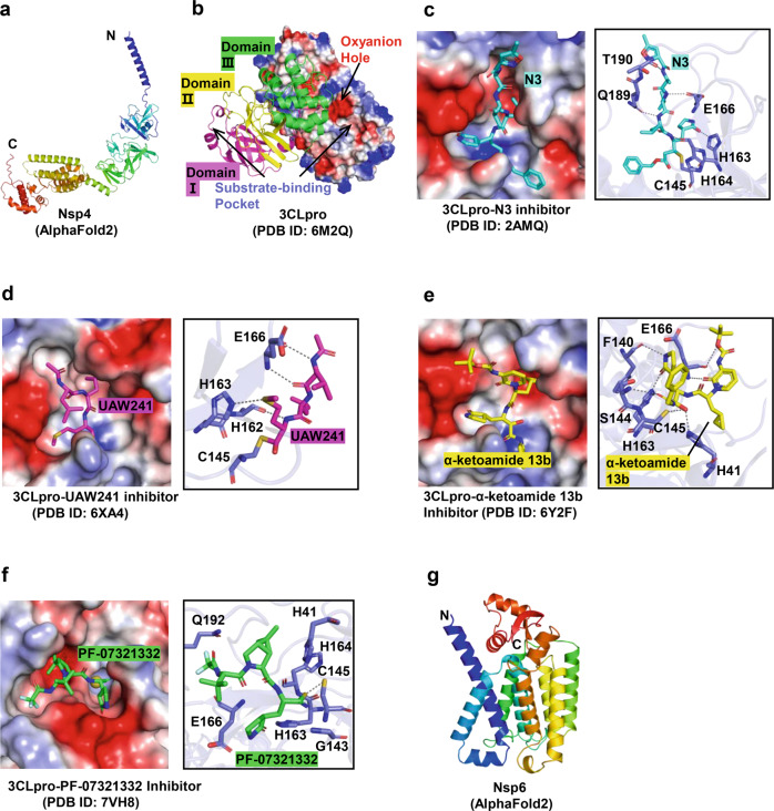
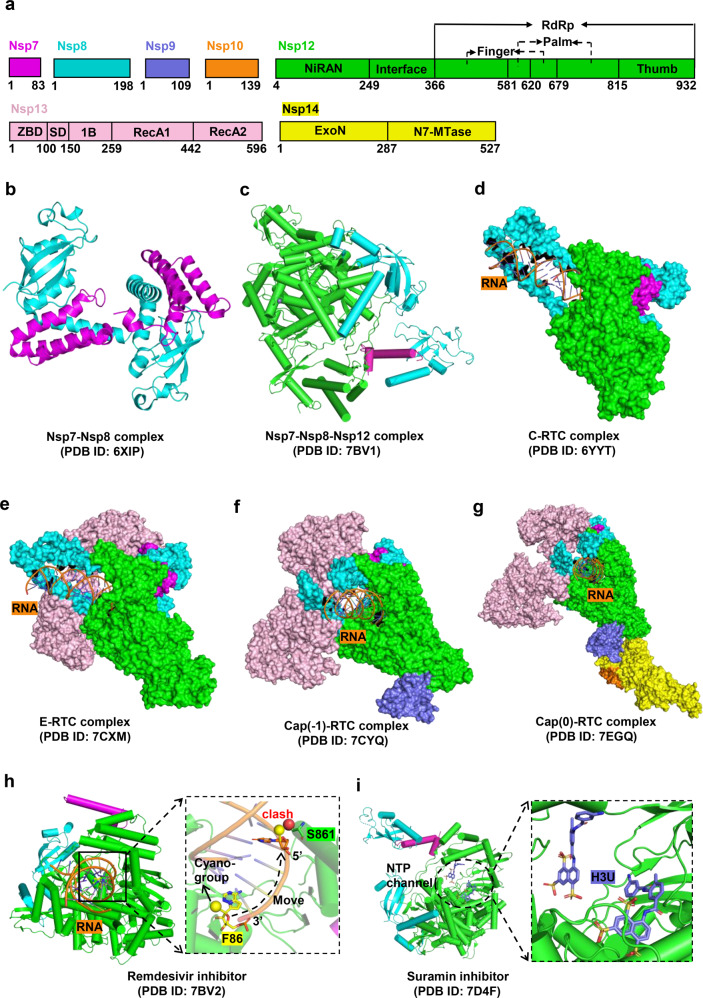
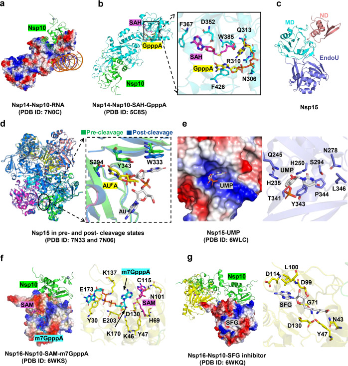
Severe Acute Respiratory Syndrome Coronavirus-2 (SARS-CoV-2) is the causative agent of the pandemic disease COVID-19, which is so far without efficacious treatment. The discovery of therapy reagents for treating COVID-19 are urgently needed, and the structures of the potential drug-target proteins in the viral life cycle are particularly important. SARS-CoV-2, a member of the Orthocoronavirinae subfamily containing the largest RNA genome, encodes 29 proteins including nonstructural, structural and accessory proteins which are involved in viral adsorption, entry and uncoating, nucleic acid replication and transcription, assembly and release, etc. These proteins individually act as a partner of the replication machinery or involved in forming the complexes with host cellular factors to participate in the essential physiological activities. This review summarizes the representative structures and typically potential therapy agents that target SARS-CoV-2 or some critical proteins for viral pathogenesis, providing insights into the mechanisms underlying viral infection, prevention of infection, and treatment. Indeed, these studies open the door for COVID therapies, leading to ways to prevent and treat COVID-19, especially, treatment of the disease caused by the viral variants are imperative.


Conflict of interest statement

The authors declare no competing interests.

Figures

Fig. 1
Fig. 1
The whole-genome composition and replication cycle of SARS-CoV-2 and potential targets. a The viral genome encodes 16 nonstructural proteins (Nsps) required for replication/transcription and structural proteins required for the assembly of new virions. b the SARS-CoV-2 mainly infects lymphatic epithelial cells and type II pneumocytes with the initiation of human body’s innate response by producing interferons (IFNs). However, IFN activates expression of ACE2 protein which acts as receptor for virus attachment to host cells. Interaction between S protein and ACE2 leads to proteolytic cleavage at the S1–S2 boundary and S2ʹ site mediated by transmembrane protease serine 2 (TMPRSS2), further inducing the viral and host cell plasma membrane fusion. The single-stranded RNA in the viral genome is translated by host machinery to produce viral polypeptides (pp1a and pp1ab), which undergo proteolytic cleavage by PLpro and Mpro proteins to synthesize Nsps. These Nsps encode replication transcription complex (RTC), which continuously replicates and produces a series of subgenomic messenger RNAs that encode the accessory and structural proteins. The viral genomic RNA and proteins are assembled to form the virus particles in the ER-Golgi intermediate compartment (ERGIC). The vesicle-containing virus then fuses with plasma membrane of the host, releasing the viral particles out of the cell The antiviral molecules with target sites are highlighted in red
Fig. 2
Fig. 2
Structures of SARS-CoV-2 Spike protein and antibody recognition. a The full-length SARS-CoV-2 S protein. NTD N terminal domain, RBD receptor-binding domain, RBM receptor-binding motif, CTD1 C-terminal domain 1, CTD2 C-terminal domain 2, FP fusion peptide, FPPR fusion-peptide proximal region, HR1 heptad repeat 1, CH central helix region, CD connector domain, HR2 heptad repeat 2, TM transmembrane segment, CT cytoplasmic tail. b Structures of S trimer in prefusion and postfusion states. Each domain is marked with a color corresponding to a. c Structure of the RBD (cyan) in complex with ACE2 (pink). Residues involved in interactions between the RBD and ACE2 are shown as sticks. d Footprints for NTD-targeted antibodies, with the NTD “supersite” outlined with a dashed line. The residue positions of important mutations and deletions are indicated in the NTD. Table 1 lists mutations represented in each variant. e Location of important emerging mutations on the RBD. The RBM can be topologically divided into three subsections: the “peak” that includes residues S477 and E484; the “valley” that includes K417, Q493, and L452; and the “mesa” includes N501. f Mutations and deleted residues affecting antibodies activity involved in significant mutants. gi footprint of a representative antibody from the Coronavirus Immunotherapeutic Consortium (CoVIC) mapped onto an RBD monomer. The ACE2 binding site is outlined with a dotted line. The website of CoVIC is at https://covic.lji.org/
Fig. 3
Fig. 3
Structures of SARS-CoV-2 Nucleocapsid, Envelope, and Membrane proteins. a Schematic representation of N protein organization. NTD N-terminal domain, CTD C-terminal domain. b Overall structures of N-NTD. c, d Overall structures of N-NTD in complex with ssRNA and dsRNA, respectively. The N-NTD is illustrated with electrostatic surface. e Overall structure of N-CTD. f Conformational changes between apo and nCoV396-bound N-NTD. The monoclonal antibody nCoV396 is illustrated with surface. The key residues of interaction between nCoV396 and N-NTD are shown on the right as sticks. g Overall structure of SARS-CoV-2 E protein shown in surface and cartoon. TMD transmembrane domain, CTD C-terminal domain, Ec18 residues 58–75 of E-CTD. h Structure of the C-terminus of E protein (Ec18) in complex with human cell junction protein PLAS1. The Ec18 is shown in yellow. i Predicted structure of M protein using alphafold 2
Fig. 4
Fig. 4
Structures of Nsp1 /Nsp2 /Nsp3 and their inhibitors. a Structure of Nsp1 in complex with 40 S ribosome. Nsp1 binds at the mRNA channel in the cleft between the head and body of the 40 S ribosome. b Overall structure of Nsp2 protein. c Schematic representation of Nsp3 protein’s domain. d Structures of Mac1 domain in complex with ADP ribose. e, f Structure of apo PLpro and its complex with interferon-stimulated gene 15 protein (ISG15), respectively. gi Structures of PLpro in complex with different inhibitors (VIR251, GRL-0617, and YM155). The PLpro is illustrated with electrostatic surface
Fig. 5
Fig. 5
Structures of Nsp4 /Nsp5 /Nsp6 and their inhibitors. a Predicted structure of Nsp4 protein. b Overall structure of 3CLpro protein with electrostatic surface and cartoon models. cf Structures of 3CLpro in complex with N3, calpain inhibitor II (UAW241), α-ketoamide 13b inhibitors, and PF-07321332, respectively. g Predicted structure of Nsp6 protein
Fig. 6
Fig. 6
Structures of SARS-CoV-2 RTC complexes and cognate inhibitors. a Schematic representations of Nsp7, Nsp8, Nsp9, Nsp10, Nsp12, Nsp13, and Nsp14 proteins organizations. bg Structures of Nsp7-Nsp8, Nsp7-Nsp8-Nsp12, the central RTC (C-RTC, Nsp12-Nsp7- Nsp8), the elongation RTC (E-RTC, Nsp12-Nsp7-Nsp8-Nsp13), cap (−1)-RTC (Nsp12-Nsp7-Nsp8-Nsp13-Nsp9), and cap (0)-RTC (Nsp12-Nsp7-Nsp8- Nsp13-Nsp9-Nsp14-Nsp10) complexes, respectively. h, i Structures of C-RTC with bound inhibitors Remdesivir (F86) can bind to the RNA strand. The schematic of the inhibition state is shown on the right (h). Two suramin (H3U) molecules occupy the catalytic cavity (i)
Fig. 7
Fig. 7
Structures of Nsp14 /Nsp15/Nsp16 and their inhibitors. a Cryo EM structure of the SARS-CoV-2 Nsp10-Nsp-14 RNA complex. Nsp14 is illustrated with electrostatic surface. Nsp10 is illustrated with cartoon in green. b The structure of the Nsp14-Nsp10 in complex with functional ligands S-adenosyl-L-homocysteine (SAH) and GpppA shown in sticks. c The overall structural of Nsp15. d Conformational changes between Nsp15 in pre- and post- cleavage states. e Structures of Nsp15 in complex with uridine-5′-monophosphate (UMP). f The structure of SARS-CoV-2 Nsp16-Nsp10 in complex with RNA cap analogue (m7GpppA) and S-adenosyl methionine (SAM). The m7GpppA and SAM are shown in sticks. Nsp16 is indicated as electrostatic surface. g The structure of Nsp16-Nsp10 heterodimer in complex with sinefungin (SFG). SFG is shown in sticks. Nsp16 is illustrated with electrostatic surface and cartoon in yellow
Fig. 8
Fig. 8
Structures of accessory proteins. ad Overall structure of ORF3a, ORF7a, ORF8, and ORF 9b, respectively. e Structure of ORF9b in complex with human protein TOM70, a subunit of the mitochondrial import receptor. ORF9b is shown as yellow cartoon. TOM70 is illustrated with electrostatic surface on the right. The ORF9b binds to the hydrophobic pocket of TOM70 and occupies its binding site

References

    1. Jin YF, et al. Virology, epidemiology, pathogenesis, and control of COVID-19. Viruses. 2020;12:372. - PMC - PubMed
    1. Mittal A, et al. COVID-19 pandemic: insights into structure, function, and hACE2 receptor recognition by SARS-CoV-2. PLoS Pathog. 2020;16:e1008762. - PMC - PubMed
    1. Chan JF, et al. A familial cluster of pneumonia associated with the 2019 novel coronavirus indicating person-to-person transmission: a study of a family cluster. Lancet. 2020;395:514–523. - PMC - PubMed
    1. Lu RJ, et al. Genomic characterisation and epidemiology of 2019 novel coronavirus: implications for virus origins and receptor binding. Lancet. 2020;395:565–574. - PMC - PubMed
    1. Wrobel A, et al. SARS-CoV-2 and bat RaTG13 spike glycoprotein structures inform on virus evolution and furin-cleavage effects. Nat. Struct. Mol. Biol. 2020;27:763–767. - PMC - PubMed

Publication types

MeSH terms