. 2022 Jan 11:15:781527.
doi: 10.3389/fnsys.2021.781527. eCollection 2021.

Cerebellar Coordination of Neuronal Communication in Cerebral Cortex

Affiliations

Cerebellar Coordination of Neuronal Communication in Cerebral Cortex

Samuel S McAfee et al. Front Syst Neurosci. .

Abstract

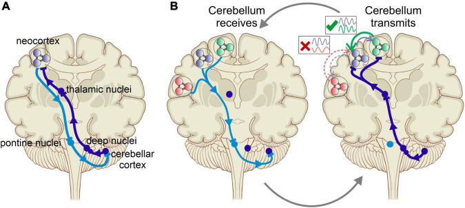
Cognitive processes involve precisely coordinated neuronal communications between multiple cerebral cortical structures in a task specific manner. Rich new evidence now implicates the cerebellum in cognitive functions. There is general agreement that cerebellar cognitive function involves interactions between the cerebellum and cerebral cortical association areas. Traditional views assume reciprocal interactions between one cerebellar and one cerebral cortical site, via closed-loop connections. We offer evidence supporting a new perspective that assigns the cerebellum the role of a coordinator of communication. We propose that the cerebellum participates in cognitive function by modulating the coherence of neuronal oscillations to optimize communications between multiple cortical structures in a task specific manner.

Keywords: cerebellum; cerebrocerebellar communication; cognition; coherence; functional connectivity.


Conflict of interest statement

The authors declare that the research was conducted in the absence of any commercial or financial relationships that could be construed as a potential conflict of interest.

Figures

FIGURE 1
FIGURE 1
(A) Cerebrocerebellar interaction via reciprocal connections between specific cerebral and cerebellar areas. Purkinje cells in the cerebellar cortex project to the neocortical areas via the cerebellar nuclei and the thalamus. In what was often described as a closed-loop projection, the neocortex in turn projects back to the cerebellar area of origin via the pontine nuclei. This one-to-one interaction scheme is the basis of most approaches to investigating cerebrocerebellar interactions. (B) Cerebellar modulation of communication between cerebral cortical areas provides a revised picture of cerebrocerebellar interactions, in which the cerebellum does not primarily modulate the activity in specific cortical areas but rather coordinates the communication between areas by augmenting the coherence of neuronal oscillations in a task specific manner. This occurs cyclically with the cerebellum areas receiving the neuronal “context” of cerebral activity from multiple regions by encoding their oscillations, comparing their timing, and then transmitting the output via the thalamus to promote synchrony between task-appropriate cerebral cortical regions.
FIGURE 2
FIGURE 2
Inactivation of cerebellar output reduces gamma coherence between S1 and M1. (A) In an experiment that involved simultaneous measurements of LFPs in S1 and M1 of awake, head fixed rats, Popa et al. (2013) demonstrated that pharmacological inactivation of the interposed nuclei selectively reduced gamma coherence between S1 and M1. The plot on the left shows a change in coherence relative to the control condition in which the interposed was kept intact. The experimental approach is illustrated on the right (from: Popa et al., 2013). (B) A similar, recent experiment showing that optogenetic inhibition of cerebellar output (via Purkinje cell excitation) significantly reduced the coherence of gamma responses evoked by whisker stimulation. The plot on the left shows estimated effect of Purkinje cell stimulation on coherence between deep layer S1 and superficial layer M1. Theta-range S1–M1 coherence was enhanced with Purkinje cell stimulation (from: Lindeman et al., 2021). *These frequencies were statistically significant (p < 0.05).
FIGURE 3
FIGURE 3
Cerebellar representations of phase and phase differences of oscillations in the mPFC and CA1. (A) Illustration of the experimental setup with recording electrodes in the mPFC and dCA1, picking up LFPs and a recording electrode in cerebellar lobulus simplex recording single unit Purkinje cell spike activity. (B) Example histogram showing Purkinje cell simple spike rate plotted against the phase of a 10 Hz oscillation recorded in the mPFC. (C) Fraction of Purkinje cells in LS (n = 32) whose simple spike activity was significantly correlated with oscillatory phase plotted as a function of mPFC oscillation frequency (plotted on a log-10 scale). The function shows two peaks at the delta frequency range (0.5–4 Hz) and the high gamma range (50–100 Hz). (D) As in (C) but showing fractions of LS Purkinje cells significantly modulated by the phase of oscillatory activity in CA1. (E) Fraction of Purkinje cells in Crus I (n = 17) whose simple spike activity was significantly correlated with the oscillatory phase in mPFC plotted as a function of mPFC oscillation frequency. The function shows a single peak at the delta frequency range (0.5–4 Hz). (F) As in (E) but showing fractions of Crus I Purkinje cells significantly modulated by the phase of oscillatory activity in CA1. D, delta; T, theta; B, beta; LG, low gamma; HG, high gamma. (G) Illustration of hypothetical oscillations at a specific frequency occurring simultaneously in the mPFC (blue traces) and CA1 (red traces) and displaying different phase relationships (4) at different times. The phase relationship 4 is defined as the phase difference relative to the mPFC oscillation. (H) Hypothetical Purkinje cell spikes recorded simultaneously with the LFP activity in the mPFC and CA1 shown in (G). The rate modulation of this hypothetical Purkinje cell shows a significant increase in spike firing when the phase difference between mPFC and CA1 oscillations reaches values around 270°. (I) Phase histogram of real Purkinje cell simple spike activity. The histogram shows spike activity as a function of mPFC-CA1 phase differences at 11 Hz. The simple spike activity of the Purkinje cell in this example was significantly modulated as a function of mPFC-CA1 phase difference, with a preference of 288.7°. (J) Same data as in (I) represented in polar coordinates. Vectors composed of the angular value 4 and the magnitude of the spikes per sample were summated to determine the angular preference of Purkinje cell activity. The resultant vector magnitude was taken to quantify the degree of modulation and tested against surrogate results for statistical significance (from McAfee et al., 2019).
FIGURE 4
FIGURE 4
Cellular and network mechanisms of oscillatory encoding and modulation in the cortico-cerebello-cortical circuit. Panel labels are color-coded according to where in the circuit a modulation of the neuronal signal occurs, corresponding to the schematics in the top-center. (A) Cortico-cerebellar signals originate in the deep layers of the neocortex, where alpha and beta oscillations predominate. (B) Pre-cerebellar neurons in the pons translate a dynamic current input into rate in a linear fashion, thereby translating oscillatory current into a rate code. (C) Deep and superficial GCs respond preferentially to different phases of the ponto-cerebellar signal, thereby encoding both phase (via time) and amplitude (via GC depth) of oscillatory input. (D) Phase and phase difference of oscillatory activity is decoded by Purkinje cells, via two potential mechanisms. Top: tidal wave theory proposes that a phase difference in a band-limited frequency range can be calculated as a time difference along slow-conducting parallel fibers. Each parallel fiber conveys information about the phase of one cerebral oscillation, and together convey information about the phase relationship of their inputs. Two inputs offset by Δt would arrive simultaneously at the Purkinje cell dendritie. Bottom: simulations show that rhythmic excitation can generate network resonance across parallel fiber beams with a phase shift, due to cross-beam inhibition from MLIs. Rhythmic excitation could augment Purkinje cell responses to input across parallel fiber beams, thereby providing a means to calculate phase differences that are too great to be accounted for in parallel fiber conduction length. (E) Feedback to the cortex conveyed via thalamocortical projections. Multi-areal matrix-type projections target superficial and deep layers in multiple cortical areas, likely inducing simultaneous beta oscillations that facilitate simultaneous gamma bursts in targeted regions. Focal matrix-type projections preferentially target the superficial layers, suggesting a role in spatially selective augmentation of gamma responses during the bottom-up flow of information.
FIGURE 5
FIGURE 5
Key functional imaging studies of cerebrocerebellar interaction. (A) Voxel-to-network mapping of cerebellar relationship to cerebral intrinsic networks. Most of the cerebellum is most-strongly linked to association and cognitive cerebral areas. (B) Co-activation pattern analysis identifies recurring spatial patterns of co-activation in the brain. Left: three unique cerebral co-activation patterns involving the intraparietal sulcus are shown. Lower panel shows unique thalamic foci associated with each pattern as well. Right: corresponding cerebellar activations. Focal activation of cerebellar cortex is linked to complex patterns of co-activation across distributed cerebral cortical networks. The non-overlapping foci suggests a voxel-to-network mapping of cerebellar activity to cortical networks is insufficient to describe the cerebellum’s role in distributed brain networks. (C) Maturation of brain networks over the course of development. Black arrow indicates cluster of cerebellar nodes at each developmental stage. Early in development, cortical areas are functionally linked to their nearest anatomical neighbors, and the cerebellum has no functional link to the cortex. Once mature however, the cerebellum acts as a hub between distributed functional networks in cortex.

Similar articles

Cited by

References

    1. Andreasen N. C., O’Leary D. S., Cizadlo T., Arndt S., Rezai K., Ponto L. L., et al. (1996). Schizophrenia and cognitive dysmetria: a positron-emission tomography study of dysfunctional prefrontal-thalamic-cerebellar circuitry. Proc. Natl. Acad. Sci. U.S.A. 93 9985–9990. 10.1073/pnas.93.18.9985 - DOI - PMC - PubMed
    1. Andreasen N. C., Paradiso S., O’Leary D. S. (1998). “Cognitive dysmetria” as an integrative theory of schizophrenia: A dysfunction in cortical-subcortical-cerebellar circuitry? Schizophr. Bull. 24 203–218. - PubMed
    1. Andreasen N. C., Pierson R. (2008). The role of the cerebellum in schizophrenia. Biol. Psychiatry 64 81–88. - PMC - PubMed
    1. Ashida R., Cerminara N. L., Edwards R. J., Apps R., Brooks J. C. W. (2019). Sensorimotor, language, and working memory representation within the human cerebellum. Hum. Brain Mapp. 40 4732–4747. 10.1002/hbm.24733 - DOI - PMC - PubMed
    1. Atallah B. V., Scanziani M. (2009). Instantaneous modulation of gamma oscillation frequency by balancing excitation with inhibition. Neuron 62 566–577. 10.1016/j.neuron.2009.04.027 - DOI - PMC - PubMed

LinkOut - more resources