. 2021 Jul 30;9(1):nwab137.
doi: 10.1093/nsr/nwab137. eCollection 2022 Jan.

H7N9 virus infection triggers lethal cytokine storm by activating gasdermin E-mediated pyroptosis of lung alveolar epithelial cells

Affiliations

H7N9 virus infection triggers lethal cytokine storm by activating gasdermin E-mediated pyroptosis of lung alveolar epithelial cells

Xiaopeng Wan et al. Natl Sci Rev. .

Abstract

The H7N9 influenza virus emerged in China in 2013, causing more than 1560 human infections, 39% of which were fatal. A 'cytokine storm' in the lungs of H7N9 patients has been linked to a poor prognosis and death; however, the underlying mechanism that triggers the cytokine storm is unknown. Here, we found that efficient replication of the H7N9 virus in mouse lungs activates gasdermin E (GSDME)-mediated pyroptosis in alveolar epithelial cells, and that the released cytosolic contents then trigger a cytokine storm. Knockout of Gsdme switched the manner of death of A549 and human primary alveolar epithelial cells from pyroptosis to apoptosis upon H7N9 virus infection, and Gsdme knockout mice survived H7N9 virus lethal infection. Our findings reveal that GSDME activation is a key and unique mechanism for the pulmonary cytokine storm and lethal outcome of H7N9 virus infection and thus opens a new door for the development of antivirals against the H7N9 virus.

Keywords: H7N9 virus; cytokine storm; gasdermin E; pyroptosis.


Figures

Figure 1.
Figure 1.
Replication and virulence of different H7N9 viruses in C57BL/6 (B6) mice. Groups of 32 B6 mice were inoculated intranasally with 105.5 EID50 of PG/S1421 or AH/1, and 16 mice were inoculated with PBS as a control. Five mice in each virus-inoculated group were euthanized on days 3 and 6 post-infection (p.i.), respectively, and (A) their organs were collected for virus titration. Six mice in each virus-inoculated group were observed for (B) body weight change and (C) death for 2 weeks. (D) Lung lavage samples from six mice in each virus-infected group and the PBS control group were collected for analysis of chemokines and cytokines on day 6 p.i.; (E) lungs of the virus-inoculated mice or PBS-inoculated control mice were collected on day 6 p.i. to test (F) the wet-to-dry ratio (n = 5), (G) total cell and (H) infiltration of immune cell quantification (n = 5). Data in panels A–D, F and G are means ± standard deviation. Statistical analysis was performed by using the Student's t-test. a, p < 0.01 compared with the values of AH/1-inoculated mice; b, p < 0.05 compared with the values of PG/S1421-inoculated mice; c, p < 0.01 compared with the values of PG/S1421-inoculated mice.
Figure 2.
Figure 2.
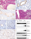
Caspase-3 and gasdermin E (GSDME) activation in the lungs of mice infected with different H7N9 viruses. Groups of 12 6-week-old B6 mice were inoculated with 105.5 EID50 of (A–C) PG/S1421 or (D–G) AH/1. Three mice in each group were euthanized on day 6 p.i. and their lungs were collected for (A and D) pathologic study and immunohistochemical (IHC) study to detect (B, E and F) the viral antigen and (C and G) activated caspase-3. Three mice in each virus-inoculated group were euthanized on days 3, 5 and 7 p.i., respectively, and their lungs were collected for the detection of (H) caspase-3 and GSDME by use of SDS-PAGE and Western blotting. Scale bars in A–C and E–G = 100 μm; scale bar in D = 200 μm. Arrows and arrowheads on F and G indicate alveolar epithelial cells and macrophages, respectively.
Figure 3.
Figure 3.
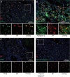
Identification of cell types infected by AH/1 virus in the lungs of mice. Dual-staining was performed on the lung sections of AH/1-infected mice to identify (A) the type II alveolar epithelial cells, (B) type I alveolar epithelial cells, (C) macrophages and (D) positive cleaved-caspase-3 staining cells that were viral antigen-positive. Scale bars: 100 μm.
Figure 4.
Figure 4.
GSDME-mediated pyroptosis of different cells after H7N9 virus infection. (A) GSDME expression levels of different cells. (B) Activation of caspase-3 and Gsdme-N in different alveolar epithelial cells after AH/1 infection. (C, D, G and K) Different alveolar epithelial cells and (J and L) Raw-GSDME/+ cells undergo pyroptosis upon AH/1 infection, and their manner of death is correlated with the abundance of cellular GSDME, as evidenced by (E, F and I) GSDME-deficient alveolar epithelial cells and (H) Raw-vector cells undergoing apoptosis upon AH/1 infection. Static bright field cell images in panels C to J were obtained using the Zeiss primovert microscope and processed using zen blue software. The time-lapse phase-contrast and fluorescent images of cells in K and L were taken at the indicated timepoints after viral infection by using a PerkinElmer UltraVIEW spinning disk confocal microscope and processed by using the Volocity software. Scale bar in K and L: 5 μm.
Figure 5.
Figure 5.
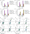
Lactate dehydrogenase (LDH) release and propidium iodide (PI) permeant of different cells after AH/1 infection. LDH release of different types of (A–C) alveolar epithelial cells and (D) macrophages were measured at the indicated timepoints after the AH/1 infection. PI permeant in different (E) A549 cells and (F) Raw246.7 cells were measured 24 h after the AH/1 infection. a, p < 0.01 compared with the values of their virus-infected control cell counterparts. All data are representative of three independent experiments.
Figure 6.
Figure 6.
Replication and virulence of H7N9 viruses in wild-type B6 and Gsdme–/– mice. Groups of 28 wild-type B6 mice and Gsdme knockout (Gsdme–/–) mice were intranasally inoculated with 10 MLD50 of AH/1 virus. (A) Five mice in each group were euthanized on days 3 and 6 p.i., respectively, and their nasal turbinates and lungs were collected for virus titration in eggs. (B) The lungs of three mice in each group that were euthanized on day 6 p.i. were collected for pathologic study; arrows indicate alveolar bronchiolization, arrowheads indicate perivasculitis. (C) Lung lavage samples from five mice from each group were collected for chemokine and cytokine analysis. The remaining 10 mice in each group were used to evaluate lethality and their (D) body weight change and (E) death were observed for 2 weeks. We repeated the lethality study one more time with groups of 10 mice, and the combined data are shown in D and E. (F–H) The replication and virulence studies in wild-type B6 mice and Gsdme/ mice were also repeated with another H7N9 virus (CK/SD008-PB2/627K). Data in panels A, C, D, F and G are means ± standard deviation. Statistical analysis was performed by using the Student's t-test. a, p < 0.01 compared with the values of wild-type mice; b, p < 0.05 compared with the values of wild-type mice. Scale bar in B: 200 μm.

References

    1. Gao R, Cao B, Hu Yet al. . Human infection with a novel avian-origin influenza A (H7N9) virus. N Engl J Med 2013; 368: 1888–97. 10.1056/NEJMoa1304459 - DOI - PubMed
    1. Wong SSY, Yuen K.. Avian influenza virus infections in humans. Chest 2006; 129: 156–68. 10.1378/chest.129.1.156 - DOI - PMC - PubMed
    1. Chen H, Yuan H, Gao Ret al. . Clinical and epidemiological characteristics of a fatal case of avian influenza A H10N8 virus infection: a descriptive study. Lancet 2014; 383: 714–21. 10.1016/S0140-6736(14)60111-2 - DOI - PubMed
    1. Song W, Qin K.. Human-infecting influenza A (H9N2) virus: a forgotten potential pandemic strain? Zoon Publ Health 2020; 67: 203–12. 10.1111/zph.12685 - DOI - PubMed
    1. Fouchier RA, Schneeberger PM, Rozendaal FWet al. . Avian influenza A virus (H7N7) associated with human conjunctivitis and a fatal case of acute respiratory distress syndrome. Proc Natl Acad Sci USA 2004; 101:1356–61. 10.1073/pnas.0308352100 - DOI - PMC - PubMed