

Plug the pit: a surgical technique for optic disc pit
- PMID: 35087980
- PMCID: PMC8764426
- DOI: 10.22336/rjo.2021.74
Plug the pit: a surgical technique for optic disc pit
Abstract
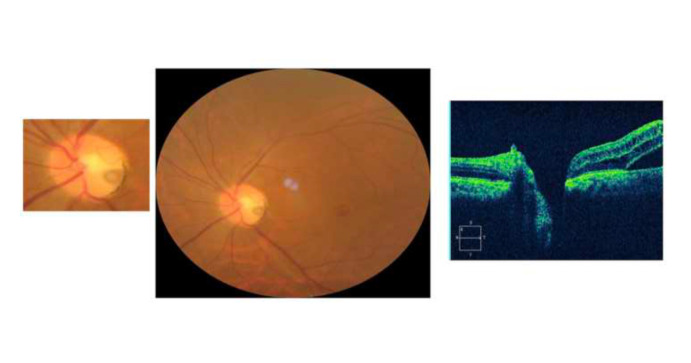
Purpose: To present a recently described surgical technique for the treatment of optic disc pit (ODP) and evaluate its outcomes. Methods: A patient presented with refractory serous macular detachment and secondary full thickness macular hole associated with ODP, for which he had already undergone pars-plana vitrectomy with internal limiting membrane peeling and autologous serum application over the optic disc pit. A recently described surgical technique was carried out to treat this case. In this procedure, a silicone punctal plug was used to close the ODP. The macular hole was closed with a human amniotic membrane graft. Endotamponade was carried out with 1000cs silicone oil. Results: Postoperatively, the serous macular detachment subsided and the punctal plug and amniotic membrane graft were in situ. Patient's visual acuity improved from counting fingers to 6/38 at one year postoperative. Conclusion: This technique appears to be safe and effective in resolving long standing serous macular detachment associated with ODP, which was refractory to the conventional intervention. However, more cases and longer follow-ups are needed to affirm the safety and efficacy of this recently described procedure.
Keywords: maculopathy; optic disc pit; punctal plug.
© The Authors.Romanian Society of Ophthalmology.
Figures


References
-
- Wiethe T. Ein fall von angeborener deformität der sehnervenpapille. Arch Augenheilkd. 1882;11:14–19.
-
- Gass JDM. Serous detachment of the macula secondary to optic disc pits. Am J Ophthalmol. 1969;67(6):821–841. - PubMed
-
- Jain N, Johnson MW. Pathogenesis and treatment of maculopathy associated with cavitary optic disc anomalies. Am J Ophthalmol. 2014;158:423–435. - PubMed
-
- Lincoff H, Yannuzzi L, Singerman L, et al. Improvement in visual function after displacement of the retinal elevations emanating from optic pits. Arch Ophthalmol. 1993;111(8):1071–1079. - PubMed
-
- Bonnet M. Serous macular detachment associated with optic nerve pits. Graefes Arch Clin Exp Ophthalmol. 1991;229(6):526–532. - PubMed
Publication types
MeSH terms
LinkOut - more resources
Full Text Sources
Medical
Miscellaneous