. 2022 Jun;110(6):1425-1438.
doi: 10.1002/jbm.b.35010. Epub 2022 Jan 28.

Surface wear in a custom manufactured temporomandibular joint prosthesis

Affiliations

Surface wear in a custom manufactured temporomandibular joint prosthesis

Nikolas De Meurechy et al. J Biomed Mater Res B Appl Biomater. 2022 Jun.

Abstract

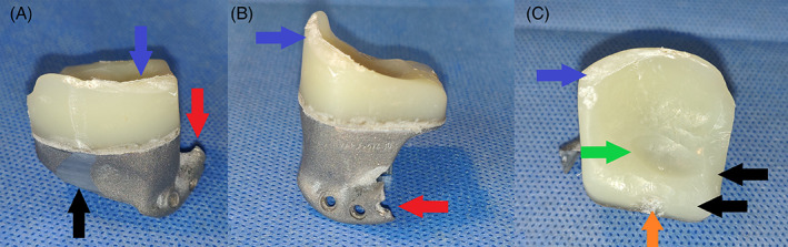
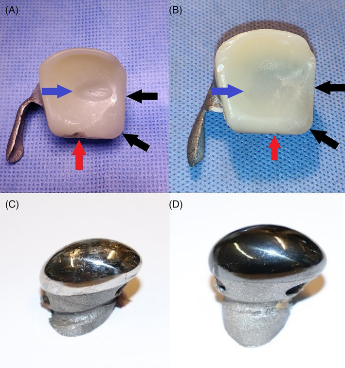
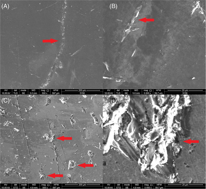
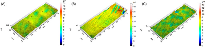
The wear of a novel temporomandibular joint (TMJ) prosthesis was evaluated in an animal model. The prosthesis consisted of an additively manufactured titanium alloy (Ti6 Al4 V) mandibular condyle and glenoid fossa created through selective laser melting, with a machined vitamin E-enriched ultra-high molecular weight polyethylene (UHMWPE) surface attached to the fossa. Thirteen TMJ prosthesis were implanted in sheep, six of which had condylar heads coated with HadSat® diamond-like carbon (H-DLC). Euthanasia took place after 288 days, equaling 22 years of human mastication. Linear and volumetric wear analysis of the fossa was performed by optical scanning. The condylar head surfaces were assessed by scanning electron and confocal laser microscopy. The average linear UHMWPE wear, when combined with the coated condyle, was 0.67 ± 0.28 mm (range: 0.34-1.15 mm), not significantly differing (p = .3765, t-test) from the non-coated combination average (0.88 ± 0.41 mm; range: 0.28-1.48 mm). The respective mean volumetric wear volumes were 25.29 ± 11.43 mm3 and 45.85 ± 22.01 mm3 , not significantly differing (p = .1448, t-test). Analysis of the coated condylar surface produced a mean Ra of 0.12 ± 0.04 μm and Sa of 0.69 ± 0.07 μm. The non-coated condylar surface measured a mean Ra of 0.28 ± 0.17 μm and Sa of 2.40 ± 2.08 μm. Both Sa (p = .0083, Mann-Whitney U test) and Ra (p = .0182, Mann-Whitney U test), differed significantly. The prosthesis exhibits acceptable wear resistance and addition of the H-DLC-coating significantly improved long-term condylar surface smoothness.

Keywords: animal; arthroplasty; models; replacement; temporomandibular joint; titanium.


Conflict of interest statement

Maurice Y. Mommaerts is co‐owner and innovation manager at CADSkills BV. Stijn Huys is R&D Officer at CADSkills BV.

Figures

FIGURE 1
FIGURE 1
Condylar head with suture threading tunnel and hook for fixation of the lateral pterygoid muscle enthesis. (A) Mesial side. Black arrow: subcondylar groove to guide enthesis' sutures. White arrow: 2.4 mm subcondylar tunnel and hook‐like extension for fixation of the enthesis. Red arrow: lattice structure for enthesis' bony ingrowth. (B) Lateral side. Black arrow: subcondylar groove to guide enthesis' sutures. White arrow: 2.4 mm subcondylar tunnel and hook‐like extension for fixation of the enthesis
FIGURE 2
FIGURE 2
Fossa component with sagittal and transversal sectional view. The titanium mesh connecting the UHWMPE to the titanium component has been removed for proprietary reasons. (A) Frontal view. Black arrow: titanium component. Red arrow: UHMWPE component. (B) Inferior view. Black arrow: titanium component. Red arrow: UHMWPE component
FIGURE 3
FIGURE 3
Macroscopic images of explanted fossa components of sheep #4473, with additional damage, having occurred during the explantation. This severe additional damage, occurred during retrieval, no longer allowed for reliable overlapping with the ‘design’‐STL. No linear, or volumetric wear could be analyzed of this fossa. (A) Posterior view. Blue arrow: damage to posterior UHMWPE ridge. Black arrow: damage to titanium part. Red arrow: damage to titanium extension for fixation onto the zygomatic arch. (B) Lateral view. Blue arrow: damage to posterior UHMWPE ridge. Red arrow: damage to titanium extension for fixation onto the zygomatic arch. (C) Inferior view. Blue arrow: damage to posterior UHMWPE ridge. Black arrow: scalpel‐reduced non‐articulating UHMWPE. Green arrow: worn out UHMWPE due to articulating with the condylar surface. Orange arrow: anteriorly worn out UHMWPE volume due to contact with the coronoid process
FIGURE 4
FIGURE 4
Representative macroscopic images of explanted components of the custom temporomandibular joint total joint replacement after 9 months of mastication and rumination in a sheep model. (A) Ultra‐high molecular weight polyethylene (UHMWPE) fossa of sheep #1724 that articulated with a non‐coated condyle. Blue arrow: worn out UHMWPE due to articulating with the condylar surface. Black arrow: scalpel‐reduced non‐articulating UHMWPE. Red arrow: anteriorly worn out UHMWPE volume due to contact with the coronoid process. (B) UHMWPE fossa of sheep #5158 that articulated with an HadSat® (H‐DLC) diamond‐like carbon coated condyle. Blue arrow: worn out UHMWPE due to articulating with the condylar surface. Black arrow: scalpel‐reduced non‐articulating UHMWPE. Red arrow: anteriorly worn out UHMWPE volume due to contact with the coronoid process. (C) Non‐coated Ti6Al4V condyle. (D) H‐DLC coated condyle
FIGURE 5
FIGURE 5
Representative 3D scans of explanted components of the custom temporomandibular joint total joint replacement after 9 months of mastication and rumination in a sheep model. (A) Ultra‐high molecular weight polyethylene (UHMWPE) fossa of sheep #1724 that articulated with a non‐coated condyle. The maximal wear depth measures 0.81 mm. Blue arrow: worn out UHMWPE due to articulating with the condylar surface. Black arrow: scalpel‐reduced non‐articulating UHMWPE. Red arrow: anteriorly worn out UHMWPE volume due to contact with the coronoid process. (B) UHMWPE fossa of sheep #5158 that articulated with an HadSat® diamond‐like carbon coated condyle. The maximal wear depth measures 0.34 mm. Blue arrow: worn out UHMWPE due to articulating with the condylar surface. Black arrow: scalpel‐reduced non‐articulating UHMWPE. Red arrow: anteriorly worn out UHMWPE volume due to contact with the coronoid process. Orange arrow: worn out sections due to post‐mortem dissection of the overlaying soft tissues
FIGURE 6
FIGURE 6
3D scans of explanted fossa component of the custom temporomandibular joint total joint replacement after 9 months of mastication and rumination in a sheep model with deviant wear patterns. (A) Ultra‐high molecular weight polyethylene (UHMWPE) fossa of sheep #2177 that articulated with a non‐coated condyle. Black arrow: worn out UHMWPE due to articulating with the condylar surface, with uneven edges. (B) UHMWPE fossa of sheep #4246 that articulated with an H‐DLC coated condyle. Black arrow: main worn out UHMWPE volume due to articulating with the condylar surface. Orange arrow: posteriorly orientated UHMWPE wear track. (C) UHMWPE fossa of sheep #8087 that articulated with an H‐DLC coated condyle. Black arrow: main worn out UHMWPE volume due to articulating with the condylar surface. Orange arrow: three additional condylar‐shaped UHMWPE wear tracks. Red arrow: UHMWPE creep deformation, in non‐articulating region. (D) UHMWPE fossa of sheep #7998 that articulated with a non‐coated condyle. Black arrow: main worn out UHMWPE volume due to articulating with the condylar surface. Orange arrow: initial, centered, UHMWPE wear volume due to articulating with the condylar surface. Red arrow: UHMWPE creep deformation, in non‐articulating region
FIGURE 7
FIGURE 7
Representative light microscopy images of the condylar surface of the custom temporomandibular joint total joint replacement. (A,B) Condylar surface of a pristine, non‐coated condyle. Red arrow: superficial, multi‐directional scratches. (C,D) Condylar surface of the non‐coated condyle of sheep #8787, explanted after 9 months of mastication and rumination in a sheep model. Red arrow: superficial, multi‐directional scratches. Blue arrow: deeper abrasive wear. (E,F) Condylar surface of the HadSat® diamond‐like carbon‐coated Ti6Al4V condyle of sheep #5158, explanted after 9 months of mastication and rumination in a sheep model. Red arrow: superficial, multi‐directional scratches
FIGURE 8
FIGURE 8
Scanning electron microscopy images of the coated condylar surfaces after explantation. (A) Sheep #5158 Intact, smooth, condylar surface without significant damage (magnification 500×). Red arrow: superficial scratch with intact coating. (B) Sheep #5158 intact, smooth, condylar surface without significant damage (magnification 5000×). Red arrow: superficial scratch with intact coating. (C) Sheep #2177 damaged condylar surface (magnification 500×). Red arrow: deep abrasive wear, penetrating the condylar coating. (D) sheep #2177 damaged condylar surface (magnification 5000×). Red arrow: deep abrasive scratches penetrating the condylar coating
FIGURE 9
FIGURE 9
Representative 3D roughness profiles of the condylar surface of the custom temporomandibular joint total joint replacement. (A) Condylar surface of pristine, non‐implanted, coated condyle. (B) Condylar surface of a non‐coated condyle of sheep #1724, explanted after 9 months of mastication and rumination in a sheep model. (C) Condylar surface of a HadSat® diamond‐like carbon coated condyle of sheep #5185, explanted after 9 months of mastication and rumination in a sheep model

References

    1. Onoriobe U, Miloro M, Sukotjo C, Mercuri LG, Lotesto A, Eke R. How many temporomandibular joint total joint alloplastic implants will be placed in the United States in 2030? J Oral Maxillofac Surg. 2016;74:1531‐1538. doi:10.1016/j.joms.2016.04.011 - DOI - PubMed
    1. De Meurechy N, Braem A, Mommaerts MY. Biomaterials in temporomandibular joint replacement: current status and future perspectives—a narrative review. Int J Oral Maxillofac Surg. 2018;47:518‐533. doi:10.1016/j.ijom.2017.10.001 - DOI - PubMed
    1. Geetha M, Singh AK, Asokamani R, Gogia AK. Ti based biomaterials, the ultimate choice for orthopaedic implants—a review. Prog Mater Sci. 2009;54:397‐425. doi:10.1016/j.pmatsci.2008.06.004 - DOI
    1. Evans JT, Walker RW, Evans JP, Blom AW, Sayers A, Whitehouse MR. How long does a knee replacement last? A systematic review and meta‐analysis of case series and national registry reports with more than 15 years of follow‐up. Lancet. 2016;393:655‐663. doi:10.1016/S0140-6736(18)32531-5 - DOI - PMC - PubMed
    1. van Loon JP, de Bont LGM, Stegenga B, Spijkervet FKL, Verkerke GJ. Groningen temporomandibular joint prosthesis. Development and first clinical application. Int J Oral Maxillofac Surg. 2002;31:44‐52. doi:10.1054/ijom.2001.0175 - DOI - PubMed

Publication types

LinkOut - more resources