. 2022 Feb 22;13(1):e0325221.
doi: 10.1128/mbio.03252-21. Epub 2022 Jan 4.

Development of a Spontaneous HPV16 E6/E7-Expressing Head and Neck Squamous Cell Carcinoma in HLA-A2 Transgenic Mice

Affiliations

Development of a Spontaneous HPV16 E6/E7-Expressing Head and Neck Squamous Cell Carcinoma in HLA-A2 Transgenic Mice

Shiwen Peng et al. mBio. .

Erratum in

Abstract

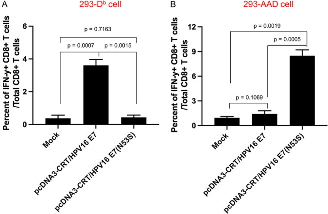
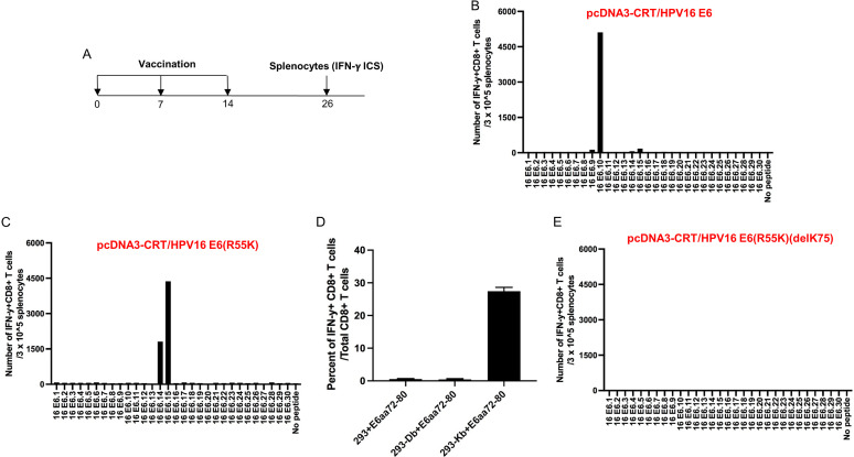
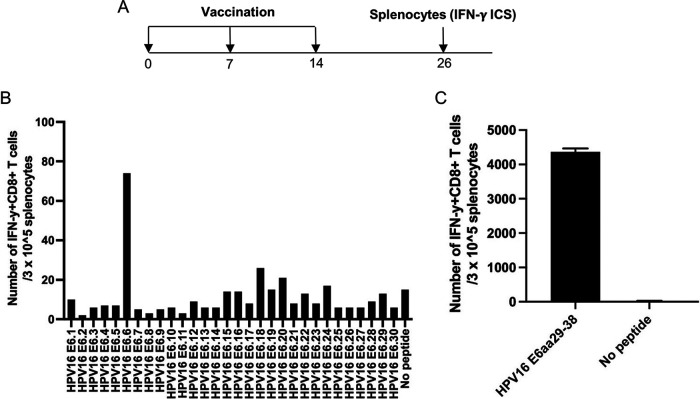
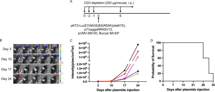
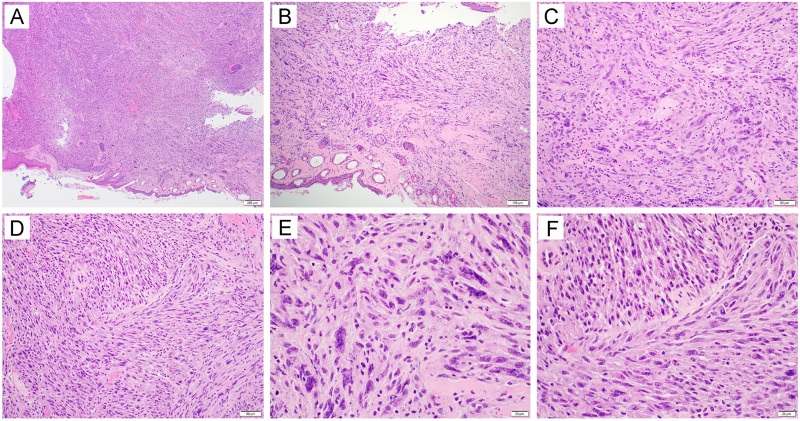
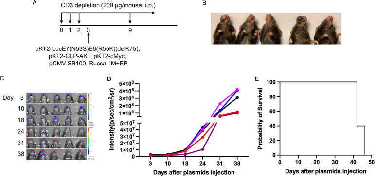
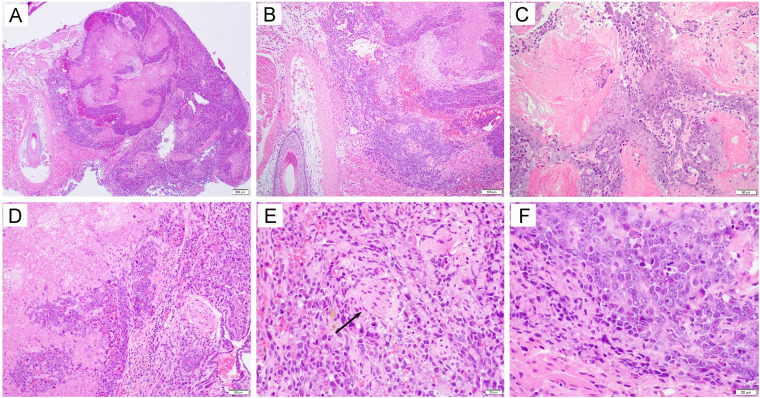
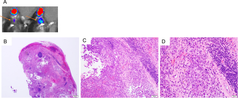
Human papillomavirus (HPV)-associated head and neck squamous cell carcinoma (HNSCC) is a growing global health problem. HPV16 has been attributed to a majority of HPV-associated HNSCCs. In order to test candidate immunotherapies, we developed a spontaneous HPV16-driven HNSCC model in HLA-A2 (AAD) transgenic mice. We sought to eliminate the confounding effects of dominant HPV antigen presentation through murine major histocompatibility complex class I (MHC-I) via epitope mutagenesis (without compromising tumorigenicity). We generated HPV16 E6(R55K)(delK75) and E7(N53S) expression constructs with mutations in known dominant H-2Db epitopes and characterized their presentation through murine and human MHC-I molecules using in vitro and in vivo activation of HPV16 E6/E7 antigen-specific CD8+ T cells. In addition, we tested the ability of E6(R55K)(delK75) and E7(N53S) for oncogenicity. The mutated E7(N53S) abolished the presentation of murine H-2Db-restricted HPV16 E7 peptide (i.e., amino acids [aa] 49 to 57) cytotoxic T lymphocyte (CTL) epitope and resulted in HLA-A2-restricted presentation of the HPV16 E7 (aa 11 to 20)-specific CTL epitope. The mutated E6(R55K)(delK75) abolished the activation of murine MHC-I-restricted E6-specific CD8+ T cell-mediated immune responses in C57BL/6 mice. In addition, the vaccination led to the activation of human HLA-A2-restricted E6-specific CD8+ T cell-mediated immune responses in HLA-A2 (AAD) transgenic mice. Injection of DNA plasmids encoding LucE7(N53S)E6(R55K)(delK75), AKT, c-Myc, and SB100 followed by electroporation results in development of squamous cell carcinoma in the oral/pharyngeal cavity of all of the HLA-A2 (AAD) transgenic mice (5/5), with 2/5 tumor-bearing mice developing metastatic carcinoma in the neck lymph nodes. IMPORTANCE Our data indicate that mutated HPV16 E6(R55K)(delK75) and mutated HPV16 E7(N53S) DNA abolishes the presentation of HPV16 E6 and E7 through murine MHC-I and results in their presentation through human HLA-A2 molecules. Additionally, the mutated HPV16 E6 and E7 remain oncogenic. Our approach is potentially applicable to different human MHC-I transgenic mice for the identification of human MHC-I restricted HPV16 E6/E7-specific CTL epitopes as well as the generation of spontaneous HPV E6/E7-expressing oral/pharyngeal carcinoma.

Keywords: E6; E7; HPV16; OPSCC; human papillomavirus; oral tumor model.


Conflict of interest statement

T.-C. Wu and Richard B. S. Roden are cofounders of and have an equity ownership interest in Papivax LLC. Also, both own Papivax Biotech, Inc., stock and are members of the Scientific Advisory Board of Papivax Biotech, Inc. All other authors declare no conflict of interest.

Figures

FIG 1
FIG 1
Comparison of CD8+ T cell epitope presentation between wild-type and mutant HPV16 E7(N53S) using 293 cells. 293-Db or 293-AAD cells were transfected with either pcDNA3-CRT/HPV16 E7 or pcDNA-CRT/HPV16E7(N53S) and then were cocultured with either HPV16 E7 (aa 49 to 57) peptide- or HPV16 E7 (aa 11 to 20) peptide-specific CD8+ T cells in the presence of brefeldin A. Peptide-specific CD8+ T cell activation was analyzed by intracellular IFN-γ staining. (A) Summary of HPV16 E7-specific CD8+ T cell activation by 293-Db cells by intracellular IFN-γ staining. (B) Summary of HPV16 E7-specific CD8+ T cell activation by 293-AAD cells by intracellular IFN-γ staining. Data are expressed as the mean percentage of IFN-γ+ CD8+ T cells out of the total number of CD8+ T cells ± SD.
FIG 2
FIG 2
Characterization of HPV16 E6-specific CD8+ T cell-mediated immune responses in C57BL/6 mice vaccinated with different DNA constructs and determination of MHC-I restriction of HPV16 E6 (aa 72 to 80) peptide. (A) Schema of the experimental regimen. Briefly, female C57BL/6 mice (5 per group) were vaccinated with 20 μg/mouse of pcDNA3-CRT/E6, pcDNA3-CRT/E6(R55K), or pcDNA3-CRT/E6(R55K)(delK75) DNA vaccine on day 0 through intramuscular injection followed by electroporation. One week later, the mice were boosted once with the same dose and regimen. One week later, the vaccinated mice were boosted with TA-HPV vaccine by skin scarification. Twelve days after the TA-HPV vaccination, the splenocytes from the mice were prepared and stimulated with HPV16 E6 overlapping peptides that span the full-length of the HPV16 E6 protein or HPV16 E6 (aa 72 to 80) peptides. The cells were then stained with PE-conjugated anti-mouse CD8a. After permeabilization and fixation, the cells were stained with FITC-conjugated anti-mouse IFN-γ. The stained cells were analyzed by flow cytometry. (B to D) Bar graphs summarizing the numbers of activated (IFN-γ+) HPV16 E6-specific CD8+ T cell responses by the various peptides with splenocytes from C57BL/6 mice after vaccination with (B) pcDNA3-CRT/E6 DNA, (C) pcDNA3-CRT/E6(R55K) DNA, or (E) pcDNA3-CRT/E6(R55K)(delK75) DNA. (D) Determination of MHC-I restriction of HPV16 E6 (aa 72 to 80) CD8+ T cell epitope.
FIG 3
FIG 3
Characterization of HPV16 E6-specific CD8+ T cell mediated immune responses in HLA-A2 (AAD) transgenic C57BL/6 mice vaccinated with the pcDNA3-CRT/E6(R55K)(delK75) DNA construct. (A) Schema of the experimental regimen. Briefly, female AAD mice (5 per group) were vaccinated with 20 μg/mouse of pcDNA3-CRT/E6(R55K)(delK75) DNA vaccine on day 0 through intramuscular injection followed by electroporation. One week later, the mice were boosted once with the same dose and regimen. One week later, the vaccinated mice were boosted with TA-HPV vaccine by skin scarification. Twelve days after the TA-HPV vaccination, the splenocytes from the mice were prepared and stimulated with HPV16 E6 overlapping peptides that span the full length of the HPV16 E6 protein or HPV16 E6 peptides (aa 29 to 38). The cells were then stained with PE-conjugated anti-mouse CD8a. After permeabilization and fixation, the cells were stained with FITC-conjugated anti-mouse IFN-γ. The stained cells were analyzed by flow cytometry. (B) Bar graph summarizing the number of activated (IFN-γ+) HPV16 E6-specific CD8+ T cell responses in vaccinated AAD mouse splenocytes against HPV16 E6 overlapping peptides. (C) Bar graph summarizing the number of activated (IFN-γ+) HPV16 E6-specific CD8+ T cell responses in vaccinated AAD mouse splenocytes against HPV16 E6 (aa 29 to 38) peptide.
FIG 4
FIG 4
Generation of spontaneous HPV16 E6E7-expressing oral sarcoma model in HLA-A2 (AAD) transgenic mice. (A) Schema of the experiment. Briefly, 6- to 8-week-old female HLA-A2 [(HLA-A*0201/Db) (AAD)] mice (n = 5) were depleted of CD3+ T cells by daily intraperitoneal (i.p.) injection of purified anti-mouse CD3 monoclonal antibody (clone 17A2; 150 μg/mouse) for 3 days. One day later, the mice were injected with plasmids encoding LucE6(R55K)(delK75)/E7(R53S), NRasG12V, and Sleeping Beauty (SB) transposase through submucosa of the oral/pharyngeal cavity followed by electroporation (EP). The expression of luciferase (tumor growth signal) was monitored by bioluminescence imaging. (B) Bioluminescence image of tumor growth caused by plasmids injected into mice. (C). Summary line graph of the bioluminescence image. Each line represents growth in one mouse. (D). Summary Kaplan-Meier survival curve for Luc-HPV16E6(R55K)(delK75)/E7(N53S)-expressing tumor-bearing AAD mice.
FIG 5
FIG 5
Histological examination of tumors formed in HLA-A2 (AAD) transgenic mice injected with plasmids encoding LucE6(R55K)(delK75)/E7(R53S), NRasG12V, and Sleeping Beauty transposase (SB100) followed by electroporation. Briefly, 6- to 8-week-old female HLA-A2 [(HLA-A*0201/Dd) (AAD)] mice (n = 5) were depleted of CD3+ T cells by daily intraperitoneal injection of purified anti-mouse CD3 monoclonal antibody (clone 17A2; 150 μg/mouse) for 3 days. One day later, the mice were injected with plasmids encoding LucE6(R55K)(delK75)/E7(R53S), NRasG12V, and SB100 through submucosa of the oral/pharyngeal cavity followed by electroporation. At week 6, mice were sacrificed, and oral tissues were harvested for histological examination. The sections showed a mass lesion (A [40×]) with entrapment of hair follicles and skeletal muscle (B [100×]). The tumor was characterized as a malignant spindle cell proliferation arranged in disordered fascicles (C [200×]) with a focal storiform growth pattern (D [200×]). The tumor cells displayed significant cytologic atypia and brisk mitotic activity (E and F [400×]).
FIG 6
FIG 6
Generation of spontaneous HPV16 E6E7-expressing oral/pharyngeal carcinoma model in HLA-A2 (AAD) transgenic mice. (A) Schematic of the experiment. Briefly, 6- to 8-week-old female HLA-A2 [(HLA-A*0201/Dd) (AAD)] mice (n = 5) were depleted of CD3+ T cells by daily intraperitoneal injection (i.p.) of purified anti-mouse CD3 monoclonal antibody (clone 17A2; 200 μg/mouse) for 3 days. One day later, the mice were injected with plasmids pKT2-LucE7(N53S)E6(R55K)(delK75), pKT2-CLP-AKT, pKT2-cMyc, and pCMV-SB100 via submucosal injection in the oral/pharyngeal cavity followed by electroporation (EP). (B) Gross image of oral/pharyngeal tumor induced by plasmids injection in AAD mice. (C) Bioluminescence image of tumor growth caused by plasmids injected into mice. (D) Summary line graph of the bioluminescence image. Each line represents growth in one mouse. (E) Survival curve of the oral tumor-bearing AAD mice.
FIG 7
FIG 7
Histological examination of tumors formed in HLA-A2 (AAD) transgenic mice injected with plasmids encoding LucE7(N53S)E6(R55K)(delK75), pKT2-CLP-AKT, pKT2-cMyc, and pCMV-SB100 followed by electroporation. Briefly, 6- to 8-week-old female HLA-A2 [(HLA-A*0201/Dd) (AAD)] mice (n = 5) were depleted of CD3+ T cells by daily intraperitoneal injection of purified anti-mouse CD3 monoclonal antibody (clone 17A2; 200 μg/mouse) for 3 days. One day later, the mice were injected with plasmids pKT2-LucE7(N53S)E6(R55K)(delK75), pKT2-CLP-AKT, pKT2-cMyc, and pCMV-SB100 via the submucosal injection in the oral/pharyngeal cavity followed by electroporation. At week 6 after oncogenic DNA transfection, mice were sacrificed and oral tissues were harvested for histological examination. The sections showed a nodular mass lesion (A [40×]) infiltrating into skeletal muscle (B [100×]). Extensive keratinization (C [200×]) and coagulative tumor cell necrosis (D [200×]) were present. On high-power magnification, the squamous cell carcinoma displayed epithelioid morphology, entrapped nerve tissue (E, arrow [400×]), and evident mitosis (F [400×]).
FIG 8
FIG 8
Histological examination of metastatic tumors of the neck lymph nodes derived from oral/pharyngeal carcinoma in HLA-A2 (AAD) transgenic mice injected with plasmids encoding LucE7(N53S)E6(R55K)(delK75), pKT2-CLP-AKT, pKT2-cMyc, and pCMV-SB100 followed by electroporation. Briefly, 6- to 8-week-old female HLA-A2 [(HLA-A*0201/Dd) (AAD)] mice (n = 5) were depleted of CD3+ T cells by daily intraperitoneal injection of purified anti-mouse CD3 monoclonal antibody (clone 17A2; 200 μg/mouse) for 3 days. One day later, the mice were injected with plasmids pKT2-LucE7(N53S)E6(R55K)(delK75), pKT2-CLP-AKT, pKT2-cMyc, and pCMV-SB100 via submucosal injection in the oral/pharyngeal cavity followed by electroporation. At week 6 after oncogenic DNA transfection, mice were sacrificed, and the metastatic tumors in the neck lymph nodes were harvested for histological examination. (A) Representative luminescence imaging of tumor-bearing mice with neck metastasis. Arrows indicate the location of the neck metastasis. (B to D) Representative histological examination of the metastatic lymph node: B, 40×; C, 200×; D, 400×. Please note that the representative neck lymph node was largely replaced by metastatic squamous cell carcinoma.

Similar articles

Cited by

References

    1. Ferlay J, Soerjomataram I, Dikshit R, Eser S, Mathers C, Rebelo M, Parkin DM, Forman D, Bray F. 2015. Cancer incidence and mortality worldwide: sources, methods and major patterns in GLOBOCAN 2012. Int J Cancer 136:E359–E386. doi:10.1002/ijc.29210. - DOI - PubMed
    1. Gillison ML, Chaturvedi AK, Anderson WF, Fakhry C. 2015. Epidemiology of human papillomavirus-positive head and neck squamous cell carcinoma. J Clin Oncol 33:3235–3242. doi:10.1200/JCO.2015.61.6995. - DOI - PMC - PubMed
    1. D'Souza G, Dempsey A. 2011. The role of HPV in head and neck cancer and review of the HPV vaccine. Prev Med 53(Suppl 1):S5–S11. doi:10.1016/j.ypmed.2011.08.001. - DOI - PMC - PubMed
    1. Ferlay J, Colombet M, Soerjomataram I, Mathers C, Parkin DM, Pineros M, Znaor A, Bray F. 2019. Estimating the global cancer incidence and mortality in 2018: GLOBOCAN sources and methods. Int J Cancer 144:1941–1953. doi:10.1002/ijc.31937. - DOI - PubMed
    1. Lewis A, Kang R, Levine A, Maghami E. 2015. The new face of head and neck cancer: the HPV epidemic. Oncology (Williston Park) 29:616–626. - PubMed

Publication types