. 2022 Apr 25;43(3):217-230.
doi: 10.1093/carcin/bgac008.

A combined treatment with melatonin and andrographis promotes autophagy and anticancer activity in colorectal cancer

Affiliations

A combined treatment with melatonin and andrographis promotes autophagy and anticancer activity in colorectal cancer

Yinghui Zhao et al. Carcinogenesis. .

Abstract

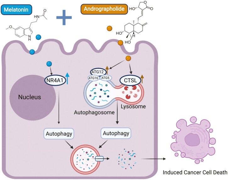
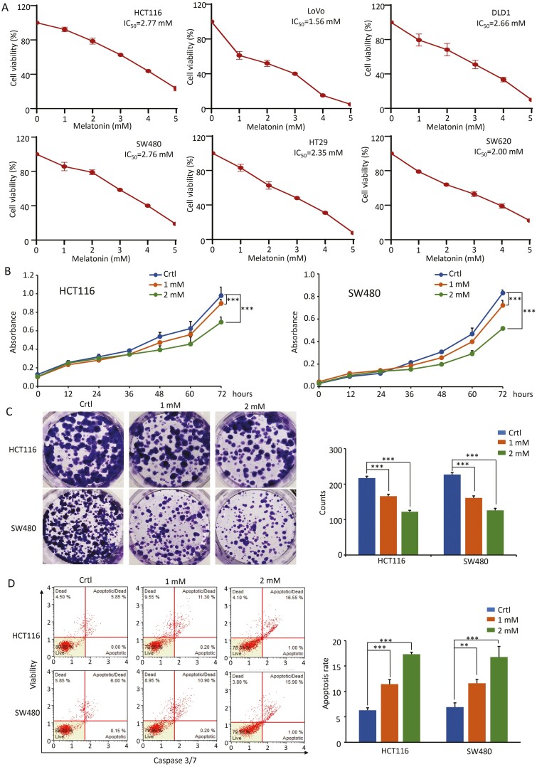
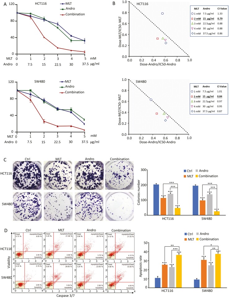
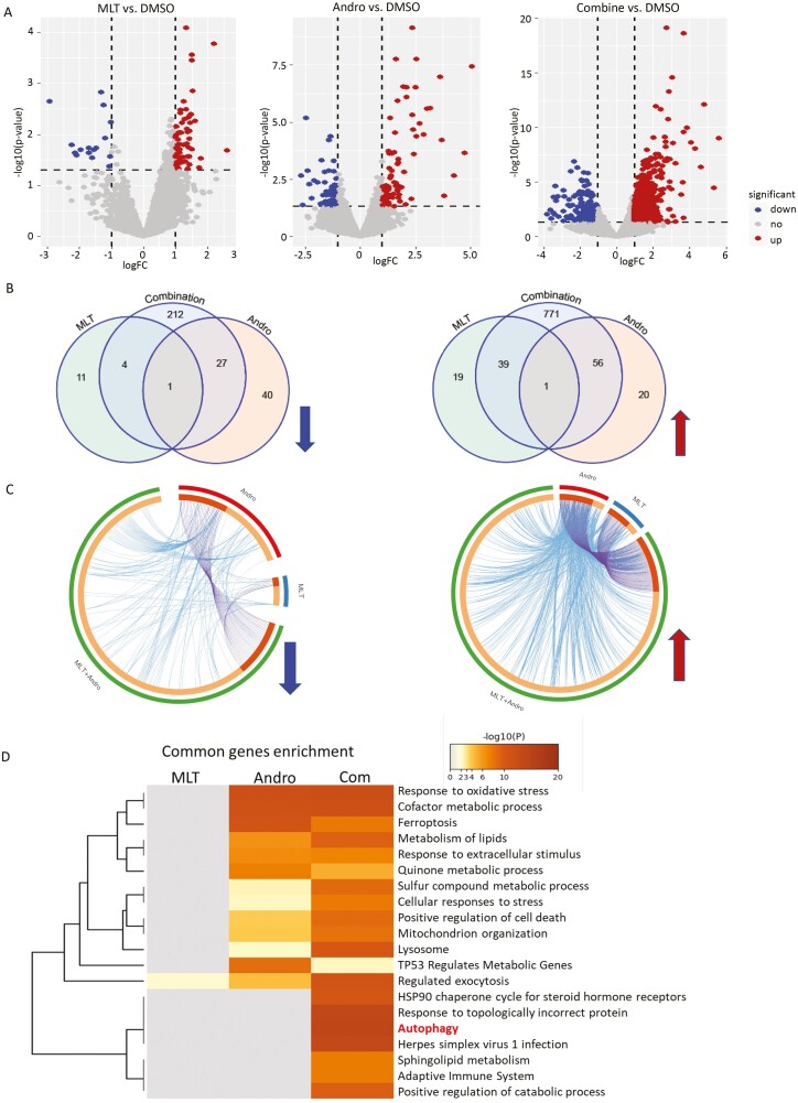
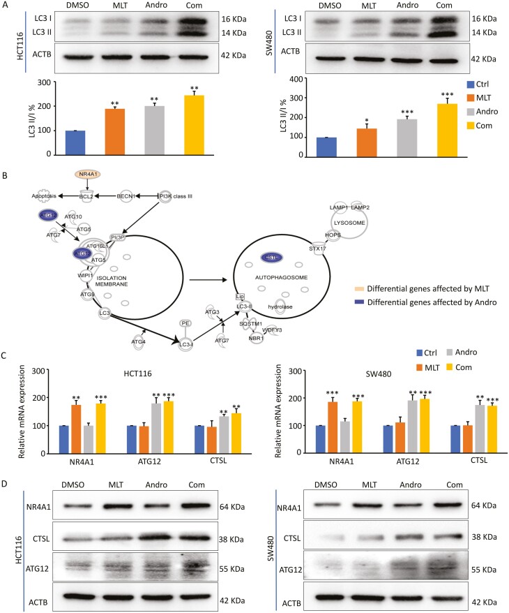
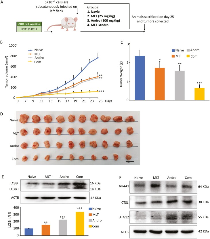
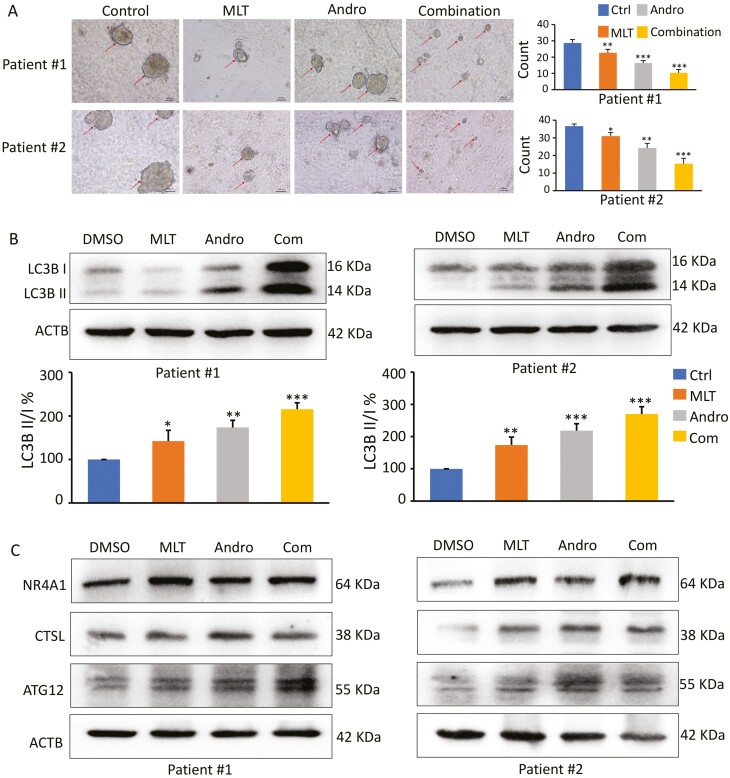
Colorectal cancer (CRC) is one of the most frequent malignancies worldwide and remains one of the leading causes of cancer-related deaths in the USA. The high degree of morbidity and mortality associated with this disease is largely due to the inadequate efficacy of current treatments as well the development of chemoresistance. In recent years, several pharmaceutical agents screened from natural products have shown the promise to offer a safe, inexpensive and synergistically multi-targeted treatment option in various cancers. Given the growing evidence of anti-carcinogenic properties of two natural compounds, melatonin (MLT) and andrographis (Andro), we aimed to evaluate their synergistic anticancer effects in CRC. We demonstrate that indeed these two compounds possessed a synergistic anticancer effect in terms of their ability to inhibit cell viability, suppression of colony-formation and induction of apoptosis (P < 0.05). In line with our in vitro findings, we were able to validate this combinatorial anticancer activity in xenograft animal models (P < 0.001) as well as tumor-derived 3D organoids (P < 0.01). RNA-sequencing analysis revealed candidate pathways and genes that mediated antitumor efficacy of MLT and Andro in CRC, among which autophagy pathway and related genes, including NR4A1, CTSL and Atg12, were found to be primarily responsible for the increased anticancer effect by the two natural products. In conclusion, our data reveal a potent and synergistic therapeutic effect of MLT and Andro in the treatment of CRC and provides a rationale for suppressing autophagy in cancer cells as a potential therapeutic strategy for CRC.


Figures

None
Graphical abstract
Figure 1.
Figure 1.
MLT inhibits the growth of CRC cells in a dose-dependent manner. (A) The cell viability of six CRC cell lines treated with different concentrations of MLT (1–5 mM) for 48 h was determined by CCK-8 assay. (B) Results of CCK-8 assay showing the effect of different MLT concentrations (1 mM, 2 mM) on CRC cell viability. Complete medium containing MLT was added at 24 h and the absorbance was measured at 450 nm after cell culture for 0, 12, 24, 36, 48, 60 and 72 h. (C) Representative results of colony formation assay in CRC cells after exposure to MLT (1 mM, 2 mM) for 7 days. Colony formation count analysis is shown in the right panel. (D) Representative results of FACS analysis using the Muse Caspase-3/7 Kit after treatment on CRC cells by MLT (1 mM, 2 mM) for 48 h. Apoptosis rate analysis is shown in the right panel. Statistical significance: ∗∗P < 0.01, ∗∗∗P < 0.001.
Figure 2.
Figure 2.
The combination of MLT and Andro results in synergistic anticancer effects in CRC cells. (A) Results of CCK-8 assay comparing cell viability following treatment with MLT, Andro and their combination for 48 h in CRC cell lines. (B) Isobologram analysis of CI values based on the results of CCK-8 assay to determine synergistic effects of MLT and Andro in CRC cell lines. (C) Representative results of colony formation assay following treatment with MLT, Andro and their combination for 48 h in CRC cell lines. Colony count analysis is shown in the right panel. (D) Representative results of FACS analysis using the Muse Caspase-3/7 Kit after treatment on CRC cells by MLT, Andro and their combination for 48 h. Apoptosis rate analysis is shown in the right panel. Statistical significance: ∗P < 0.05, ∗∗P < 0.01, ∗∗∗P < 0.001.
Figure 3.
Figure 3.
Genomewide transcriptomic profiling changes in CRC cells following treatment with MLT, Andro and their combination. (A) The volcano plot obtained from genomewide transcriptomic analyses in HCT116 and SW480 cells in each treatment group (MLT, Andro and their combination) relative to vehicle-treated controls. (B) The Venn diagrams representing the overlap of DEGs among various treatment groups. The up- and down-regulated genes were analyzed and represented separately. (C) The circos diagram depicting the gene correlations among different treatment groups in the up- and down-regulated DEGs using Metascape software. The outermost ring depicts different treatment groups of MLT, Andro and their combination. The inner ring represents the genes that are common in multiple groups (darker orange) and that are unique to each group (lighter orange). (D) Comparative heatmap representing the convergence and difference among different treatment groups by biological functions and pathway enrichment using Metascape software.
Figure 4.
Figure 4.
Andro sensitizes MLT-induced anticancer effects by promoting autophagy in CRC cells. (A) Representative images of LC3B expression in the extracts of CRC cells in each treatment group by western blot analysis. The ratios of LC3-II to LC3-I based on the quantification of the bands in the immunoblot were shown in the lower panel. (B) Pattern diagram representing the significantly DEGs in the autophagy pathway after MLT and ANDRO treatment, respectively, by IPA analysis. (C) Bar graph showing relative mRNA expression of NR4A1, ATG12 and CTSL in each treatment group by qPCR analysis. (D) Representative images of NR4A1, ATG12 and CTSL expression in the extracts of CRC cells in each treatment group by western blot analysis. Statistical significance: ∗P < 0.05, ∗∗P < 0.01, ∗∗∗P < 0.001.
Figure 5.
Figure 5.
The combination of MLT and Andro synergistically inhibit xenograft growth in an animal model. (A) Schematic diagram of HCT116 cell-derived xenograft model in nude mice and the treatment schedule of MLT, Andro and their combination. (B) Tumor volume alterations in each treatment group were measured at different time points after inoculation. (C) The measurement of tumor weights within each treatment group 25 days post-inoculation. (D) Representative images of harvested tumors surgically removed from nude mice 25 days post-inoculation. (E) Representative images of LC3B expression in the extracts of xenografts in each treatment group by western blot analysis. The ratios of LC3-II to LC3-I based on the quantification of the bands in the immunoblot were shown in the lower panel. (F) Representative images of NR4A1, ATG12 and CTSL expression in the extracts of xenografts in each treatment group by western blot analysis. Statistical significance: ∗P < 0.05, ∗∗P < 0.01, ∗∗∗P < 0.001.
Figure 6.
Figure 6.
A combination of MLT and Andro synergistically inhibit patient-derived tumor organoid growth. (A) Microscopic images of patient-derived tumor organoids derived from two patients cultured in each treatment group (20× magnification); the organoid count analysis are shown in the right panel. (B) Representative images of LC3B expression in the extracts of organoids in each treatment group by western blot analysis. The ratios of LC3-II to LC3-I based on the quantification of the bands in the immunoblot were shown in the lower panel. (C) Representative images of NR4A1, ATG12 and CTSL expression in the extracts of organoids in each treatment group by western blot analysis. Statistical significance: ∗P < 0.05, ∗∗P < 0.01, ∗∗∗P < 0.001.

References

    1. Siegel, R.L. et al. (2021) Cancer Statistics, 2021. CA. Cancer J. Clin., 71, 7–33. - PubMed
    1. Arnold, M. et al. (2017) Global patterns and trends in colorectal cancer incidence and mortality. Gut, 66, 683–691. - PubMed
    1. Mlecnik, B. et al. (2020) Multicenter International Society for Immunotherapy of Cancer Study of the Consensus Immunoscore for the Prediction of Survival and Response to Chemotherapy in Stage III Colon Cancer. J. Clin. Oncol., 38, 3638–3651. - PMC - PubMed
    1. Modest, D.P. et al. (2018) Surgical treatment options following chemotherapy plus cetuximab or bevacizumab in metastatic colorectal cancer—central evaluation of FIRE-3. Eur. J. Cancer, 88, 77–86. - PubMed
    1. Xie, Y.H. et al. (2020) Comprehensive review of targeted therapy for colorectal cancer. Signal Transduct. Target. Ther., 5, 22. - PMC - PubMed

Publication types