. 2022 Jan 28;8(4):eabi7711.
doi: 10.1126/sciadv.abi7711. Epub 2022 Jan 28.

Modulating environmental signals to reveal mechanisms and vulnerabilities of cancer persisters

Affiliations

Modulating environmental signals to reveal mechanisms and vulnerabilities of cancer persisters

Xiaoxiao Sun et al. Sci Adv. .

Abstract

Cancer persister cells are able to survive otherwise lethal doses of drugs through nongenetic mechanisms, which can lead to cancer regrowth and drug resistance. The broad spectrum of molecular differences observed between persisters and their treatment-naïve counterparts makes it challenging to identify causal mechanisms underlying persistence. Here, we modulate environmental signals to identify cellular mechanisms that promote the emergence of persisters and to pinpoint actionable vulnerabilities that eliminate them. We found that interferon-γ (IFNγ) can induce a pro-persistence signal that can be specifically eliminated by inhibition of type I protein arginine methyltransferase (PRMT) (PRMTi). Mechanistic investigation revealed that signal transducer and activator of transcription 1 (STAT1) is a key component connecting IFNγ's pro-persistence and PRMTi's antipersistence effects, suggesting a previously unknown application of PRMTi to target persisters in settings with high STAT1 expression. Modulating environmental signals can accelerate the identification of mechanisms that promote and eliminate cancer persistence.


Figures
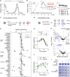
Fig. 1.
Fig. 1.. Sequential search for persistence modulators in PC9 cells.
(A) Reversibility of cancer persistence [solid/dashed lines, on/off erlotinib (Erl)]. Relative cell viability normalized to day 0 (n = 3). (B) Left: Relative cell viability in − Erl (gray; n = 6) or + Erl (black; n = 8). Inset: Zoom-in of + Erl curve highlighting the emergence of persisters (red). Right: reproducibility across experimental batches. (C) Workflow of pro-persistence screen. Red: Pro-persistence factor. (D) Pro-persistence screen. Relative persistence normalized to controls (Ctrl). Pert_L/H, low/high-dose perturbations. Red arrow: IFNγ. TFGβ, transforming growth factor–β; TNFα, tumor necrosis factor–α; IL6, interleukin-6; GH, growth hormone; FSH, follicle stimulating hormone; LTH, luterotropic hormone; PGE2, prostaglandin E2; NAD+, nicotinamide adenine dinucleotide; LPA, lysophosphatidic acid; sDLL1, secreted extracellular domain of Notch receptor ligand DLL-1; SAM, S-(5’-adenosyl)-L-methionine. (E) Relative Erl persistence in ± IFNγ, normalized to − IFNγ (n = 6). (F) Relative Erl persistence in ± IFNγ + DMSO or ruxolitinib (200 nM), normalized to − IFNγ + DMSO (n = 3). (G) Workflow of persister vulnerability screen. Blue, context-dependent vulnerability. (H) Vulnerability screen. Relative persistence normalized to DMSO controls horizontal dashed line: P < 0.05. Compounds with no significant change in the counter screen (top) are shown (bottom). Blue, type I PRMT inhibitor MS023. (I) Crystal violet staining of cells in ± Erl ± IFNγ (0.5 ng/ml) ± MS023 (0.5 μM). Dashed box, antipersistence efficacy of MS023 in IFNγ. (A to I) Erl: 2.5 μM unless otherwise indicated. Error bar: SD. Unpaired, two-tailed t tests: ns, P > 0.05; *P ≤ 0.05; **P ≤ 0.01; ****P ≤ 0.0001.
Fig. 2.
Fig. 2.. IFNγ’s pro-persistence and PRMTi’s antipersistence effects in PC9 cells depend on STAT1.
(A) Relative Erl persistence of STAT1+/+, STAT1+/−, or STAT1−/− cells in ± IFNγ, normalized to − IFNγ (n = 5). (B) Relative Erl persistence of STAT1+/+ (left) or STAT1−/− (right) cells in ± MS023 ± IFNγ, normalized to − MS023 ± IFNγ (n = 3). STAT1 protein level (C) and mRNA level (D) of cells in ± GSK3368715 (GSK715) for 3 days, followed by ± IFNγ (5 ng/ml) for 1 day. (E) Relative intensity of unmodified, monomethylated, and dimethylated PABP2 peaks in mass spectrometry with cells in ± MS023 (5 μM) for 3 days. (F) STAT1 protein level of cells with ± PABP2 knockdown (siPABP2) for 3 days, followed by ± IFNγ (5 ng/ml) for 1 day. (G) P-STAT1 intensity ratio (N/C: nuclear versus whole cell) of cells in ± GSK715 for 3 days, followed by ± IFNγ (2 ng/ml) for 1 day. Shown are averages of single cell results in replicate wells (n = 3; >1000 cells per well). IRF1 mRNA level (H) and protein level (I) of cells in the same treatment as in (D) and (G), respectively. IRF1 intensity in whole cell was measured. (J) Relative Erl persistence of cells with (siIRF1) or without (siNC) knockdown for 2 days, followed by persister assays in ± IFNγ; normalized to − IFNγ with siNC (n = 5). (A to J) Erl: 2.5 μM. Error bar: SD. Unpaired, two-tailed t tests: ns, P > 0.05; *P ≤ 0.05; **P ≤ 0.01; ***P ≤ 0.001; ****P ≤ 0.0001. (C and F) Loading controls: Glyceraldehyde-3-phosphate dehydrogenase (GAPDH).
Fig. 3.
Fig. 3.. Type I PRMT inhibition reduces EGFRi persistence with STAT1-high cancer cells.
(A) Baseline protein levels of NSCLC cells without IFNγ stimulation. Loading control: GAPDH. (B) Relative persistence of endogenously STAT1-high cells with EGFRi ± MS023 ± IFNγ (2 ng/ml), normalized to − MS023 ± IFNγ (n = 3). Erlotinib, 0.5 μM; osimertinib, 50 nM. No significant differences between ± IFNγ curves in each of the three models (two-tailed, paired t test; P > 0.05). (C) Crystal violet staining of H1975 cells in ± osimertinib (Osi, 100 nM) ± IFNγ (0.5 ng/ml) ± GSK715 (0.5 μM). (D) Effects of type I PRMT inhibition as a sequential treatment in PC9 cells. Top: Treatment schedules. Solid lines, + Erl (2.5 μM) for 6 days; dotted line, − Erl for 12 days; arrow, +MS023 for 9 days. Bottom left: STAT1 mRNA levels, normalized to Erl-naïve cells (n = 2). Unpaired, two-tailed t tests: *P ≤ 0.05. Bottom right: relative viability of cells, normalized to no MS023 controls (n = 5). (B and D) Error bar: SD.

References

    1. Sharma S. V., Lee D. Y., Li B., Quinlan M. P., Takahashi F., Maheswaran S., McDermott U., Azizian N., Zou L., Fischbach M. A., Wong K.-K., Brandstetter K., Wittner B., Ramaswamy S., Classon M., Settleman J., A chromatin-mediated reversible drug-tolerant state in cancer cell subpopulations. Cell 141, 69–80 (2010). - PMC - PubMed
    1. Ramirez M., Rajaram S., Steininger R. J., Osipchuk D., Roth M. A., Morinishi L. S., Evans L., Ji W., Hsu C.-H., Thurley K., Wei S., Zhou A., Koduru P. R., Posner B. A., Wu L. F., Altschuler S. J., Diverse drug-resistance mechanisms can emerge from drug-tolerant cancer persister cells. Nat. Commun. 7, 10690 (2016). - PMC - PubMed
    1. Hata A. N., Niederst M. J., Archibald H. L., Gomez-Caraballo M., Siddiqui F. M., Mulvey H. E., Maruvka Y. E., Ji F., Bhang H. C., Radhakrishna V. K., Siravegna G., Hu H., Raoof S., Lockerman E., Kalsy A., Lee D., Keating C. L., Ruddy D. A., Damon L. J., Crystal A. S., Costa C., Piotrowska Z., Bardelli A., Iafrate A. J., Sadreyev R. I., Stegmeier F., Getz G., Sequist L. V., Faber A. C., Engelman J. A., Tumor cells can follow distinct evolutionary paths to become resistant to epidermal growth factor receptor inhibition. Nat. Med. 22, 262–269 (2016). - PMC - PubMed
    1. Shen S., Vagner S., Robert C., Persistent cancer cells: The deadly survivors. Cell 183, 860–874 (2020). - PubMed
    1. Russo M., Crisafulli G., Sogari A., Reilly N. M., Arena S., Lamba S., Bartolini A., Amodio V., Magrì A., Novara L., Sarotto I., Nagel Z. D., Piett C. G., Amatu A., Sartore-Bianchi A., Siena S., Bertotti A., Trusolino L., Corigliano M., Gherardi M., Lagomarsino M. C., Nicolantonio F. D., Bardelli A., Adaptive mutability of colorectal cancers in response to targeted therapies. Science 366, 1473–1480 (2019). - PubMed

Publication types

Substances