. 2022 Mar;603(7903):919-925.
doi: 10.1038/s41586-022-04466-x. Epub 2022 Jan 28.

Memory B cell repertoire from triple vaccinees against diverse SARS-CoV-2 variants

Affiliations

Memory B cell repertoire from triple vaccinees against diverse SARS-CoV-2 variants

Kang Wang et al. Nature. 2022 Mar.

Abstract

Omicron (B.1.1.529), the most heavily mutated SARS-CoV-2 variant so far, is highly resistant to neutralizing antibodies, raising concerns about the effectiveness of antibody therapies and vaccines1,2. Here we examined whether sera from individuals who received two or three doses of inactivated SARS-CoV-2 vaccine could neutralize authentic Omicron. The seroconversion rates of neutralizing antibodies were 3.3% (2 out of 60) and 95% (57 out of 60) for individuals who had received 2 and 3 doses of vaccine, respectively. For recipients of three vaccine doses, the geometric mean neutralization antibody titre for Omicron was 16.5-fold lower than for the ancestral virus (254). We isolated 323 human monoclonal antibodies derived from memory B cells in triple vaccinees, half of which recognized the receptor-binding domain, and showed that a subset (24 out of 163) potently neutralized all SARS-CoV-2 variants of concern, including Omicron. Therapeutic treatments with representative broadly neutralizing monoclonal antibodies were highly protective against infection of mice with SARS-CoV-2 Beta (B.1.351) and Omicron. Atomic structures of the Omicron spike protein in complex with three classes of antibodies that were active against all five variants of concern defined the binding and neutralizing determinants and revealed a key antibody escape site, G446S, that confers greater resistance to a class of antibodies that bind on the right shoulder of the receptor-binding domain by altering local conformation at the binding interface. Our results rationalize the use of three-dose immunization regimens and suggest that the fundamental epitopes revealed by these broadly ultrapotent antibodies are rational targets for a universal sarbecovirus vaccine.


Conflict of interest statement

Y.H., Lin Wang and M.L. are employees of Sinovac Biotech Ltd. Y.J., P.G. and Y.C. are employees of Acrobiosystems Inc. The other authors declare no competing interests.

Figures

Fig. 1
Fig. 1. Evolution and neutralization characteristics of Omicron variant.
a, A linear representation of Omicron S with mutations indicated. The replacements are in red, deletions are in grey and insertions are in purple. b, Distribution of Omicron S mutations on the cryo-EM structure of pre-fusion S trimer determined at pH 7.5 (Protein Data Bank (PDB) ID 7WG6). The mutations listed in a are indicated in the ‘up’ protomer shown in cartoon, with mutated residues highlighted as spheres and coloured as in a. The RBD, NTD, SD1 and S2 domains of this subunit are marked with arrows and coloured green, blue, magenta and yellow, respectively; the other two protomers are in the ‘down’ state and shown in surface representation in pale cyan and pale yellow. Alpha, B.1.1.7; Gamma, P.1; Lambda, C.37. c, The neutralizing antibody response against WT and Omicron SARS-CoV-2 authentic virus for sera from healthy vaccinees who received two (n = 60 volunteers) or three (n = 60 volunteers) doses of Coronavac. Data are geometric mean ± s.d. of technical triplicates. The dotted line represents the detection limit. NT50 values of less than 4 were plotted as 2. Fold difference in neutralizing antibody titre Delta or Omicron over WT for each group of sera is shown above each set of points.
Fig. 2
Fig. 2. Characteristics of a subset of broadly neutralizing antibodies from recipients of a booster immunization.
a, Vertical slice chart shows the gross distribution of binding epitopes of monoclonal antibodies isolated from individuals who received three doses of inactivated SARS-CoV-2 vaccine. The total number of antibodies and the percentage of antibodies that recognize the RBD, NTD and S2 domain are indicated. b, Heat map representation of 41 selected representative monoclonal antibodies against pseudotyped viruses expressing WT or variant SARS-CoV-2 S. The colour bar on the right shows IC50 values for the indicated monoclonal antibodies against pseudotyped viruses in c. Antibodies marked with star were selected for structural analysis. c, Heat map showing the competition ability of selected monoclonal antibodies with human ACE2. Competition ability is represented by the AUC, ranging from 1 (weakest) to 24 (strongest). d, Neutralization curves for the selected antibodies towards pseudotyped viruses expressing Omicron S. Data represent three groups of antibodies shown in b. Yellow indicates antibodies with high neutralizing activity against all five VOCs; green indicates antibodies with high neutralizing activity against four VOCs and intermediate neutralizing activity against Omicron; red indicates antibodies with high neutralizing activity against four VOCs and weak neutralizing activity against Omicron. XGv347, XGv282 and XGv265 were selected as representatives of each group. All experiments were performed in duplicate.
Fig. 3
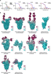
Fig. 3. Structural basis of the broad and potent neutralization of representative antibodies.
a, Side and top views of Cryo-EM maps of Omicron S trimer in complex with XGv347 (S states 1–3), XGv289, XGv282 and XGv265. State 1, one up RBD and one down RBD; state 2, three down RBDs; state 3, two up RBDs. b, Cartoon representations of the structures of Omicron S RBD in complex with XGv347 (top left), XGv289 (top right), XGv282 (bottom left) and XGv265 (bottom right). Two views are shown to illustrate the binding modes of the four antibodies. RBD is shown in cyan. c, Interactions between the four antibodies and Omicron S RBD. The CDRs of the four antibodies that interact with the RBD are shown as cartoon over the light green surface of RBD. The mutation sites on Omicron S RBD are in red; the epitopes of antibodies are in deep green and the overlap of mutation sites and epitopes are in blue. Residues of each epitope are indicated in the corresponding regions. d, Superposition of Omicron and WT S trimers. Omicron S trimer is in cyan and WT S trimer is in yellow.
Fig. 4
Fig. 4. Protection against challenge by SARS-CoV-2 Beta and Omicron variants in mice.
a, Experimental design for protection assay against Beta variant challenge. n = 4 mice in XGv347, XGv052 and XGv052 + XGv289 groups; n = 5 mice in other groups. bd, Lung tissues of mice challenged with Beta variant, collected at 5 dpi: virus titre (b), immunostaining (c) and haematoxylin and eosin staining (d). b, Viral subgenomic (sg) RNA loads in the lungs at 5 dpi were measured by quantitative PCR with reverse transcription. Data are mean ± s.d. Dashed line represents the limit of detection. c, In situ hybridization with a SARS-CoV-2 specific probe. Brown staining indicates the presence of SARS-CoV-2 genomic RNA. d, Histopathological analysis of lung samples at 5 dpi. e, f, Weight change (e) and viral RNA in lung tissues (f) of K18-hACE2 mice challenged with Omicron variant of concern. n = 5 mice in each group. e, The weight of each mouse in both groups was monitored and recorded daily following infection. Data are mean ± s.d. f, Viral RNA loads in the lungs at 5 dpi were measured as in b. Data are mean ± s.d. Dashed line represents the limit of detection. g, Histopathological analysis of lung tissues from infected mice treated with XGv347 or PBS. Micrographs in c, d, g are representative of two experiments.
Extended Data Fig. 1
Extended Data Fig. 1. Antibody-hACE2 competition ELISA assay.
Data shown are the curves of 31 antibodies used to compete with ACE2. All experiments were performed in duplicate.
Extended Data Fig. 2
Extended Data Fig. 2. Characteristics of representative antibodies against pseudotyped viruses.
a, Heatmap representation of five therapeutic mAbs approved or in clinical trials against pseudotyped viruses with the S proteins of wild-type or variants of concern or interst (Alpha, Beta, Gamma, Delta, Lambda and Omicron). b, Neutralization curves for these mAbs in correspondence with a. Mean of two experiments is shown.
Extended Data Fig. 3
Extended Data Fig. 3. Heatmap representation of representative mAbs against WT and variants of concern.
Color bar on the right showed the gradient of IC50 of different antibodies against the authentic WT and variants of concern. All experiments were performed in duplicate.
Extended Data Fig. 4
Extended Data Fig. 4. Data sheets of ELISA assay of representative mAbs against Omicron RBD.
Different Classes of mAbs (Class I-VI) are colored by yellow, green, red, blue, brown and magenta, respectively. Values are filled with black (>75), grey (50–75), silver (25–50) and white (<25). Each data is the mean of three values from three independent experiments.
Extended Data Fig. 5
Extended Data Fig. 5. Flowcharts for cryo-EM data processing.
Flowcharts for Omicron S protein in complex with a, XGv347, b, XGv289, c, XGv282 and d, XGv265 are shown. Scala bar in micrographs, 100 nm.
Extended Data Fig. 6
Extended Data Fig. 6. Resolution estimation of the EM maps.
a, The gold-standard FSC curves of overall maps of Omicron S trimer in complex with Fab XGv347, XGv289, XGv282 and XGv265 and local maps of interfaces. b, Local resolution assessments of cryo-EM maps using ResMap are shown.
Extended Data Fig. 7
Extended Data Fig. 7. Density maps and atomic models.
Cryo-EM density maps of Omicron S trimer in complex with XGv347, XGv289, XGv282 and XGv265 and their interfaces are shown. Color scheme is the same as in Fig. 3a. Residues are shown as sticks with oxygen colored in red, nitrogen colored in blue and sulfurs colored in yellow.
Extended Data Fig. 8
Extended Data Fig. 8. Multiple sequence alignment of XGv347, CoV2-2196 and A23-58.1.
Multiple sequence alignments of heavy chains and light chains of XGv347, CoV2-2196 and A23-58.1 were performed, respectively. Paratopes of XGv347 binding to Omicron variant RBD are highlighted by green boxes.
Extended Data Fig. 9
Extended Data Fig. 9. Mechanism of XGv347 binding to 3 closed RBD.
a, Superimposition of A23-58.1 onto WT S trimer. b, Superimposition of XGv347 onto WT S trimer. c, complex of XGv347 and Omicron S trimer. All complexes are in the same orientation with close-ups of Fab-RBD binding modes showing potential clashes.
Extended Data Fig. 10
Extended Data Fig. 10. Binding modes of XGv289, 282 and 265.
Binding modes of XGv289, XGv282 and XGv265. RBD is colored in light cyan and color scheme of XGv289, XGv282 and XGv265 is the same as in Fig. 3a. LY-CoV1404, BD-812 and REGN10987 are colored in purple, deep pink and blue, respectively.
Extended Data Fig. 11
Extended Data Fig. 11. Structural fitting.
XGv265, XGv282 and XGv289 are superimposed onto XGv347 and all structure are shown as surface.
Extended Data Fig. 12
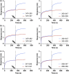
Extended Data Fig. 12. BLI assay for XGv347 competing with XGv289, XGv282 and XGv265.
Affinity curves of XGv347 to Omicron S protein competing with a, XGv265, b, XGv282 and c, XGv289. In each panel, (left) XGv347 was first injected, followed by the XGv265, XGv282 and XGv289 in ac, respectively. (right) Also, XGv265 in a, XGv282 in b and XGv289 in c, was injected first and competed with the second injection of XGv347. Each curve is a representative of three independent experiments.
Extended Data Fig. 13
Extended Data Fig. 13. Interactions details between antibodies (XGv347, XGv289, XGv282 and XGv265) and SARS-CoV-2 WT (left) and Omicron RBD (right).
All the WT structures are predicted with GROMACS. Hydrophobic patches and hydrogen bonds are denoted by surface and dash lines. Color scheme is the same as in Fig. 3a. For hydrophobic patches of XGv289, XGv282 and XGv265, G446 and S446 are colored in magenta. The dash lines marked out the hydrophobic patches only found in WT RBD.
Extended Data Fig. 14
Extended Data Fig. 14. Histopathological analysis of lung samples from XGv282 treatment group at 5 dpi.
Shown here are the H&E staining of lung samples from each of the remaining four mice in XGv282 group. Each micrograph is representative of two separate experiments.

Comment in

References

    1. Carreno, J. M. et al. Activity of convalescent and vaccine serum against a B. 1.1. 529 variant SARS-CoV-2 isolate. Nature10.1038/s41586-022-04399-5 (2021).
    1. Cao, Y. R. et al. B. 1.1. 529 escapes the majority of SARS-CoV-2 neutralizing antibodies of diverse epitopes. Nature10.1038/s41586-021-04385-3 (2021).
    1. Mlcochova P, et al. SARS-CoV-2 B. 1.617. 2 Delta variant replication and immune evasion. Nature. 2021;599:114–119. doi: 10.1038/s41586-021-03944-y. - DOI - PMC - PubMed
    1. Wang G-L, et al. Susceptibility of circulating SARS-CoV-2 variants to neutralization. N. Engl. J. Med. 2021;384:2354–2356. doi: 10.1056/NEJMc2103022. - DOI - PMC - PubMed
    1. Altmann DM, Boyton RJ, Beale R. Immunity to SARS-CoV-2 variants of concern. Science. 2021;371:1103–1104. doi: 10.1126/science.abg7404. - DOI - PubMed

MeSH terms

Supplementary concepts