

Learning white matter subject-specific segmentation from structural MRI
- PMID: 35090192
- PMCID: PMC9053869
- DOI: 10.1002/mp.15495
Learning white matter subject-specific segmentation from structural MRI
Abstract
Purpose: Mapping brain white matter (WM) is essential for building an understanding of brain anatomy and function. Tractography-based methods derived from diffusion-weighted MRI (dMRI) are the principal tools for investigating WM. These procedures rely on time-consuming dMRI acquisitions that may not always be available, especially for legacy or time-constrained studies. To address this problem, we aim to generate WM tracts from structural magnetic resonance imaging (MRI) image by deep learning.
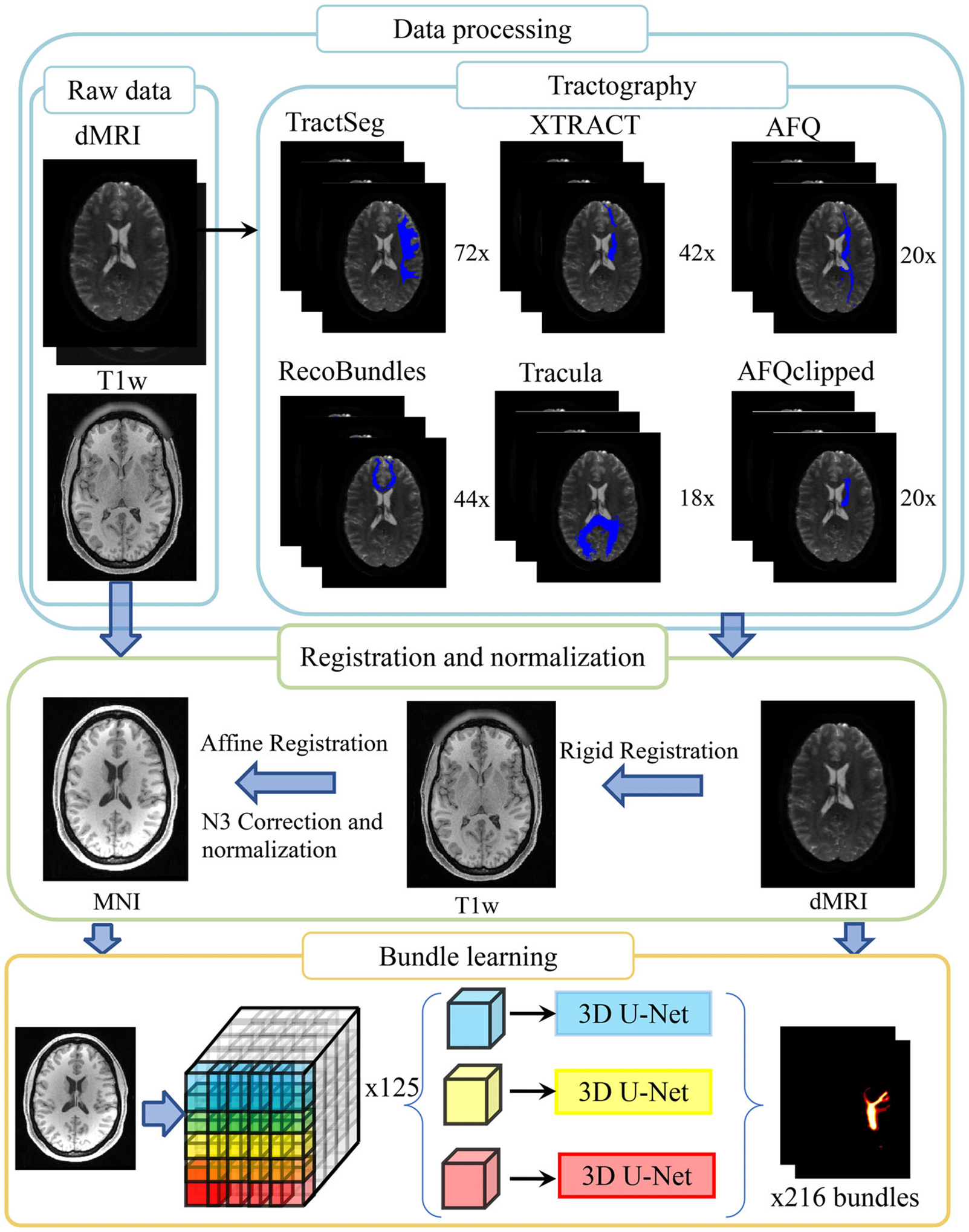
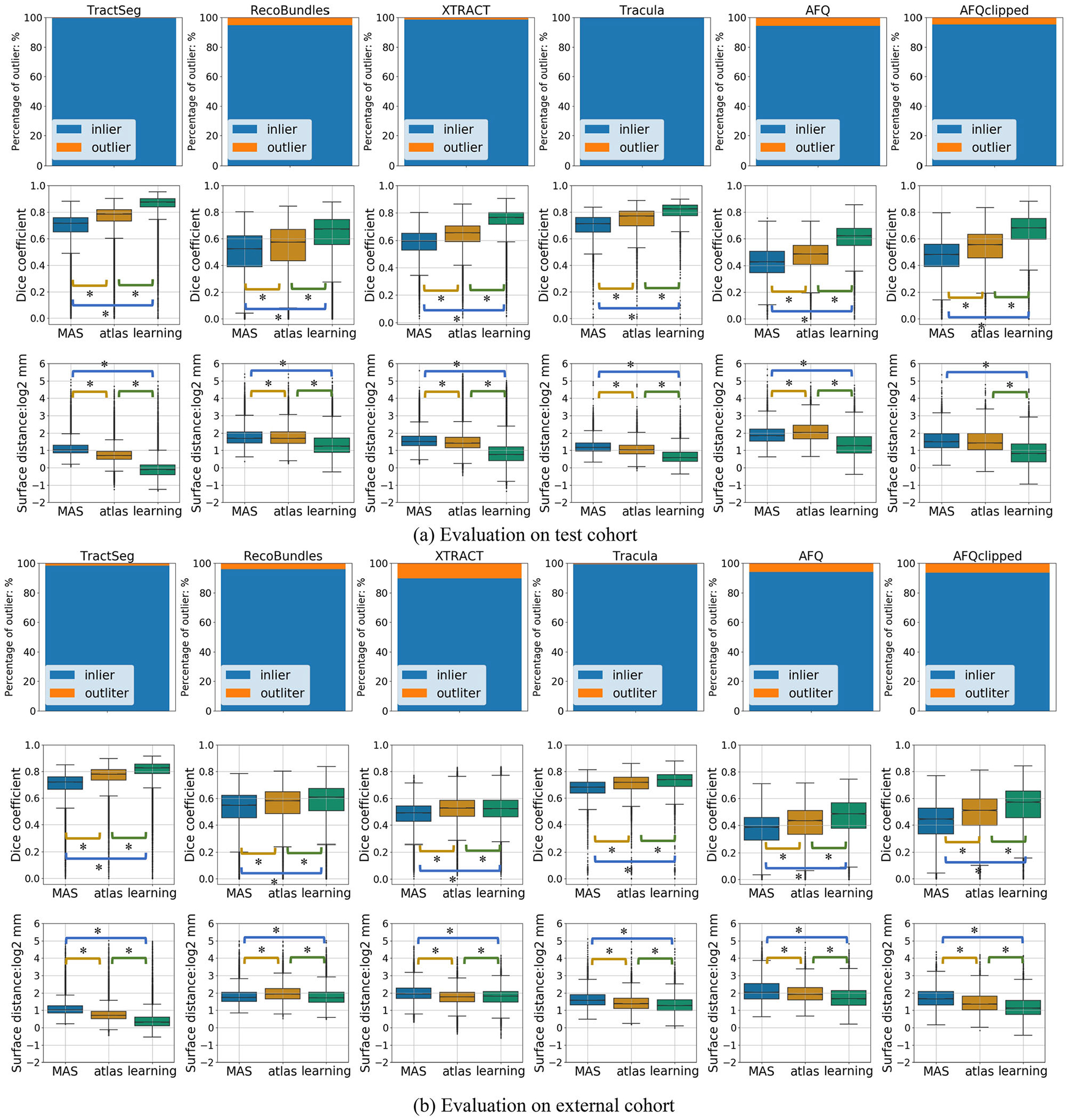
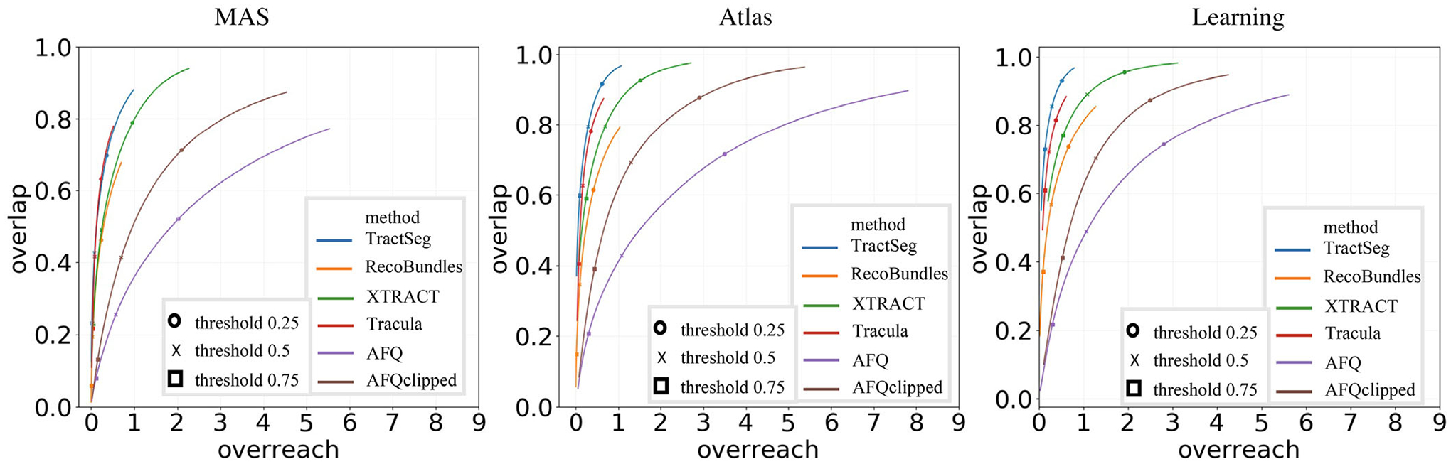
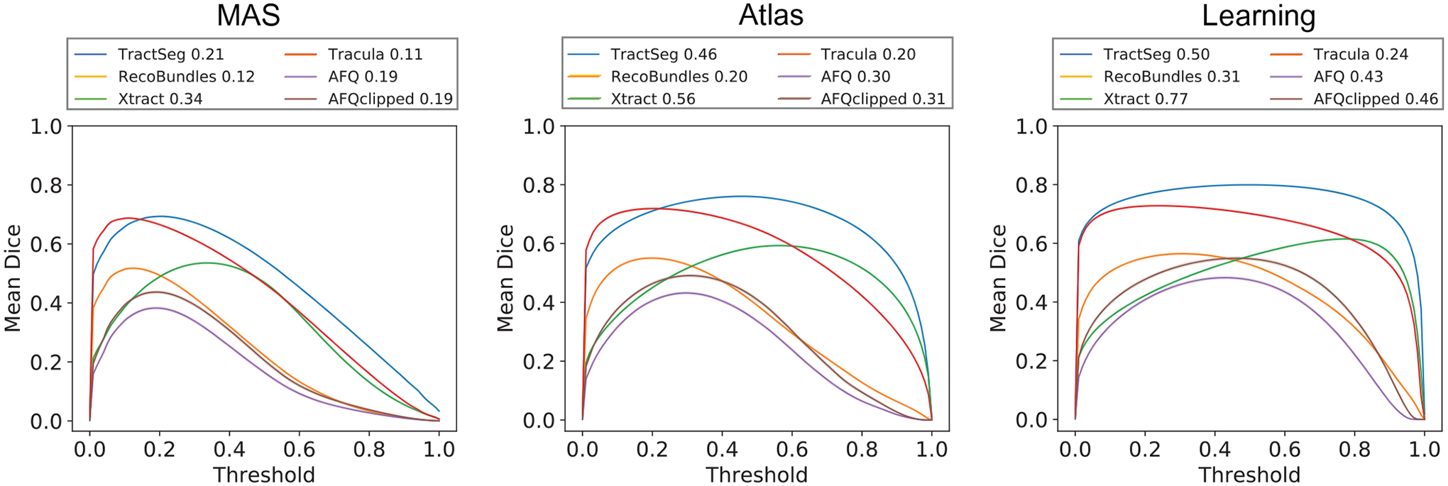
Methods: Following recently proposed innovations in structural anatomical segmentation, we evaluate the feasibility of training multiply spatial localized convolution neural networks to learn context from fixed spatial patches from structural MRI on standard template. We focus on six widely used dMRI tractography algorithms (TractSeg, RecoBundles, XTRACT, Tracula, automated fiber quantification (AFQ), and AFQclipped) and train 125 U-Net models to learn these techniques from 3870 T1-weighted images from the Baltimore Longitudinal Study of Aging, the Human Connectome Project S1200 release, and scans acquired at Vanderbilt University.
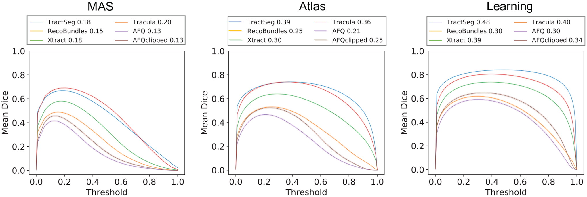
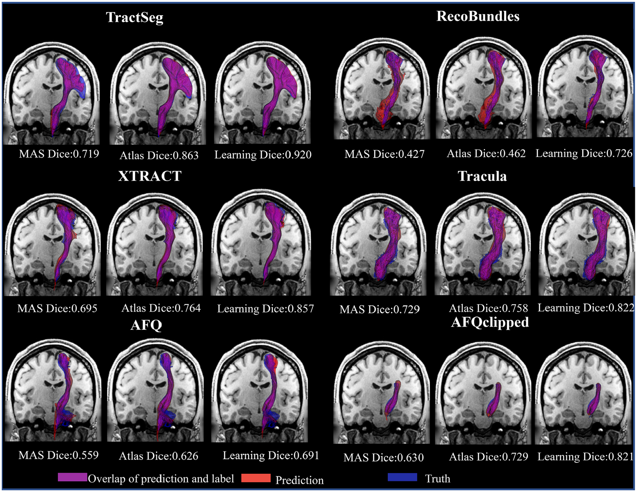
Results: The proposed framework identifies fiber bundles with high agreement against tractography-based pathways with a median Dice coefficient from 0.62 to 0.87 on a test cohort, achieving improved subject-specific accuracy when compared to population atlas-based methods. We demonstrate the generalizability of the proposed framework on three externally available datasets.
Conclusions: We show that patch-wise convolutional neural network can achieve robust bundle segmentation from T1w. We envision the use of this framework for visualizing the expected course of WM pathways when dMRI is not available.
Keywords: T1 weight MRI; learning methods and patch-wise deep neural network; tractography algorithms; white matter.
© 2022 American Association of Physicists in Medicine.
Conflict of interest statement
CONFLICT OF INTEREST
The authors have no conflict of interest to disclose.
Figures
References
-
- Jeurissen B, Descoteaux M, Mori S, Leemans A. Diffusion MRI fiber tractography of the brain. NMR Biomed. 2019;32(4):e3785. - PubMed
-
- Basser PJ,Mattiello J,LeBihan D. Estimation of the effective self-diffusion tensor from the NMR spin echo. J Magn Reson Ser B. 1994;103(3):247–254. - PubMed
-
- Alexander DC. Multiple-fiber reconstruction algorithms for diffusion MRI. White Matter Cogn Neurosci Adv Diffus Tensor Imaging Its Appl. 2005;1064:113–133. - PubMed
MeSH terms
Grants and funding
- R01NS058639/National Institutes of Health under award numbers
- T32 EB001628/EB/NIBIB NIH HHS/United States
- R01 EB017230/EB/NIBIB NIH HHS/United States
- R01EB017230/NH/NIH HHS/United States
- T32EB001628/NH/NIH HHS/United States
- R01 NS058639/NS/NINDS NIH HHS/United States
- R01EB017230/National Institutes of Health under award numbers
- R01EB027585/National Institutes of Health under award numbers
- R01 EB027585/EB/NIBIB NIH HHS/United States
- P50 HD103537/HD/NICHD NIH HHS/United States
- R01EB027585/NH/NIH HHS/United States
- R01NS058639/NH/NIH HHS/United States
- T32EB001628/National Institutes of Health under award numbers
LinkOut - more resources
Full Text Sources
