Review
. 2022 Apr:135:104544.
doi: 10.1016/j.neubiorev.2022.104544. Epub 2022 Jan 25.

Opioid-induced microglia reactivity modulates opioid reward, analgesia, and behavior

Affiliations
Review

Opioid-induced microglia reactivity modulates opioid reward, analgesia, and behavior

Jacob M Green et al. Neurosci Biobehav Rev. 2022 Apr.

Abstract

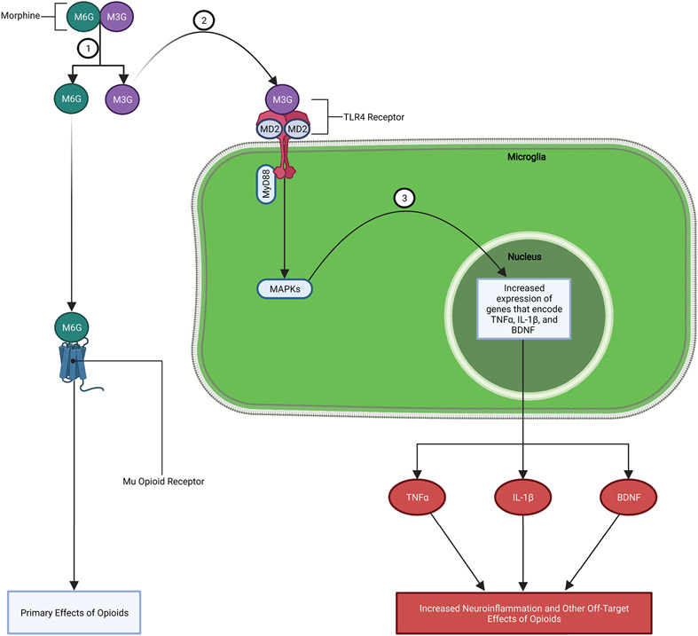
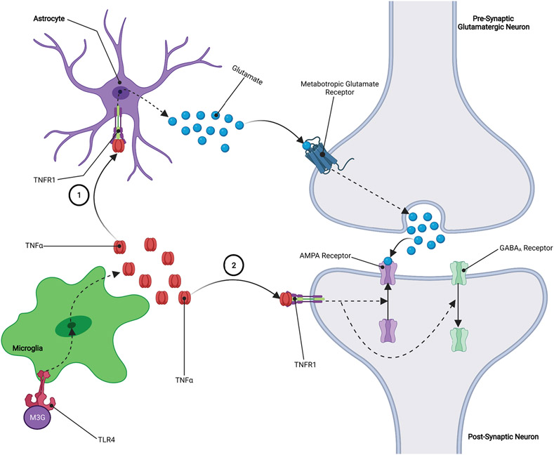
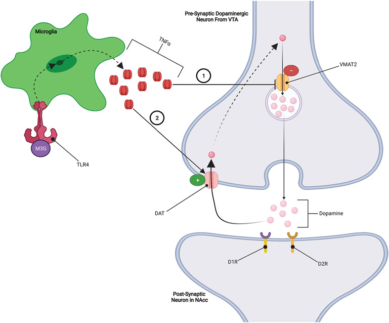
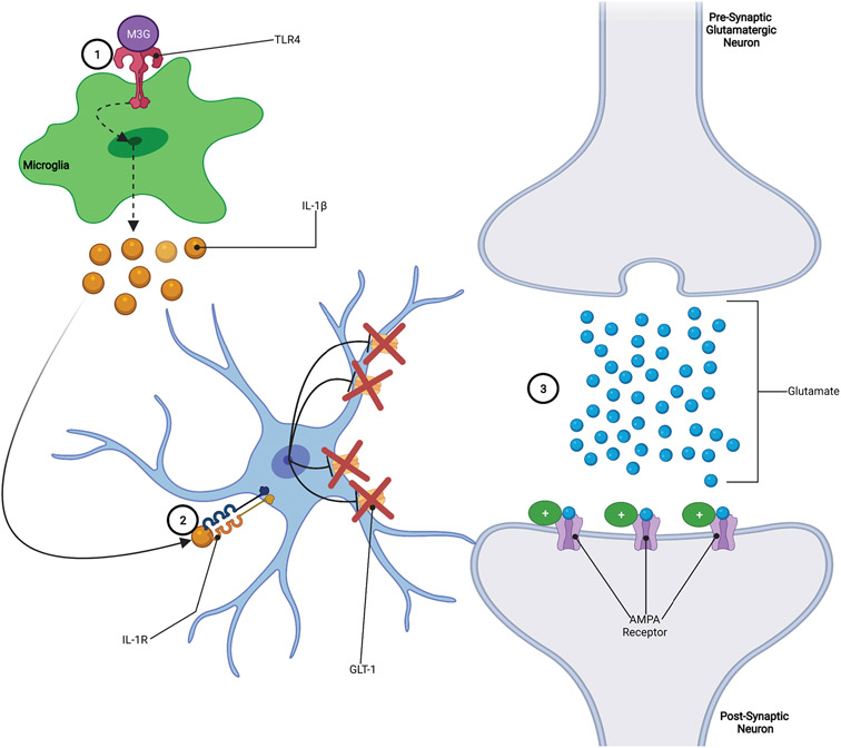
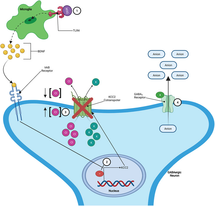
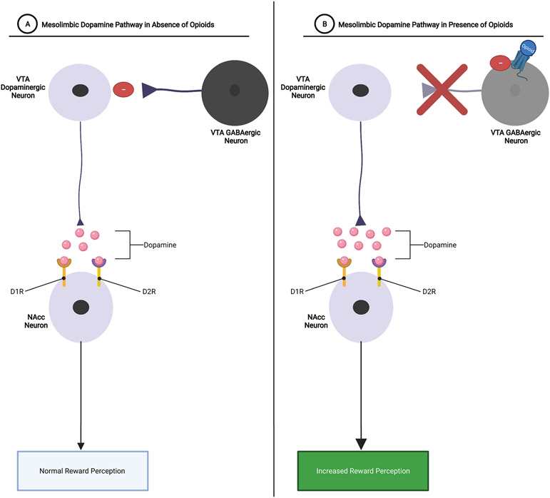
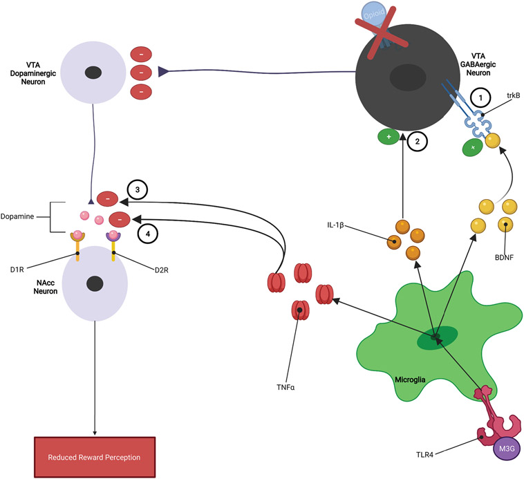
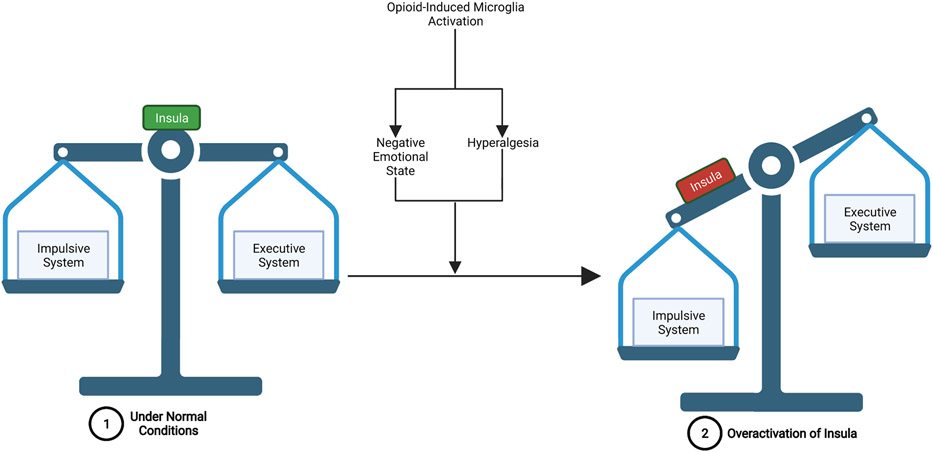
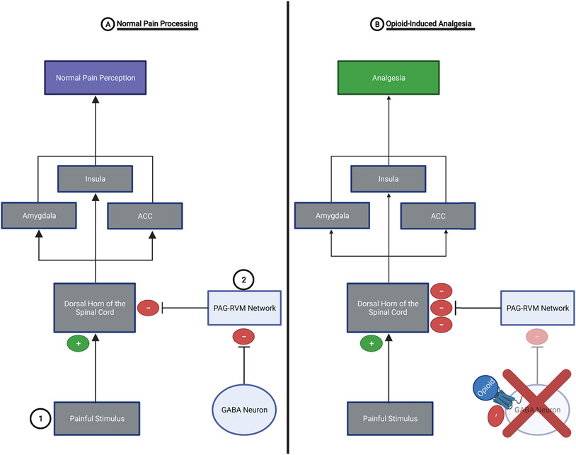
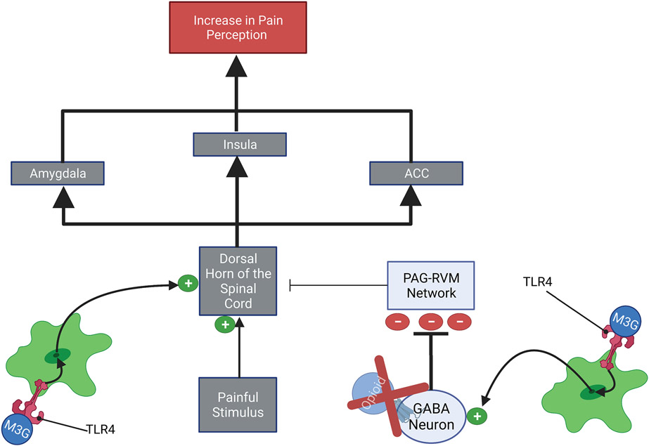
Opioid-induced microglia reactivity affects opioid reward and analgesic processes in ways that may contribute to the neurocognitive impairment observed in opioid addicted individuals. Opioids elicit microglia reactivity through the actions of opioid metabolites at TLR4 receptors, that are located primarily on microglia but are also present on astrocytes. Specifically, the M3G metabolite, which has no affinity for opioid receptors, exerts off-target effects on TLR4 receptors that can trigger downstream immunologic consequences. This off-target microglial reactivity, and the subsequent increase in microglial release of TNFα, IL-1β, and BDNF, have been suggested to modulate both opioid-induced reward and opioid-induced analgesia. Despite occurring independently of each other, these neuro-immune effects could converge and result in overactivation of the insula. This would produce an imbalance between the "impulsive system" and the "executive system", such that the impulsive system's influence over behavior becomes dominant. This state, derived from changes in microglial reactivity, could contribute to impairment in a range of neurocognitive domains that are intricately involved in addiction and lead to increases in addiction-related behaviors.

Keywords: Analgesia; BDNF; Cognitive impairment; Hyperalgesia; IL-1β; Microglia reactivity; Neuroinflammation; Opioids; Reward; TNFα.


Conflict of interest statement

Declaration of Interest

None

Figures

Figure 1:
Figure 1:. Mechanism of Opioid-induced Microglia Reactivity:
1.) Morphine is broken down into two main metabolite forms: M3G and M6G. 2.) M3G binds to TLR4-MD2 complex on microglia causing an increase in MAPK signaling. 3.) Increased MAPK signaling leads to increased expression and release of proinflammatory factors (e.g., TNFα, IL-1β, etc.), chemokines, BDNF, and other substances.
Figure 2:
Figure 2:. TNFα Increases Glutamatergic Tone:
1.) TNFα binds to TNFR1 receptors located on astrocytes leading to increased release of astrocytic glutamate. Astrocytic glutamate binds to metabotropic glutamate receptors located on the presynaptic neuron, leading to increased release of glutamate into the synapse. 2.) TNFα binds to TNFR1 receptors located on the postsynaptic neuron, resulting in an upregulation of AMPA receptors and a downregulation of GABAA receptors on the surface of the postsynaptic neuron. The overall effect of these processes is an increase in glutamatergic signaling which causes an increase in neuroactivity at the synapse.
Figure 3:
Figure 3:. TNFα Inhibition of Dopaminergic Signaling:
1.) TNFα inhibits VMAT2 expression and/or function, resulting in less dopamine being packed into vesicles leading to reduced dopamine release. 2.) TNFα increases the expression and/or function of DAT dopamine transporters, which results in increased reuptake of dopamine from the synapse reducing the effects of dopamine.
Figure 4:
Figure 4:. IL-1β Increases Glutamatergic Tone:
1.) Opioids bind to TLR4 receptors, increasing the release of IL-1β from microglia. 2.) IL-1β binds to IL-1R receptors on astrocytes, leading to the downregulation of GLT-1 glutamate transporters. 3.) Downregulation of GLT-1 on astrocytes results in less uptake of glutamate from the synapse, allowing glutamate to remain in the synapse longer and exert stronger effects on the postsynaptic neuron.
Figure 5:
Figure 5:. BDNF Increases GABAergic Activity:
1.) Opioid metabolite (e.g., M3G) binding to TLR4 induces increased release of BDNF. 2.) Elevated levels of BDNF downregulates KCC2 potassium-chloride cotransporters located on GABAergic neurons. 3.) The downregulation of the KCC2 cotransporters results in disruption of chloride (Cl) homeostasis by increasing the intracellular concentration of Cl. 4.) In such conditions, the activation of GABAA receptors on the GABAergic neurons results in the efflux of anions which leads to a more positive/depolarized state, increasing the excitability of the GABAergic neurons.
Figure 6:
Figure 6:. Mesolimbic Dopaminergic Signaling with and without Opioids:
A.) Mesolimbic Dopamine Pathway in Absence of Opioids: VTA dopaminergic neurons release dopamine into the NAcc. Dopamine then binds to D1R and/or D2R dopamine receptors on NAcc neurons, resulting in reward perception. GABAergic neurons function as a reward “brake system” by inhibiting dopaminergic signaling within the mesolimbic pathway, thereby mediating how rewarding a particular stimulus is. B.) Mesolimbic Dopamine Pathway in Presence of Opioids: Opioids bind to Mu-receptors on VTA GABAergic neurons, thereby removing the reward brake system and allowing for larger amounts of dopamine to be released into the NAcc. Increased release of dopamine within the NAcc results in increased reward perception.
Figure 7:
Figure 7:. Microglia Inhibition of Opioid Reward:
1.) Elevated levels of BDNF results in increased GABAergic tone within the VTA, 2.) IL-1β may increase neurotransmission of GABAergic neurons within the mesolimbic pathway, 3.) TNFα reduces VMAT2 activity which reduces the amount of dopamine released from VTA dopaminergic neurons, and 4.) TNFα increases DAT reuptake of dopamine within the NAcc, reducing the ability of dopamine to exert rewarding effects.
Figure 8:
Figure 8:. Overactivation of the Insula Leads to an Imbalance Between the Impulsive and Executive Systems:
1.) Under normal conditions-the insula functions to maintain the balance between the impulsive system and the executive system, such that the executive system is able to exert control over the impulsive system when required. 2.) Opioid-induced microglia reactivity- contributes to the development of a negative emotional state and hyperalgesia, both of which lead to increased activation of the insula. This overactivation of the insula can then lead to impulsive system dominance over the executive system and an inability of the executive system to exert self-control over drug-related, impulsive behaviors.
Figure 9:
Figure 9:. Pain Processing with and without the Presence of Opioids:
A.) Normal Pain Processing: 1.) Spinothalamic pain pathway- painful stimuli activate nociceptors which propagate a pain signal to the dorsal horn of the spinal cord. The pain signal is then sent to the contralateral thalamus and then to the pain perception areas of the brain (e.g., insula, amygdala, and ACC). 2.) PAG-RVM network- exerts analgesic effects by inhibiting the pain signal at the dorsal horn of the spinal cord. However, the PAG-RVM network is inhibited by GABAergic neurons in the PAG and RVM. B.) Opioid-Induced Analgesia: Opioids inhibit the activity of GABAergic neurons within the PAG and RVM, thereby removing the “brake” from the PAG-RVM network. This allows this analgesic system to exert potent inhibition of the pain signal at the dorsal horn of the spinal cord. This results in a weaker pain signal reaching the brain and a substantial reduction in pain perception (i.e., analgesia).
Figure 10:
Figure 10:. Opioid-Induced Microglia Reactivity Opposes Analgesia:
1.) At the dorsal horn of the spinal cord- Microglia derived TNFα, IL-1β, and BDNF may increase the neuroactivity within the dorsal horn of the spinal cord, this leads to an amplification of the pain signal that will be propagated to the pain processing centers of the brain. 2.) At the PAG-RVM network- Microglia derived TNFα, IL-1β, and BDNF may increase GABAergic tone within the PAG-RVM network. This results in increased GABAergic inhibition of the PAG-RVM network, thereby reducing the ability of this analgesic system to inhibit the pain signal at the dorsal horn of the spinal cord. The effects of microglia reactivity at both the PAG-RVM network and the dorsal horn of the spinal cord would lead to a stronger pain signal reaching pain processing centers of the brain, thereby increasing the perception of pain (i.e., hyperalgesia).

References

    1. Baldacchino A, Armanyous M, Balfour DJK, Humphris G, & Matthews K (2017). Neuropsychological functioning and chronic methadone use: A systematic review and meta-analysis. Neuroscience and Biobehavioral Reviews, 73, 23–38. 10.1016/j.neubiorev.2016.11.008 - DOI - PubMed
    1. Baldacchino A, Balfour DJK, Passetti F, Humphris G, & Matthews K (2012). Neuropsychological consequences of chronic opioid use: A quantitative review and meta-analysis. Neuroscience and Biobehavioral Reviews, 36(9), 2056–2068. 10.1016/j.neubiorev.2012.06.006 - DOI - PubMed
    1. Baskar Jesudasan SJ, Todd KG, & Winship IR (2014). Reduced inflammatory phenotype in microglia derived from neonatal rat spinal cord versus brain. PLoS ONE, 9(6). 10.1371/journal.pone.0099443 - DOI - PMC - PubMed
    1. Bechara A (2005). Decision making, impulse control and loss of willpower to resist drugs: A neurocognitive perspective. Nature Neuroscience, 8(11), 1458–1463. 10.1038/nn1584 - DOI - PubMed
    1. Bechara A, Berridge KC, Bickel WK, Morón JA, Williams SB, & Stein JS (2019). A neurobehavioral approach to addiction: Implications for the opioid epidemic and the psychology of addiction. Psychological Science in the Public Interest, 20(2), 96–127. 10.1177/1529100619860513 - DOI - PMC - PubMed

Publication types

Substances