. 2022 Jan 28;5(5):e202101088.
doi: 10.26508/lsa.202101088. Print 2022 May.

Refining the domain architecture model of the replication origin firing factor Treslin/TICRR

Affiliations

Refining the domain architecture model of the replication origin firing factor Treslin/TICRR

Pedro Ferreira et al. Life Sci Alliance. .

Abstract

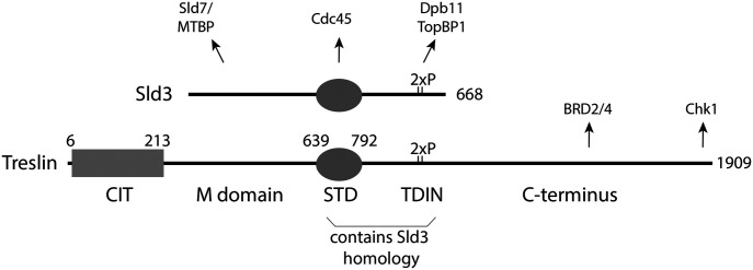
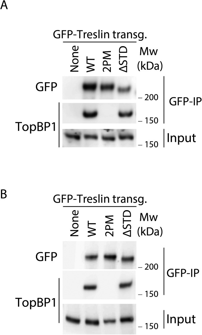
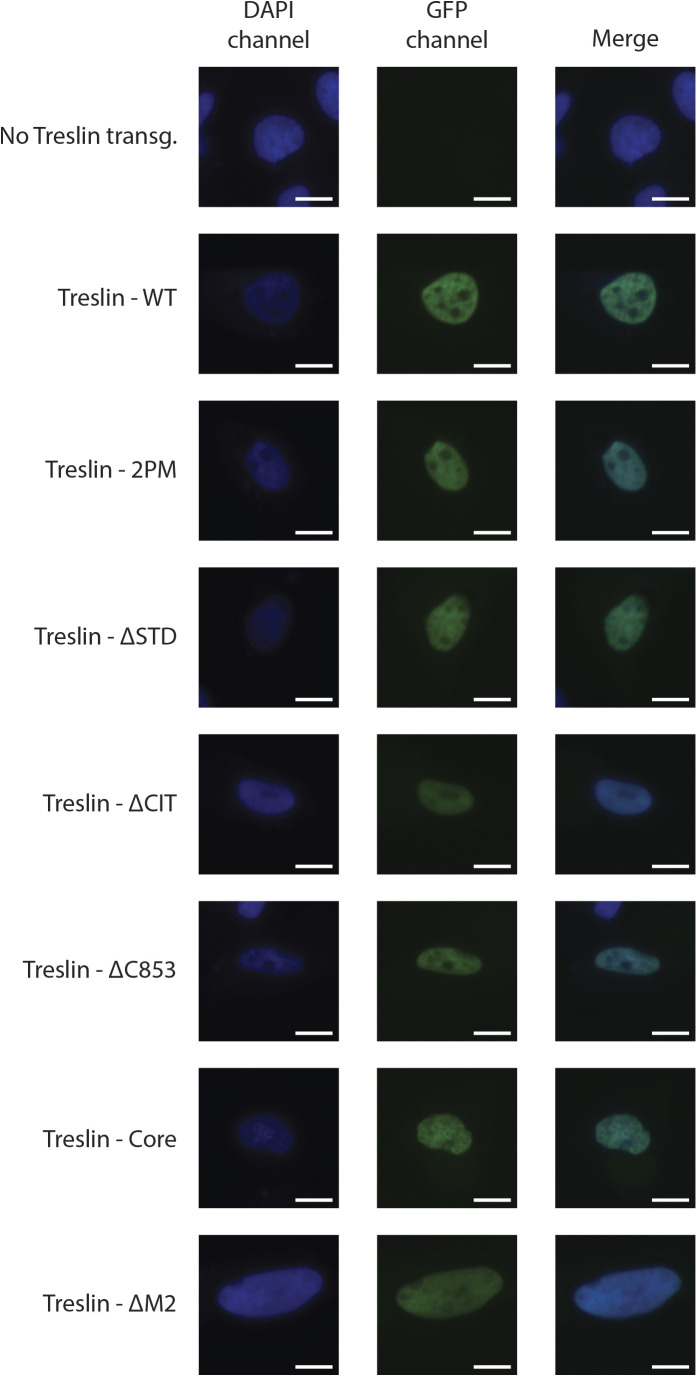
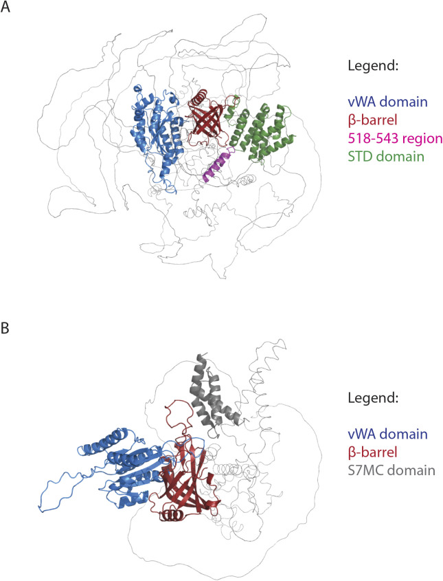
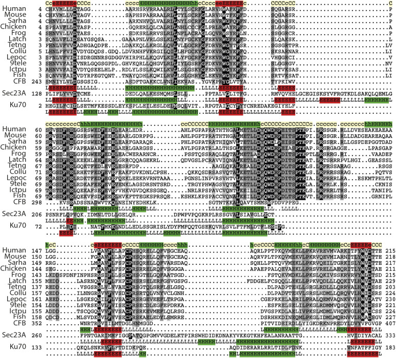
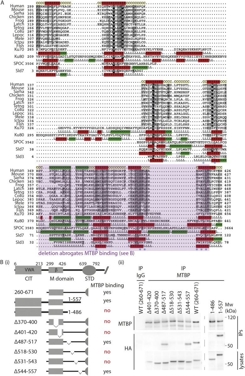
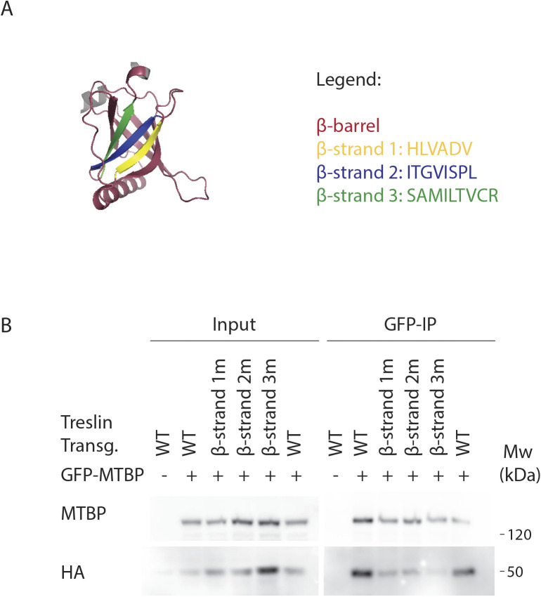
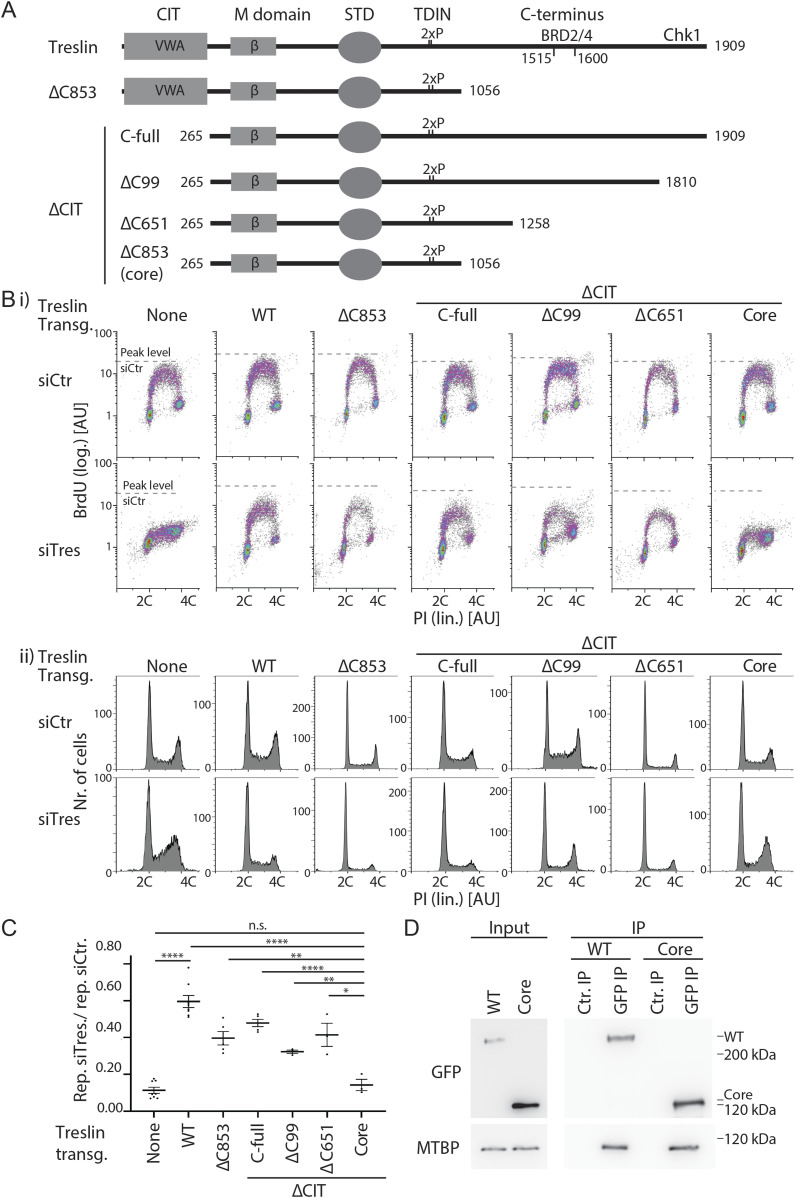
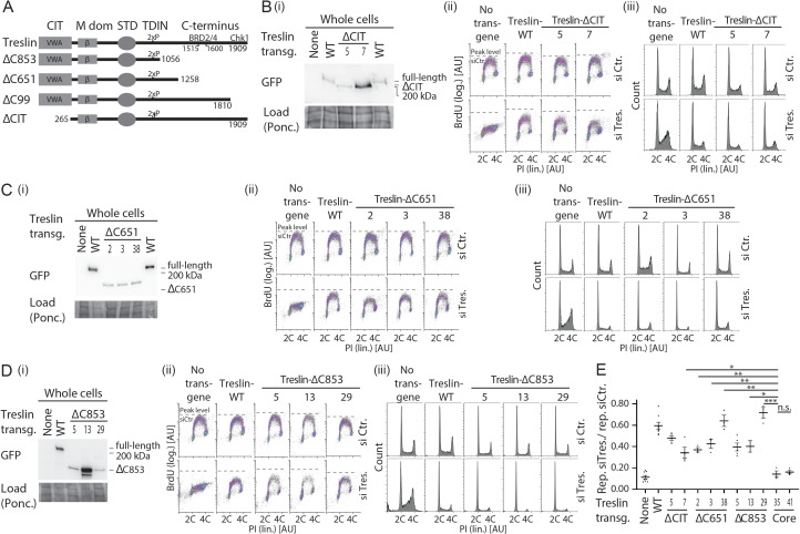
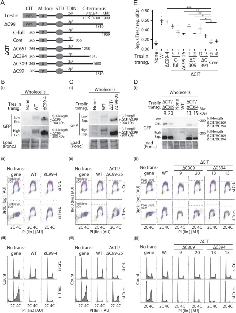
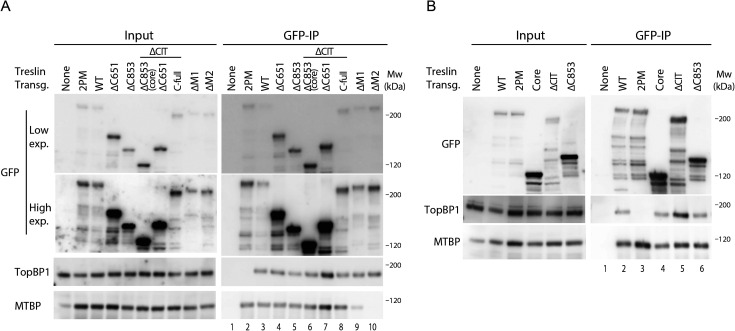
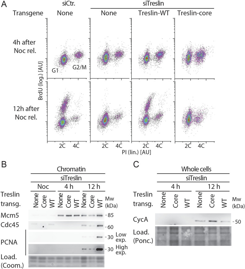
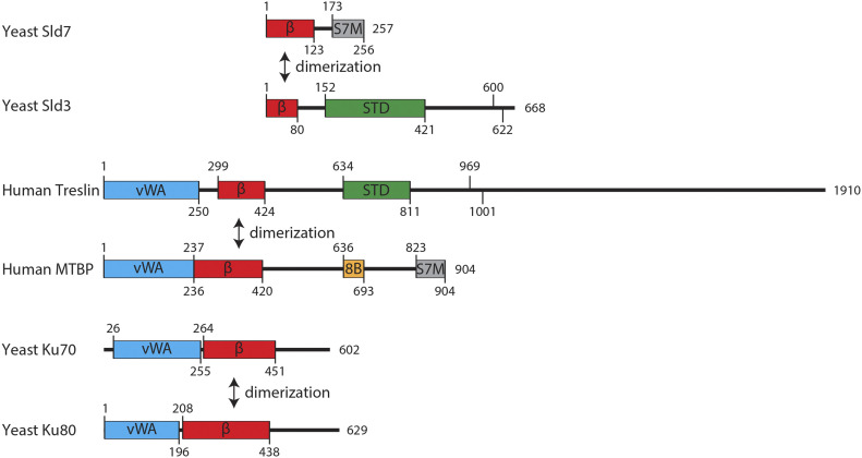
Faithful genome duplication requires appropriately controlled replication origin firing. The metazoan origin firing regulation hub Treslin/TICRR and its yeast orthologue Sld3 share the Sld3-Treslin domain and the adjacent TopBP1/Dpb11 interaction domain. We report a revised domain architecture model of Treslin/TICRR. Protein sequence analyses uncovered a conserved Ku70-homologous β-barrel fold in the Treslin/TICRR middle domain (M domain) and in Sld3. Thus, the Sld3-homologous Treslin/TICRR core comprises its three central domains, M domain, Sld3-Treslin domain, and TopBP1/Dpb11 interaction domain, flanked by non-conserved terminal domains, the CIT (conserved in Treslins) and the C terminus. The CIT includes a von Willebrand factor type A domain. Unexpectedly, MTBP, Treslin/TICRR, and Ku70/80 share the same N-terminal domain architecture, von Willebrand factor type A and Ku70-like β-barrels, suggesting a common ancestry. Binding experiments using mutants and the Sld3-Sld7 dimer structure suggest that the Treslin/Sld3 and MTBP/Sld7 β-barrels engage in homotypic interactions, reminiscent of Ku70-Ku80 dimerization. Cells expressing Treslin/TICRR domain mutants indicate that all Sld3-core domains and the non-conserved terminal domains fulfil important functions during origin firing in human cells. Thus, metazoa-specific and widely conserved molecular processes cooperate during metazoan origin firing.


Conflict of interest statement

The authors declare that they have no conflict of interest.

Figures

Figure 1.
Figure 1.. Treslin/TICRR domain structure.
CIT, Conserved in Treslins; M, middle domain; STD, Sld3-Treslin domain; TDIN, TopBP1/Dpb11 interaction domain. Numbers indicate amino acid position in human Treslin/TICRR or budding yeast Sld3. Arrows point to interacting proteins: MTBP binds to the Treslin/TICRR M domain, Cdc45 binds to the Sld3-Treslin domain of Sld3 (unknown for Treslin/TICRR), TopBP1 binds to a region containing the two CDK phospho-serine (2xP) residues T969 and S1001 (Boos et al, 2011; Kumagai et al, 2011), Chk1 binds to the very C-terminal 99 amino acids of Treslin (Guo et al, 2015), and Brd2/4 binds to the Treslin/TICRR region 1560–1580 (Sansam et al, 2018).
Figure 2.
Figure 2.. The Sld3-Treslin domain (STD) domain of Treslin/TICRR is required for DNA replication in cultured human cells.
(A) Whole cell lysates of stable U2OS cell lines carrying siRNA-resistant transgenes of Treslin/TICRR-WT, Treslin/TICRR-2PM (threonine 969 and serine 1001 double alanine mutant that cannot interact with TopBP1 [Boos et al, 2011]), or three clones of Treslin/TICRR with a deletion of the STD (amino acids 717–792 deleted) were immunoblotted with rabbit anti-Treslin/TICRR (amino acids 1566–1909) antibodies. Ponceau (Ponc.) staining controlled for loading (Load.). (B) Cells described in (A) were treated with control or Treslin/TICRR siRNAs (siCtr/siTres) before analysis by flow cytometry detecting BrdU (logarithmic [log.] scale) and PI (propidium iodide; linear [lin.] scale). Density plots (i) and PI profiles (ii) are shown. Dashed lines indicating peak level of maximal BrdU incorporation in each cell line upon siCtr-treatment allow visual comparison with level upon siTres treatment. PI profiles histograms show relative cell count. (C) Quantification of relative overall DNA replication in cells described in (A) based on flow cytometry experiments described in (B). Averages of BrdU-replication signals of two experiments. Replication signals of siTreslin-treated cells were normalised to replication signals of the same cell line upon siCtr-treatments. (D) Stable U2OS cell lines expressing siTreslin-resistant Treslin/TICRR-ΔSTD, WT, or 2PM were released from a double thymidine arrest before treatment with siTreslin and nocodazole. After nocodazole-release for 4 or 12 h cells chromatin was isolated for immunoblotting with goat anti-Mcm2, rat anti-Cdc45, and mouse anti-PCNA antibodies. Whole cell lysates from the same samples were immunoblotted using mouse anti-cyclin A and goat anti-Mcm2 antibodies. For each antibody, crops are from the same immunoblot exposure. Coomassie (Coom.) staining of low molecular weight part including histones controlled for loading. Clone Treslin-ΔSTD -11 was used.
Figure S1.
Figure S1.. RNAi-replacement of endogenous Treslin/TICRR in U2OS-Flip-In cell lines.
(A) siRNA against Treslin/TICRR (siTres) specifically eliminates endogenous, but not siRNA-resistant GFP-Flag-Treslin/TICRR transgenes (Boos et al, 2011, 2013). Whole cell lysates of U2OS cells or U2OS cells expressing RNAi-resistant Treslin/TICRR-WT or Treslin/TICRR-ΔSTD-clone11 were treated with no siRNA (−), control siRNA (siCtr), or siRNA against Treslin/TICRR (siTres) as indicated and analysed by immunoblotting using anti-Treslin/TICRR (148) and Ponceau (Pon.) staining. Note that endogenous Treslin/TICRR and GFP-Flag-Treslin/TICRR-WT migrate very similarly on SDS polyacrylamide gels. (B) Whole-cell lysates of U2OS cells expressing RNAi-resistant Treslin/TICRR-WT, Treslin/TICRR-core-clone35, Treslin/TICRR-ΔCIT-clone5, or Treslin/TICRR-ΔC853-clone5 were treated and analysed as described in (A), except that immunoblotting was done using anti-Treslin/TICRR (30 × 107) because the antibody anti-Treslin/TICRR (148) used in (A) recognizes a region of Treslin/TICRR not present in the core and ΔC853 mutants. Treslin/TICRR-WT samples are the same immunoblotted in (A). All samples shown in (A, B) were processed in parallel. Treslin/TICRR-core runs as a more distinct band than WT because of the absence of the highly posttranslationally modified C terminus.
Figure S2.
Figure S2.. Gating and data processing strategy for BrdU-propidium iodide flow cytometry.
(A) Forward and side scatter plot and BrdU-propidium iodide profiles of ungated data from a sample of U2OS cells treated with siCtr. The indicated threshold was used in the forward scatter channel to eliminate small debris. (B) Strategy used for cell doublet (aggregates of two cells) discrimination of sample shown in (A). (C) Gates used to discriminate between the S-phase (BrdU+) and non–S-phase (BrdU−) cells. Same gates were used within individual experiments. Average BrdU signal intensity was then calculated for each cell population based on the geometric mean of the signal intensities in the BrdU channel. (D) To calculate BrdU-dependent replication signal, the BrdU signal intensity of the S-phase cell population was background-subtracted using the signal intensity of the non–S-phase population. (E) Replication of siTreslin treated cells was normalised to siCtr-treated cells to calculate the relative replication rescue.
Figure S3.
Figure S3.. Treslin/TICRR-ΔSTD and Treslin/TICRR-core expressing cells progress slower through S-phase.
(A) Stable U2OS cell lines expressing no transgene or siTreslin-resistant GFP-Flag-Treslin/TICRR-WT, 2PM, ΔSTD, or core were arrested in a double thymidine block and treated with siTreslin or siCtr 8 h after release from the first thymidine arrest, so that the siRNA would take effect only after the genome had been replicated. After release for 0, 6, or 10 h from the second thymidine block, cells were analysed by propidium iodide flow cytometry. Histograms show overlays between the samples for the three time points and the gates used to calculate amount of cells in the early S phase and the late S phase, respectively. Propidium iodide histograms show relative cell count. Clones Treslin/TICRR-ΔSTD-11 and Treslin/TICRR-core-35 were used. (B) Quantification of number of cells in early S-phase or late S-phase, 0 and 10 h after a double thymidine release of samples described in (A), using the gates show in (A). At 0 h after release, all samples show around 70% of cells in early the S phase, consistent with similar synchronisation by the double thymidine arrest. 10 h after release, U2OS cells treated with siCtr and Treslin/TICRR-WT expressing cells treated with siTreslin show almost 60% of cells had progressed to late S-phase, with 10–20% remaining in the early S phase. In contrast, only around 30% U2OS cells treated with siTreslin and cells expressing Treslin/TICRR-2PM, ΔSTD, or core had progressed to the late S phase by 10 h. This shows that Treslin/TICRR-ΔSTD or core expressing cells replicated at similar rates as cells expressing the inactive Treslin/TICRR-2PM mutant or cells lacking Treslin/TICRR, indicating that Treslin/TICRR-ΔSTD and core do not support normal S-phase replication. (C, E) Chromatin of cells shown in (A) was isolated for immunoblotting with goat anti-Mcm2 and mouse anti-PCNA antibodies. Coomassie (Coom.) staining of low molecular weight part including histones controlled for loading. Samples shown in (C, E) are the same shown in (A) and were processed in parallel. 10 h after double thymidine release, U2OS cells treated with siCtr and Treslin/TICRR-WT expressing cells treated with siTreslin show that pre-RCs became largely cleared from chromatin, and replisomes (PCNA on chromatin) were also severely decreased, consistent with genome replication being nearly complete at 10 h. In contrast, in U2OS cells treated with siTreslin and in cells expressing Treslin/TICRR-2PM, ΔSTD, or core, pre-RCs and replisomes were cleared from chromatin at much slower rates, consistent with slow replication. (D) Quantification of Mcm2 (i) and PCNA (ii) signals of immunoblots shown in (C). (F) Quantification of Mcm2 (i) and PCNA (ii) signals of immunoblots shown in (E).
Figure S4.
Figure S4.. Treslin/TICRR-ΔSTD is proficient in binding TopBP1.
(A) GFP-Flag-Treslin/TICRR-WT, 2PM, or ΔSTD were transiently transfected into 293T cells. Native lysates were used for anti-GFP nanobody immunoprecipitation in the presence of recombinant Cdk2-cyclin A to promote interaction with TopBP1. Lysates and bead-bound material were analysed by immunoblotting using mouse anti-GFP and rabbit anti-antibodies. Treslin/TICRR-2PM did not bind TopBP1, as expected because the relevant CDK sites in the TopBP1/Dpb11 interaction domain are mutated to alanine. Treslin/TICRR-ΔSTD was able to bind to TopBP1. (B) Independent experimental replicate of (A).
Figure S5.
Figure S5.. GFP-Treslin/TICRR-WT and mutants used in this work retain their nuclear localisation.
U2OS cells were transiently transfected with the indicated GFP-Flag-tagged Treslin/TICRR constructs used across this work (2PM, ΔSTD, ΔCIT, ΔC853, Core, and ΔM2) to ascertain their cellular localisation. Cells were fixed with 2% PFA for 20 min and stained with DAPI. Representative pictures of transfected cells are shown for each transfection. As can be seen by the juxtaposition with the DAPI channel, GFP signal in all conditions except the empty line localised to the nucleus, indicating that Treslin/TICRR-WT and all mutants tested retained nuclear localisation. Scale bar: 10 μM.
Figure S6.
Figure S6.. Alphafold 2 models of Treslin/TICRR and MTBP.
The Alphafold 2 structural models of Treslin/TICRR and MTBP suggest that their N-terminal regions share the same domain architecture, a vWA domain followed by a Ku70-like β-barrel, suggesting a common ancestry. (A) Treslin/TICRR full length Alphafold 2 model showing the predicted vWA domain (blue), Ku70/80–like β-barrel (red), Sld3-Treslin domain (green), and the 518–543 region whose deletion abrogated MTBP binding (pink; Alphafold 2 prediction score <70%). (B) MTBP full length Alphafold 2 model showing the predicted vWA (blue), Ku70-like β-barrel (red) and S7MC domain (grey).
Figure S7.
Figure S7.. The conserved in Treslins domain of Treslin/TICRR contains a vWA fold.
Representative multiple sequence alignment of VWA domain in Treslin/TICRR family. Secondary structure prediction using PsiPred was performed for the Treslin/TICRR family, shown in the first lane; this prediction is consistent with the secondary structure of VWA domains, shown below each of the selected proteins with known structure (Complement factor B, PDB: 3HRZD; Sec23, PDB: 2NUTA; Ku70, PDB: 5Y58E). For figure methods and abbreviations see Fig 3A legend.
Figure 3.
Figure 3.. Treslin/TICRR, Sld3, and Sld7 contain a Ku70/80–like β-barrel that are required for Treslin/Sld3-MTBP/Sld7 dimerization.
(A) Representative multiple sequence alignment of Ku70-like β-barrel domain in the Treslin/TICRR family. The alignment generated with the program T-Coffee (Notredame et al, 2000) using default parameters and slightly refined manually. The final alignment was obtained using a combination of profile-to-profile comparisons (Söding et al, 2005) and sequence alignments derived from structural super-positions of a selection of Ku70-like β-barrel domains whose tertiary structure is known (Holm & Sander, 1995). The limits of the protein sequences included in the alignment are indicated by flanking residue positions. Secondary structure prediction using PsiPred (Jones, 1999) was performed for the Treslin family, shown in the first lane; this prediction is consistent with the secondary structure of Ku70-like β-barrel domains, shown below each of the proteins with known structure (Ku70, PDB: 5Y58E; Ku80, PDB: 5Y58F; SPOC, PDB: 1OW1A; Sld7, PDB: 3X37B; Sld3, PDB: 3X37A). α-helices and β-strands are indicated by H and E, respectively. The alignment was presented with the program Belvu using a colouring scheme indicating the average BLOSUM62 scores (which are correlated with amino acid conservation) of each alignment column: black (>3), grey (between 3 and 1.5) and light grey (between 1.5 and 0.5) (Sonnhammer & Hollich, 2005). Sequences are named according to their specie common name or abbreviation corresponding as follow to their UniProt identification and specie name (Wu et al, 2006): Human, Q7Z2Z1_HUMAN, Homo sapiens; Mouse, Q8BQ33_MOUSE, Mus musculus; Sarha, G3WMD4_SARHA; Sarcophilus harrisii; Chicken, E1BU88_CHICK; Gallus gallus; Frog, D3IUT5_XENLA, Xenopus laevis; Latch, H3BCK8_LATCH, Latimeria chalumnae; Tetng, H3CYF8_TETNG, Tetraodon nigroviridis; Collu, A0A4U5UGV6_COLLU, Collichthys lucidus; Lepoc, W5ND48_LEPOC, Lepisosteus oculatus; 9tele, A0A3B3T1X9_9TELE, Paramormyrops kingsleyae; Ictpu, A0A2D0SG01_ICTPU, Ictalurus punctatus; Fish, Q6DRL4_DANRE, Danio rerio. Blue asterisks: amino acid positions in Sld3 that mediate Sld7 interaction (Itou et al, 2015). (B) Schematic representation of Treslin/TICRR mutants (i) used for interaction studies (ii). For (ii), the indicated N-terminally 3HA-tagged Treslin/TICRR fragments were transiently transfected into 293T cells before immunoprecipitation (IP) from cell lysates using control IgG (IgG IP) or rabbit anti-MTBP (amino acids 1–284) (MTBP-IP). Lysates and precipitates were immunoblotted with detection by rat anti-MTBP (12H7) and anti-HA antibodies. VWA, von Willebrand A domain; β, β-barrel.
Figure S8.
Figure S8.. Structural similarities among Treslin/TICRR, MTBP, and Ku70 proteins.
(A) Contact maps of Ku70-like β-barrel domains of Treslin/TICRR (Alphafold 2 model), Ku70 (PDB: 5Y58_A), Sld7 (PDB: 3x37_B), and Sld3 (PDB: 3x37_A). Contact maps were generated using the Cocomaps server (cut-off distance value = 7 Å) (Vangone et al, 2011). β-strands are labeled 1–7 and coloured in red, orange, yellow, green, cyan, violet, and purple, respectively. β-strand contact pairs are labeled, showing the identical arrangement of the seven β-strands conserved among these Ku70-like β-barrel domains. In Sld3 the β-barrel is incomplete, missing β strands 1 and 2 (Ku70 barrel numbering). (B) Dali structural superposition of the vWA domains of human Treslin/TICRR (Treslin/TICRR Alphafold 2 model positions 1–250 in green), human MTBP (MTBP Alphafold 2 model positions 1–236 in cyan; Z-score 9.6 and RMSD 3.6 Å versus Treslin/TICRR), and yeast Ku70 (PDB: 5y58-A, positions 28–263 in purple; Z-score 10 and RMSD 3.8 Å versus Treslin/TICRR). (C) Dali structural superposition of the β-barrel domains of human Treslin/TICRR (Treslin/TICRR Alphafold 2 model position 299–424 in green), human MTBP (MTBP Alphafold 2 model positions 237–420 in cyan; Z-score 10.1 and RMSD 3.4 Å versus Treslin/TICRR), and yeast Ku70 (PDB: 5y58-A, positions 264–451 in purple; Z-score 13.3 and RMSD 2.6 Å versus Treslin/TICRR).
Figure S9.
Figure S9.. Mutating individual β-strands from Treslin/TICRR β-barrel compromises binding to MTBP.
(A) Treslin/TICRR Alphafold 2 model showing the β-barrel (red), including the three β-strands mutated in (B). β-strand 1 (amino acids 391–396, HLVADV, replaced with amino acids SGELRL, labeled in yellow), β-strand 2 (amino acids 405–412, ITGVISPL, replaced with amino acids SGELRLPS, labeled in blue), or β-strand 3 (amino acids 415–423, SAMILTVCR, replaced with amino acids LLCIKVEAF, labeled in green). (B) N-terminally 3HA-tagged Treslin/TICRR fragments (from amino acid 260–671) were transiently transfected into 293T together with C-terminally GFP-tagged MTBP before immunoprecipitation from cell lysates using anti-GFP nanobody immunoprecipitation. The Treslin/TICRR fragments used were WT, β-strand 1m (labeled in yellow in A), β-strand 2m (labeled in blue in A), or β-strand 3m (labeled in green in A). The Treslin/TICRR β-strand amino acids were replaced by unrelated β-strand forming sequences, to try to change the amino acid sequence without disrupting the overall structure. Results show that each β-strand mutation weakened but did not abrogate binding to MTBP, indicating that each individual β-strand may contribute to the MTBP interaction surface.
Figure 4.
Figure 4.. The conserved in Treslins and the region between amino acids 1057–1257 of Treslin/TICRR cooperate to support replication in human cells.
(A) Schematic representation of Treslin/TICRR mutants used in this figure. Δ, deletion; C99, 651, 853: C-terminal 99, 651, or 853 amino acids, Chk1 kinase binding requires the C-terminal 99 amino acids, BRD2/4 binds to a region between amino acids 1515 and 1600 that were deleted in Treslin/TICRR-ΔC651, -ΔC853, -ΔC394, and -ΔC309 (latter two mutants shown in Fig S5), respectively. ΔCIT, amino acids 1–264 deleted. (B) Flow cytometry density plots (i) and propidium iodide profiles (ii) of experiments as described in Fig 2B using the stable U2OS cell lines expressing siTreslin-resistant Treslin/TICRR mutants described in (A). Propidium iodide profiles histograms show relative cell count. Cell clones: ΔC853-5, ΔCIT(-C-full)-5; ΔCIT-ΔC99-25; ΔCIT-ΔC651-61; core-35. (C) Quantification of relative overall replication as described in Fig 2C of several independent experiments as described in (B). Cell clones as in (B); Error bars: SEM; sample numbers (n): 8 (none; WT), 5 (ΔCIT[-C-full]; ΔC853), 3 (ΔCIT-ΔC99; ΔCIT-ΔC651; core); significance tests: parametric, unpaired, two tailed t test, *P ≤ 0.05. (D) Immunoblot with mouse anti-GFP or rat anti-MTBP (12H7) antibodies of co-immunoprecipitation (IP) experiment using 293T cells transiently transfected with GFP-Flag-Treslin/TICRR-WT or core. Native lysates were immunoprecipitated with anti-GFP nanobodies (GFP-IP) or empty control beads (Ctr. IP).
Figure S10.
Figure S10.. Analysis of several stable U2OS clones expressing Treslin/TICRR-ΔCIT or various C-terminal truncation mutants.
(A) Schematic giving an overview over the Treslin/TICRR mutants used in this figure. (B, C, D) Immunoblots (i) to assess transgene expression levels and BrdU-flow cytometry (ii) and propidium iodide profiles (iii) to determine overall DNA replication of the indicated Treslin/TICRR mutants shown in (A). The following U2OS clones were used for main Fig 4: Treslin/TICRR-ΔCIT-5, ΔC853-5. Immunoblots of whole cell lysates used mouse anti-GFP and Ponceau staining (as a loading control). Flow cytometry was done after replacing endogenous Treslin/TICRR against the indicated siRNA-resistant transgenes using RNAi. Density plots are shown. Parental U2OS cells and a line expressing Treslin/TICRR-WT served to control the experiment. Dashed lines show BrdU peak level of the respective control siRNA-treated cell line in the same experiment. Clones were picked that expressed the Treslin/TICRR transgenes at similar or higher levels than Treslin/TICRR-WT to avoid under-estimating the capability of the mutants to support replication. For Treslin/TICRR-ΔC651, only low-expressing clones were found. The results are still conclusive, though, because all clones were capable to support replication. (E) Quantification of overall replication in mutant Treslin/TICRR U2OS cell lines described in (A, B, C, D), based on BrdU-propidium iodide flow cytometry experiments as described in (B, C, D). For comparison, the Treslin/TICRR-core clones are shown in addition to the usual control lines. The quantifications indicate that ΔCIT, ΔC651, and ΔC853 mutants were active. It also shows the clonal variability that did not clearly correlate with expression levels, as indicated by the Treslin/TICRR-ΔC651 clones 1–3. Error bars: SEM; sample numbers (n): 8 (none; WT), 5 (ΔCIT-5; ΔC853-5), 4(ΔCIT-7); 3 (ΔC651-2; ΔC651-3; ΔC651-38; ΔC853-29, core-35; core-41); 2 (ΔC853-13); significance tests: parametric, unpaired, two tailed t test, *P ≤ 0.05.
Figure S11.
Figure S11.. Analysis of several stable U2OS clones expressing Treslin/TICRR-core and Treslin/TICRR-ΔCIT/ΔC651.
(A) Schematic giving an overview over the Treslin/TICRR mutants used in (B, C, D, E). (B, C, D) Immunoblots (B) and BrdU-Flow cytometry/propidium iodide profiles (C, D) of stable U2OS cell lines expressing indicated Treslin/TICRR-mutants shown in (A). Immunoblots of whole cell lysates using mouse anti-GFP and Ponceau staining (as a loading control) served to assess transgene expression levels relative to each other and Treslin/TICRR-WT. The following U2OS clones were used for main figures: Treslin/TICRR-ΔCIT/ΔC651-61 (Fig 4), core-35 (Figs 4 and 5). Clones were picked that expressed the Treslin/TICRR transgenes at similar or higher levels than Treslin/TICRR-WT to avoid under-estimating the capability of the mutants to support replication. For BrdU-flow cytometry, density plots show overall DNA replication of stable U2OS clones shown in (A). Flow cytometry was done after replacing endogenous Treslin/TICRR against the indicated siRNA-resistant transgenes using RNAi. Parental U2OS cells and a line expressing Treslin/TICRR-WT served to control the experiment. Dashed lines show BrdU peak level of the respective control siRNA-treated cell line in the same experiment. (E) Quantification of overall replication in mutant Treslin/TICRR U2OS cell lines described in (A), based on BrdU-propidium iodide flow cytometry experiments as described in (B). For comparison, Treslin/TICRR-ΔCIT containing the full C terminus and Treslin/TICRR-core are shown in addition to the usual control lines. Treslin/TICRR-ΔCIT/ΔC651 supports replication to levels comparable with Treslin/TICRR-ΔCIT. The exact level of replication depended on the clone used. No Treslin/TICRR-ΔCIT/ΔC651 clone, however, supported replication as poorly as Treslin/TICRR-core that showed replication similar to control U2OS cells not expressing a siRNA-resistant transgene. Error bars: SEM; sample numbers (n): 8 (none; WT), 5 (ΔCIT(-C-full)-5), 4 (ΔCIT(-C-full)-7), and 3 (ΔCIT-ΔC651-57; ΔCIT-ΔC651-61; core-35; core-41); significance tests: parametric, unpaired, two tailed t test, *P ≤ 0.05.
Figure S12.
Figure S12.. Analysis of several stable U2OS clones expressing various C-terminal truncations in combination with deletion of the conserved in Treslins or Treslin/TICRR-ΔC99.
(A) Schematic giving an overview over the Treslin/TICRR mutants used in (B, C, D, E). (B, C, D) Immunoblots (i) to assess transgene expression levels of stable U2OS clones expressing Treslin/TICRR-ΔCIT/ΔC651 and ΔCIT/ΔC853. Immunoblots of whole cell lysates used mouse anti-GFP and Ponceau staining (as a loading control). Clones were picked that expressed the Treslin/TICRR transgenes at similar or higher levels than Treslin/TICRR-WT to avoid under-estimating the capability of the mutants to support replication. Density plots (ii) of BrdU-flow cytometry and propidium iodide profiles (iii) to determine overall DNA replication of stable U2OS clones described in (A, B). Flow cytometry was done after replacing endogenous Treslin/TICRR against the indicated siRNA-resistant transgenes using RNAi. Parental U2OS cells and a line expressing Treslin/TICRR-WT served to control the experiment. Dashed lines show BrdU peak level of the respective control siRNA-treated cell line in the same experiment. (E) Quantification of overall replication in mutant Treslin/TICRR U2OS cell lines described in (A), based on BrdU-propidium iodide flow cytometry experiments as described in (B). For comparison, Treslin/TICRR-ΔCIT containing the full C terminus and Treslin/TICRR-core are shown in addition to the usual control lines. Treslin/TICRR-ΔC99 supports replication to similar levels as Treslin/TICRR-WT. Treslin/TICRR-ΔCIT/ΔC99 supports replication to levels comparable with Treslin/TICRR-ΔCIT, but much better than Treslin/TICRR-core. Also here, the exact level of replication depended on the clone used. Error bars: SEM; sample numbers (n): 8 (none; WT), 5 (ΔCIT(-C-full)-5), 4 (ΔCIT(-C-full)-7), 3 (ΔC-99-4; ΔCIT-ΔC99-25; ΔCIT-ΔC309-9; ΔCIT-ΔC309-20; ΔCIT-ΔC394-13; ΔCIT-ΔC394-15; core-35; core-41); significance tests: parametric, unpaired, two tailed t test, *P ≤ 0.05.
Figure S13.
Figure S13.. Treslin/TICRR-core is proficient in binding MTBP and TopBP1.
(A) The indicated GFP-Flag-Treslin/TICRR mutants were transiently transfected into 293T cells together with MTBP. Native lysates were used for anti-GFP nanobody immunoprecipitation (IP) in the presence of recombinant Cdk2-cyclin A to promote interaction with TopBP1. Lysates and bead-bound material were analysed by immunoblotting using mouse anti-GFP, rabbit anti-TopBP1, and rat anti-MTBP antibodies. Controls for IP specificity were made: Treslin/TICRR-ΔM1 and ΔM2 show decreased (M1) or absent (M2) MTBP signals, as expected. Treslin/TICRR-2PM did not bind TopBP1, as expected because the relevant CDK sites in the TopBP1/Dpb11 interaction domain are mutated to alanine. IP capabilities using (near) full-length Treslin/TICRR versions are hard to compare by immunoblotting with those containing larger deletions because of the often weak blotting efficiency of the 210 kD full-length Treslin/TICRR. However, the smaller C-terminal truncations are better comparable. Treslin/TICRR-ΔC853 and Δ651 bound similar amounts of TopBP1 and MTBP, whether they contained conserved in Treslins or not. In some experiments, however, deletion of the conserved in Treslins seemed to have a minor effect on the amount of MTBP bound (Fig 4D). (B) Independent experimental replicate of A, containing only some key samples.
Figure 5.
Figure 5.. Treslin/TICRR-core does not support replisome formation.
(A) Stable U2OS cell lines expressing no transgene or siTreslin-resistant Treslin/TICRR-WT or core were released from a thymidine arrest before treatment with siTreslin or siCtr and nocodazole. After nocodazole release for 4 or 12 h cells were analysed by BrdU-propidium iodide flow cytometry. Clone Treslin/TICRR-core-35 was used. (B) Chromatin of cells treated as described in (A) was isolated for immunoblotting with rabbit anti-Mcm5, rat anti-Cdc45 and mouse anti-PCNA antibodies. Coomassie (Coom.) staining of low molecular weight part including histones controlled for loading. In the high exposure (exp.) the strongest band is saturated. (C) Whole cell lysates of cells treated as described in (A) were immunoblotted using mouse anti-cyclin A antibody.
Figure 6.
Figure 6.. Common domain architecture of Treslin/TICRR/Sld3, MTBP and Ku70/Ku80 proteins.
Domain models of the indicated proteins. vWA, von Willebrand factor type A domain; β, Ku70/80–like β-barrel; STD, Sld3-Treslin domain; 8B, Cdk8/19-cyclin C binding domain; S7M, Sld7/MTBP C-terminal domain; Numbers indicate amino acids position and protein length. In Sld3 and Treslin/TICRR are indicated two conserved CDK phosphorylated S/TP sites (Sld3, position 600 and 622; Treslin/TICRR, position 669, 1001).

References

    1. Abid Ali F, Douglas ME, Locke J, Pye VE, Nans A, Diffley JFX, Costa A (2017) Cryo-em structure of a licensed DNA replication origin. Nat Commun 8: 2241. 10.1038/s41467-017-02389-0 - DOI - PMC - PubMed
    1. Berezney R, Dubey DD, Huberman JA (2000) Heterogeneity of eukaryotic replicons, replicon clusters, and replication foci. Chromosoma 108: 471–484. 10.1007/s004120050399 - DOI - PubMed
    1. Boos D, Ferreira P (2019) Origin firing regulations to control genome replication timing. Genes (Basel) 10: 199. 10.3390/genes10030199 - DOI - PMC - PubMed
    1. Boos D, Sanchez-Pulido L, Rappas M, Pearl LH, Oliver AW, Ponting CP, Diffley JFX (2011) Regulation of DNA replication through Sld3-dpb11 interaction is conserved from yeast to humans. Curr Biol 21: 1152–1157. 10.1016/j.cub.2011.05.057 - DOI - PubMed
    1. Boos D, Yekezare M, Diffley JF (2013) Identification of a heteromeric complex that promotes DNA replication origin firing in human cells. Science 340: 981–984. 10.1126/science.1237448 - DOI - PubMed

MeSH terms

LinkOut - more resources