. 2022 Jan 29;19(1):26.
doi: 10.1186/s12974-022-02382-5.

TRPV4 contributes to ER stress and inflammation: implications for Parkinson's disease

Affiliations

TRPV4 contributes to ER stress and inflammation: implications for Parkinson's disease

Na Liu et al. J Neuroinflammation. .

Abstract

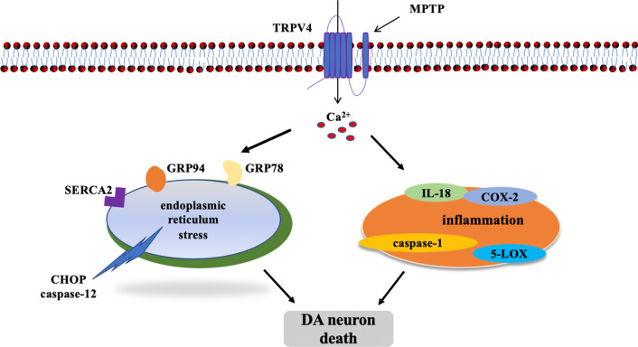
Background: Parkinson's disease (PD) is a progressive neurodegenerative disorder. Its molecular mechanism is still unclear, and pharmacological treatments are unsatisfactory. Transient receptor potential vanilloid 4 (TRPV4) is a nonselective Ca2+ channel. It has recently emerged as a critical risk factor in the pathophysiology of neuronal injuries and cerebral diseases. Our previous study reported that TRPV4 contributed to endoplasmic reticulum (ER) stress in the MPP+-induced cell model of PD. In the present study, we detected the role and the mechanism of TRPV4 in 1-Methyl-4-phenyl-1,2,3,6-tetrahydropyridine (MPTP)-induced PD mice.

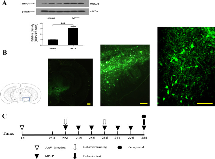
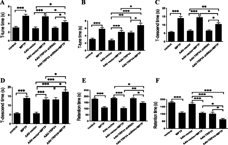
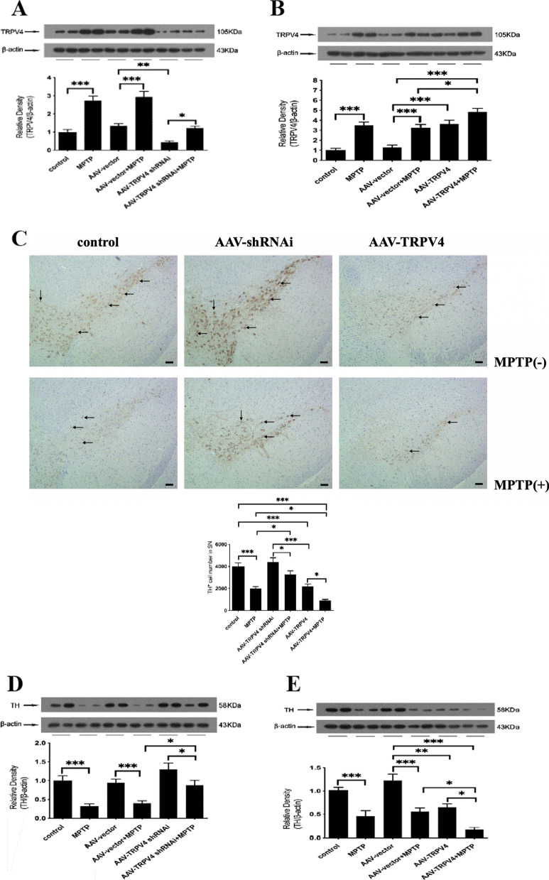
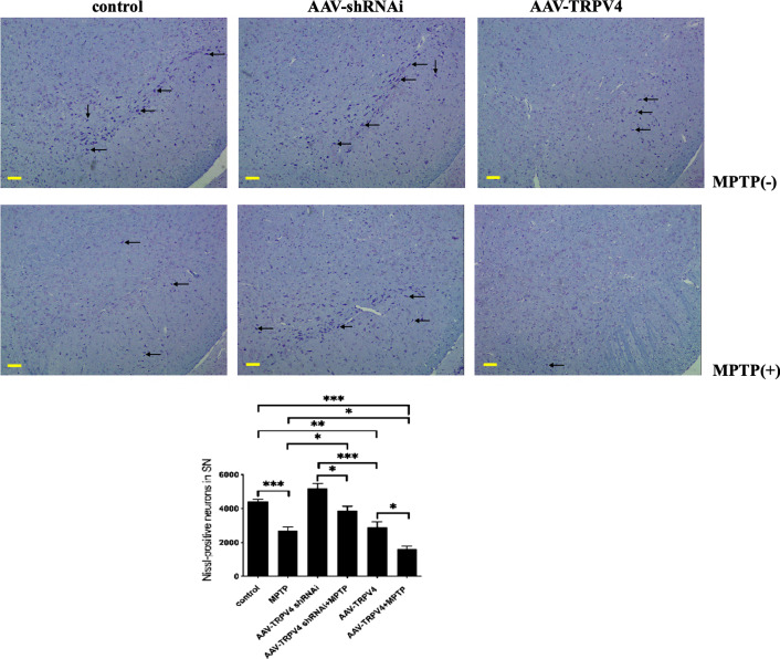
Methods: Intracerebral injection of an adeno-associated virus (AAV) into the substantia nigra (SN) of mice was used to knockdown or upregulate the expression of TRPV4 and intraperitoneal injection of MPTP. Rotarod and pole tests were used to evaluate the locomotor ability of mice. We used immunohistochemistry, Nissl staining and Western blot to detect the alterations in the number of tyrosine hydroxylase (TH)-positive neurons, Nissl-positive neurons, the levels of ER stress-associated molecules and proinflammatory cytokines in the SN.

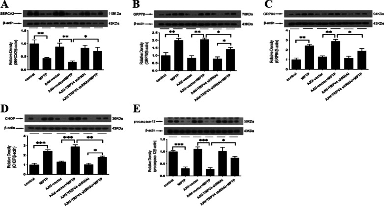
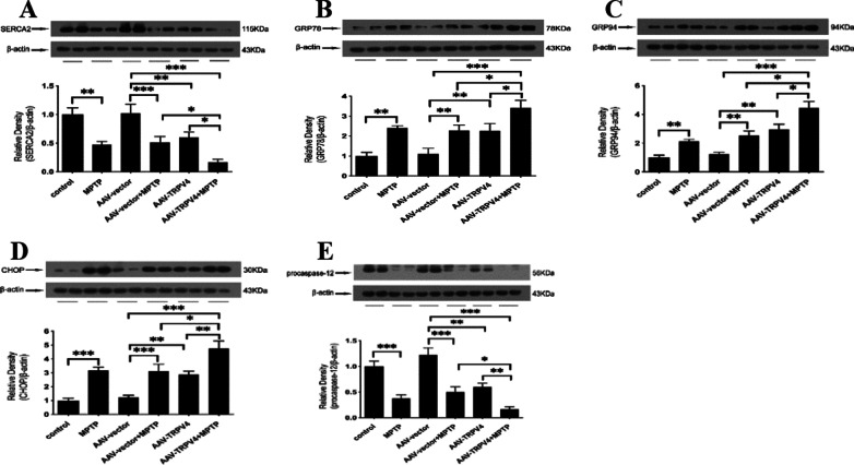
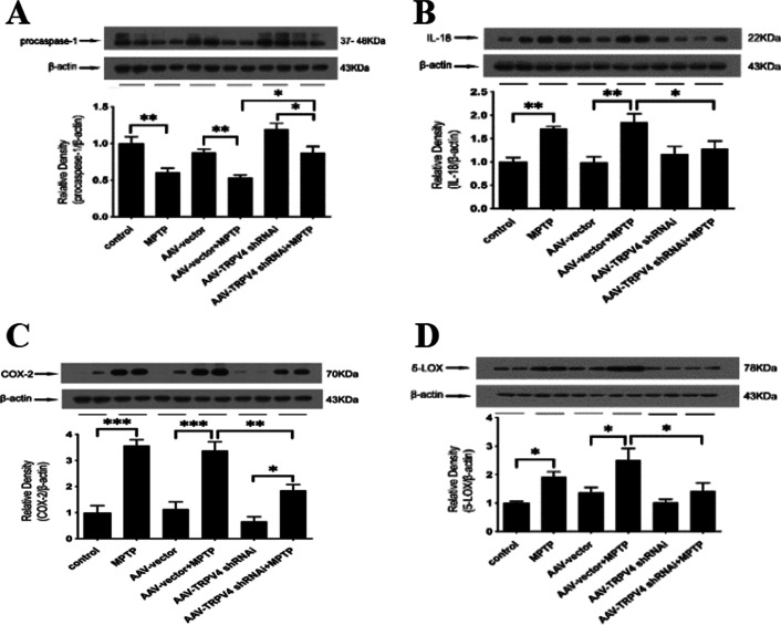
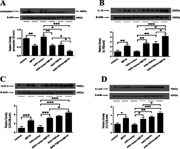
Results: The SN was transfected with AAV for 3 weeks and expressed the target protein with green fluorescence. Knockdown of TRPV4 via injection of a constructed AAV-TRPV4 shRNAi into the SN alleviated the movement deficits of PD mice. Upregulation of TRPV4 via injection of a constructed AAV-TRPV4 aggravated the above movement disorders. The expression of TRPV4 was upregulated in the SN of MPTP-treated mice. Injection of AAV-TRPV4 shRNAi into the SN rescued the number of TH-positive and Nissl-positive neurons in the SN decreased by MPTP, while injection of AAV-TRPV4 induced the opposite effect. Moreover, MPTP-decreased Sarco/endoplasmic reticulum Ca2+-ATPase 2 (SERCA2) and pro-cysteinyl aspartate specific proteinase-12 (procaspase-12), MPTP-increased Glucose-regulated protein 78 (GRP78), Glucose-regulated protein 94 (GRP94) and C/EBP homologous protein (CHOP) were inhibited by AAV-TRPV4 shRNAi infection, and enhanced by AAV-TRPV4. In the same way, MPTP-decreased procaspase-1, MPTP-increased Interleukin-18 (IL-18), Cyclooxgenase-2 (COX-2) and 5-Lipoxygenase (5-LOX) were inhibited by AAV-TRPV4 shRNAi, or further exacerbated by AAV-TRPV4.

Conclusions: These results suggest that TRPV4 mediates ER stress and inflammation pathways, contributing to the loss of dopamine (DA) neurons in the SN and movement deficits in PD mice. Moreover, this study provides a new perspective on molecular targets and gene therapies for the treatment of PD in the future.

Keywords: ER stress; Inflammation; MPTP; Parkinson’s disease; SN; TRPV4.


Conflict of interest statement

All the authors declared no competing financial interests.

Figures

Fig. 1
Fig. 1
Expression of TRPV4 in the SN after treatment with MPTP and AAV injection. A MPTP was administrated intraperitoneally at 30 mg/kg/day for 7 days, mice were sacrificed after the behavioral tests by cervical vertebra dislocation, the SN was rapidly dissected out. The expression of TRPV4 in the SN was assessed by Western blot (n = 10). Compared to that in the control group, TRPV4 was upregulated by MPTP. Data are presented as mean ± SEM. Statistical significance: ***P < 0.001. B AAV 0.5 μl was stereotaxically injected bilaterally into the SN at a rate of 250 nl/min, 21 days later the brains were dissected and cut into coronal slices at 25 μm thickness, fluorescence images of coronal frozen sections at different magnifications were presented by GFP in neurons in the SN region. Scale bar = 100 μm. C Schematic illustration of the experimental schedule
Fig. 2
Fig. 2
TRPV4 affected the behavioral performances of PD mice. After infused with 0.5 μl AAV-TRPV4 shRNAi or 0.3 μl AAV-TRPV4 bilaterally into the SN for 21 days and administrated with MPTP at 30 mg/kg/day for 7 days, mice were subjected to movement behavioral tests, including the pole test and rotarod test. A Knockdown of TRPV4 restored the T-tune time of PD mice (n = 12). B Upregulation of TRPV4 further prolonged the T-tune time of PD mice (n = 12). C Knockdown of TRPV4 restored the T-descend time of PD mice (n = 12). D Upregulation of TRPV4 further prolonged the T-descend time of PD mice (n = 12). E Knockdown of TRPV4 restored the retention time of PD mice (n = 12). F Upregulation of TRPV4 further shortened the retention time of PD mice (n = 12). For each condition, data were collected from three trials for each mouse. All data are presented as mean ± SEM. Significant differences: *P < 0.05, **P < 0.01, ***P < 0.001
Fig. 3
Fig. 3
TRPV4 affected the number of TH-positive neurons in the SN. After infused with 0.5 μl AAV-TRPV4 shRNAi or 0.3 μl AAV-TRPV4 bilaterally into the SN of mice for 21 days and administrated with MPTP at 30 mg/kg/day for 7 days, the SN was rapidly dissected out. The expressions of TRPV4 and TH in the SN were detected by Western blot. A AAV-TRPV4 shRNAi downregulated the expression of TRPV4 in the SN, and significantly inhibited the MPTP-induced increase in TRPV4 (n = 6). B AAV-TRPV4 upregulated the expression of TRPV4 in the SN, and further increased the MPTP-induced high levels of TRPV4 (n = 6). C Representative sample of brain slices with immunohistochemistry staining of TH-positive neurons in the SN of mice. Knockdown of TRPV4 rescued the number of TH-positive neurons, while upregulation of TRPV4 exacerbated the already reduction in the number of TH-positive neurons in the SN of PD mice (n = 3). D Knockdown of TRPV4 restored the expression of TH in the SN of PD mice (n = 6). E Upregulation of TRPV4 further decreased the expression of TH in the SN of PD mice (n = 6). All data are presented as mean ± SEM. Significant differences: *P < 0.05, **P < 0.01, ***P < 0.001
Fig. 4
Fig. 4
TRPV4 affected the number of Nissl-positive neurons in the SN. After infused with 0.5 μl AAV-TRPV4 shRNAi or 0.3 μl AAV-TRPV4 bilaterally into the SN of mice for 21 days and administrated with MPTP at 30 mg/kg/day for 7 days, representative sample of brain slices with Nissl staining showed Nissl-positive neurons in the SN. Knockdown of TRPV4 rescued the number of Nissl-positive neurons in the SN of PD mice, while upregulation of TRPV4 exacerbated the already reduction in the number of Nissl-positive neurons in the SN of PD mice (bar: 100 μm, n = 3). Data are presented as mean ± SEM. Significant differences: *P < 0.05, **P < 0.01, ***P < 0.001
Fig. 5
Fig. 5
TRPV4 knockdown attenuated MPTP-induced ER stress in the SN. After infused with 0.5 μl AAV-TRPV4 shRNAi bilaterally into the SN of mice for 21 days and administrated with MPTP at 30 mg/kg/day for 7 days, the SN was rapidly dissected out. The protein levels of SERCA2, GRP78, GRP94, CHOP and procaspase-12 in the SN of mice were detected by Western blot. A Knockdown of TRPV4 restored the MPTP-induced decrease in SERCA2 (n = 6). B Knockdown of TRPV4 inhibited the MPTP-induced upregulation of GRP78 (n = 6). C Knockdown of TRPV4 inhibited the MPTP-induced upregulation of GRP94 (n = 6). D Knockdown of TRPV4 inhibited the MPTP-induced upregulation of CHOP (n = 6). E Knockdown of TRPV4 partly inhibited the MPTP-induced decrease in procaspase-12 (n = 6). All data are presented as mean ± SEM. Statistical significance: *P < 0.05, **P < 0.01, ***P < 0.001
Fig. 6
Fig. 6
Effects of TRPV4 upregulation on MPTP-induced ER stress in the SN. After infused with 0.3 μl AAV-TRPV4 bilaterally into the SN of mice for 21 days and administrated with MPTP at 30 mg/kg/day for 7 days, the SN was rapidly dissected out. The protein levels of SERCA2, GRP78, GRP94, CHOP and procaspase-12 in the SN of mice were detected by Western blot. A Upregulation of TRPV4 deteriorated the MPTP-induced reduction in SERCA2 in the SN (n = 6). B Upregulation of TRPV4 exacerbated the MPTP-induced activation of GRP78 in the SN (n = 6). C Upregulation of TRPV4 exacerbated the MPTP-induced activation of GRP94 in the SN (n = 6). D Upregulation of TRPV4 exacerbated the MPTP-induced activation of CHOP in the SN (n = 6). E Upregulation of TRPV4 accelerated the MPTP-induced decrease in procaspase-12 in the SN (n = 6). All data are presented as mean ± SEM. Statistical significance: *P < 0.05, **P < 0.01, ***P < 0.001
Fig. 7
Fig. 7
Knockdown of TRPV4 in the SN attenuated inflammation in PD mice. After infused with 0.5 μl AAV-TRPV4 shRNAi bilaterally into the SN of mice for 21 days and administrated with MPTP at 30 mg/kg/day for 7 days, the SN was rapidly dissected out. The protein levels of procaspase-1, IL-18, COX-2 and 5-LOX in the SN of mice were detected by Western blot. A Knockdown of TRPV4 in the SN restored the MPTP-induced the reduction in procaspase-1 (n = 6). B Knockdown of TRPV4 in the SN decreased the MPTP-induced high level of IL-18 (n = 6). C Knockdown of TRPV4 in the SN decreased the MPTP-induced high level of COX-2 (n = 6). D Knockdown of TRPV4 in the SN decreased the MPTP-induced high level of 5-LOX (n = 6). All data are presented as mean ± SEM. Statistical significance: *P < 0.05, **P < 0.01, ***P < 0.001
Fig. 8
Fig. 8
Upregulation of TRPV4 in the SN aggravated inflammation in PD mice. After infused with 0.3 μl AAV-TRPV4 bilaterally into the SN of mice for 21 days and administrated with MPTP at 30 mg/kg/day for 7 days, the SN was rapidly dissected out. The protein levels of procaspase-1, IL-18, COX-2 and 5-LOX in the SN of mice were detected by Western blot. A Upregulation of TRPV4 further decreased the MPTP-induced the reduction in procaspase-1 in the SN (n = 6). B Upregulation of TRPV4 further increased the MPTP-induced high level of IL-18 in the SN (n = 6). C Upregulation of TRPV4 further increased the MPTP-induced high level of COX-2 in the SN (n = 6). D Upregulation of TRPV4 further increased the MPTP-induced high level of 5-LOX in the SN (n = 6). All data are presented as mean ± SEM. Statistical significance: *P < 0.05, **P < 0.01, ***P < 0.001
Fig. 9
Fig. 9
MPTP-induced activation of TRPV4 mediating ER stress and inflammation caused DA neuron death in the SN

References

    1. Lai F, Jiang R, Xie W, et al. Intestinal pathology and gut microbiota alterations in a methyl-4-phenyl-1,2,3,6-tetrahydropyridine (MPTP) mouse model of Parkinson's disease. Neurochem Res. 2018;43(10):1986–1999. - PubMed
    1. Pupyshev AB, Tikhonova MA, Akopyan AA, et al. Therapeutic activation of autophagy by combined treatment with rapamycin and trehalose in a mouse MPTP-induced model of Parkinson's disease. Pharmacol Biochem Behav. 2019;177:1–11. - PubMed
    1. Zhu J, Dou S, Jiang Y, Chen J, et al. Apelin-13 protects dopaminergic neurons in MPTP-induced Parkinson's disease model mice through inhibiting endoplasmic reticulum stress and promoting autophagy. Brain Res. 2019;1715:203–212. - PubMed
    1. Li Y, Liu Z, Wang D, et al. Ucf-101 protects in vivoandin vitro models of PD against 6-hydroxydopamine toxicity by alleviating endoplasmic reticulum stress via the Wnt/β-catenin pathway. J Clin Neurosci. 2020;71:217–225. - PubMed
    1. Liu N, Liu J, Wen X, et al. TRPV4 contributes to ER stress: relation to apoptosis in the MPP+-induced cell model of Parkinson's disease. Life Sci. 2020;261:118461. - PubMed

MeSH terms

Substances