. 2022 Jan 29;17(1):12.
doi: 10.1186/s13024-022-00518-y.

Islet amyloid polypeptide cross-seeds tau and drives the neurofibrillary pathology in Alzheimer's disease

Affiliations

Islet amyloid polypeptide cross-seeds tau and drives the neurofibrillary pathology in Alzheimer's disease

Guoxin Zhang et al. Mol Neurodegener. .

Abstract

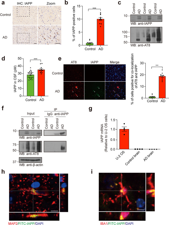
Background: The pathologic accumulation and aggregation of tau is a hallmark of tauopathies including Alzheimer's disease (AD). However, the molecular mechanisms mediating tau aggregation in AD remain elusive. The incidence of AD is increased in patients with type 2 diabetes (T2DM), which is characterized by the amyloid deposition of islet amyloid polypeptide (IAPP) in the pancreas. However, the molecular mechanisms bridging AD and T2DM remain unknown.

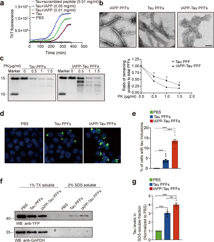
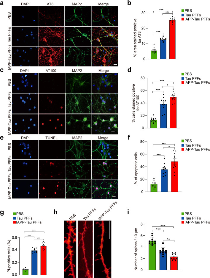
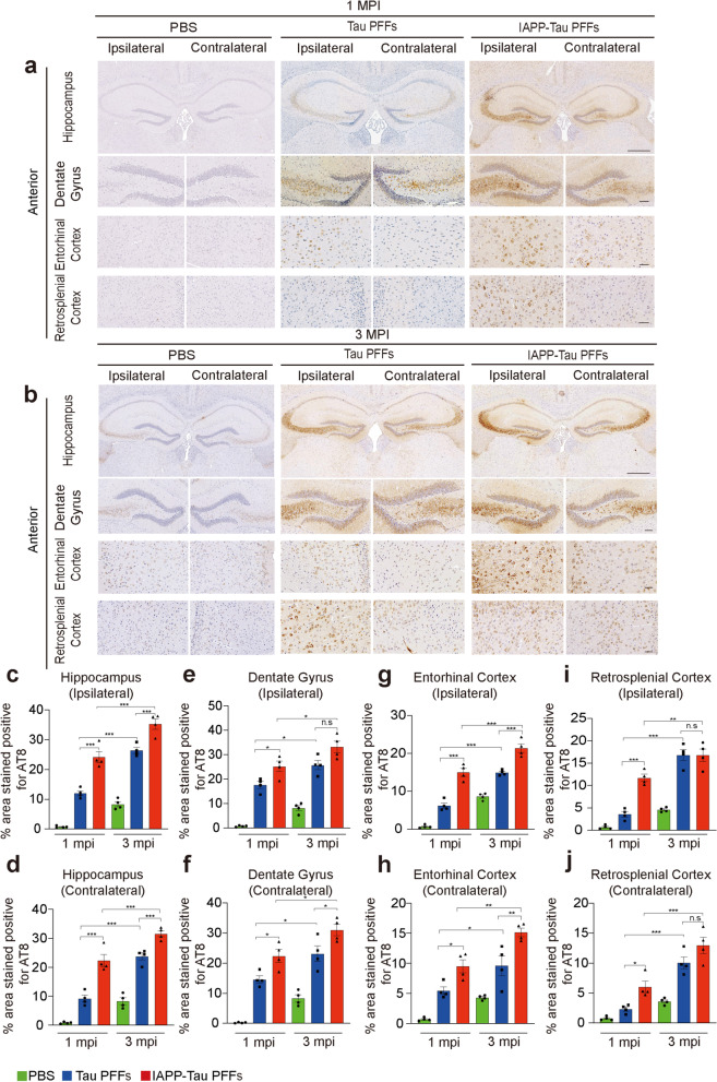
Methods: We first examined the presence of IAPP in the neurofibrillary tangles of AD patients. Then we tested the effect of IAPP on tau aggregation. The biochemical and biological characteristics of the IAPP-tau fibrils were tested in vitro. The seeding activity and neurotoxicity of the IAPP-tau fibrils were confirmed in cultured neurons. Lastly, the effect of IAPP on tau pathology and cognitive impairments was determined by injecting the IAPP-tau fibrils and IAPP fibrils into the hippocampus of tau P301S mice.

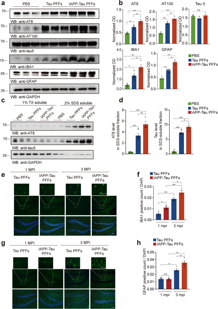
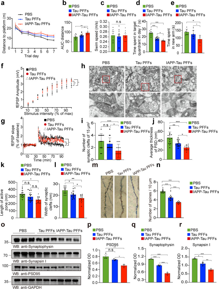
Results: We found that IAPP interacts with tau and accelerates the formation of a more toxic strain, which shows distinct morphology with enhanced seeding activity and neurotoxicity in vitro. Intrahippocampal injection of the IAPP-tau strain into the tau P301S transgenic mice substantially promoted the spreading of tau pathology and induced more severe synapse loss and cognitive deficits, when compared with tau fibrils. Furthermore, intracerebral injection of synthetic IAPP fibrils initiated tauopathy in the brain of tau P301S transgenic mice.

Conclusions: These observations indicate that IAPP acts as a crucial mediator of tau pathology in AD, and provide a mechanistic explanation for the higher risk of AD in individuals with T2DM.

Keywords: Cross-seeding; Islet amyloid polypeptide; Tau; Tauopathies; Type 2 diabetes.


Conflict of interest statement

The authors declare no competing financial interests.

Figures

Fig. 1
Fig. 1
IAPP deposits in the brain of AD patients. a Representative images of IAPP deposition in the hippocampal sections of AD patients and age-matched controls. b Quantification of the percentage of IAPP-positive cells. Scale bars, 50 μm. Bars represent means ± SEM. Unpaired Student’s t-test. n = 10 slides from 10 AD patients and 10 slides from 10 control subjects. ***P < 0.001. c Western bolt assay detects the presence of IAPP in the formic acid fraction from AD brains. n = 3 AD patients and 3 controls. d Concentrations of IAPP in the CSF of AD patients (n = 11) and healthy controls (n = 22). Bars represent means ± SEM. Unpaired Student’s t-test, ***P < 0.001. e Co-immunostainings of IAPP and phosphorylated tau (p-tau) in brain sections of AD patients. n = 10 AD patients and 10 controls. Scale bars, 20 μm. f Co-immunoprecipitation showing the interaction between IAPP and p-tau in AD human brains. n = 3 AD patients and 3 controls. g RT-PCR analysis found no IAPP mRNA expression in both AD and control brains. n = 4 AD patients and 4 controls. (h, i) Uptake of intravenously injected pure IAPP PFFs by neurons (g) and microglia (h) in tau P301S mice. Scale bars, 20 μm
Fig. 2
Fig. 2
IAPP accelerates tau fibrillization in vitro. a ThT fluorescence assay of tau fibrillization in the presence of different concentrations of IAPP monomers or scrambled peptide (n = 3 independent experiments. The mean ThT fluorescence are shown). b Representative images of transmission electron microscopy (TEM) for IAPP PFFs, Tau PFFs, and IAPP-tau PFFs. Scale bar, 200 nm. c PK digestion assay. Tau PFFs or IAPP-tau PFFs were incubated with increasing concentrations of PK (0 to 1.5 mg/ml) and analyzed by Coomassie blue staining (left). Quantification represents the ratio of remaining protein to total PFFs (right). Data are means ± SEM (n = 3 independent experiments). d Tau PFFs and IAPP-tau PFFs induce tau aggregation in HEK293 cells stably expressing YFP-tagged tau RD. Scale bars, 20 μm. e The percentage of cells with tau inclusions induced by tau PFFs and IAPP-tau PFFs. Data are presented as means ± SEM. One-way ANOVA followed by Tukey’s post hoc test (n = 10 slices from 3 independent experiments). ***P < 0.001. f, g Cells transduced with tau PFFs and IAPP-tau PFFs were sequentially extracted with 1% Triton X-100 (TX-100) (TX soluble) and 2% SDS (TX insoluble). Lysates were subjected to Western blot to show the presence of tau in different fractions (n = 4 independent samples). Data are presented as means ± SEM. One-way ANOVA followed by Tukey’s post hoc test. **P < 0.01, ***P < 0.001. DAPI, 4′,6-diamidino-2-phenylindole
Fig. 3
Fig. 3
IAPP-tau PFFs induce tau phosphorylation and neuronal apoptosis in vitro. a-d Representative immunostaining and quantification of AT8 (a, b) and AT100 p-tau (c, d) in primary neurons transduced with tau PFFs or IAPP-tau PFFs. Scale bar, 20 μm. e Representative images of TUNEL staining of primary cortical neurons transduced with tau PFFs or IAPP-tau PFFs. f, g Quantification of TUNEL staining (f) and Hoechst/PI staining (g). Scale bars, 30 μm. h, i Representative images and quantification of Dil staining of dendritic spines in neurons. Experiments were independently performed three times. Fifty visual fields (Fig. 3b, d), 150 cells (Fig. 3f, g), and 50 dendrites (Fig. 3i) from 10 slices were counted in each group. Scale bar, 20 μm. Bars represent means ± SEM. One-way ANOVA followed by Tukey’s post hoc test (n = 10 slices). *P < 0.05, **P < 0.01, ***P < 0.001
Fig. 4
Fig. 4
IAPP-Tau PFFs promote the propagation of tau pathology in vivo. a, b Representative images of AT8 p-tau pathology in tissue sections (anterior hippocampal level) from the hippocampus, dentate gyrus, retrosplenial cortex, and entorhinal cortex of tau P301S mice 1 month (a) and 3 months (b) after the injection of PBS, tau PFFs or IAPP-tau PFFs. Scale bar, 200 μm in (a) and (b) upper panel, 50 μm in (a) and (b) lower panels. c-j Quantification of p-tau pathology of ipsilateral and contralateral sides in the hippocampus (c, d), dentate gyrus (e, f), entorhinal cortex (g, h), and retrosplenial cortex (i, j). n = 4 mice per group. Data are presented as means ± SEM. One-way ANOVA followed by Tukey’s post hoc test. *P < 0.05, **P < 0.01, ***P < 0.001, n.s. not significant
Fig. 5
Fig. 5
IAPP-Tau PFFs promote tau phosphorylation and neuroinflammation in vivo. a Representative immunoblots of p-tau (AT8 and AT100), total-tau (tau5), IBA-1, and GFAP in the ipsilateral hippocampus of mice injected with PBS, tau PFFs or IAPP-tau PFFs. b Statistical analysis shows significantly increased expression of AT8, AT100, IBA1, and GFAP in mice injected with IAPP-Tau PFFs, compared to mice injected with PBS and tau PFFs. c, d Ipsilateral hippocampal samples were sequentially extracted with 1% Triton X-100 (TX-soluble) and 2% SDS (TX-insoluble) from mice injected with either PBS, tau PFFs, or IAPP-tau PFFs. Quantification is presented in (d). e-h Immunostaining and quantification of microglia marker IBA1 (e, f) and astrocyte marker GFAP (g, h) in mice 3 months after injection with tau PFFs or IAPP-tau PFFs. Scale bar, 50 μm for lower panel, 20 μm for magnification. n = 4 mice per group. Data are presented as means ± SEM. One-way ANOVA followed by Tukey’s post hoc test. *P < 0.05, **P < 0.01, ***P < 0.001
Fig. 6
Fig. 6
IAPP-tau PFFs induce cognitive impairment and synaptic dysfunction in vivo. Three-month-old male mice were injected with PBS, tau PFFs or IAPP-tau PFFs, respectively. The mice were sacrificed three months after PFF injection. a Spatial memory was assessed by the Morris water maze test. Shown are the distance traveled to the platform by mice injected with PBS, Tau PFFs, or IAPP-Tau PFFs. b Integrated time traveled in Morris water maze test. AUC, area under the curve. c Average swim speed of mice in three groups. d Probe trial results. n = 9 mice in PBS group, n = 12 mice in Tau PFFs group, n = 7 mice in IAPP-tau PFFs group. e Time spent in the novel arm in the Y-maze test. n = 7 mice per group. f Long-term potentiation (LTP) of fEPSPs was induced by 3 × TBS (4 pulses at 100 Hz, repeated three times with a 200-ms interval). The magnitude of LTP and synaptic transmission was assessed by input/output (I/O). g Representative fEPSPs of hippocampal slices prepared from mice in three groups. n = 3 mice per group. h Electron microscopy of synapses (top) and magnification (below). Scale bar, 1 μm in the top panel, 200 nm in the lower panel. i The number of synaptic clefts. j Postsynaptic density. k Length of the active zone. l Width of synaptic clefts. n = 10–15 slices per group. m, n Golgi staining of dendritic spines of hippocampal slides. Scale bar, 20 μm. n = 10 slices per group. o Western blot analysis of synaptic markers in the ipsilateral hippocampus of tau P301S mice. p-r Quantification for synaptophysin (p), synapsin I (q), and PSD95 (r), n = 3 mice per group. Data are presented as means ± SEM. One-way ANOVA followed by Tukey’s post hoc test. *P < 0.05, **P < 0.01, ***P < 0.001. n.s. not significant

References

    1. Peng C, Trojanowski JQ, Lee VM. Protein transmission in neurodegenerative disease. Nat Rev Neurol. 2020;16:199–212. - PMC - PubMed
    1. Narasimhan S, Guo JL, Changolkar L, Stieber A, McBride JD, Silva LV, He Z, Zhang B, Gathagan RJ, Trojanowski JQ, Lee VMY. Pathological tau strains from human brains recapitulate the diversity of Tauopathies in nontransgenic mouse Brain. J Neurosci. 2017;37:11406–11423. - PMC - PubMed
    1. Kaufman SK, Sanders DW, Thomas TL, Ruchinskas AJ, Vaquer-Alicea J, Sharma AM, Miller TM, Diamond MI. Tau prion strains dictate patterns of cell pathology, progression rate, and regional vulnerability in vivo. Neuron. 2016;92:796–812. - PMC - PubMed
    1. Biessels GJ, Staekenborg S, Brunner E, Brayne C, Scheltens P. Risk of dementia in diabetes mellitus: a systematic review. Lancet Neurol. 2006;5:64–74. - PubMed
    1. Janson J, Laedtke T, Parisi JE, O'Brien P, Petersen RC, Butler PC. Increased risk of type 2 diabetes in Alzheimer disease. Diabetes. 2004;53:474–481. - PubMed

Publication types

LinkOut - more resources