

Stroke population-specific neuroanatomical CT-MRI brain atlas
- PMID: 35094103
- PMCID: PMC9271109
- DOI: 10.1007/s00234-021-02875-9
Stroke population-specific neuroanatomical CT-MRI brain atlas
Abstract
Purpose: Development of a freely available stroke population-specific anatomical CT/MRI atlas with a reliable normalisation pipeline for clinical CT.
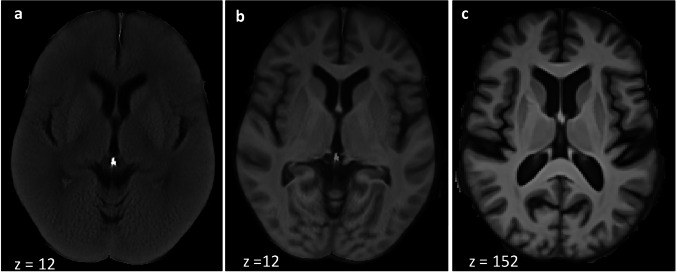
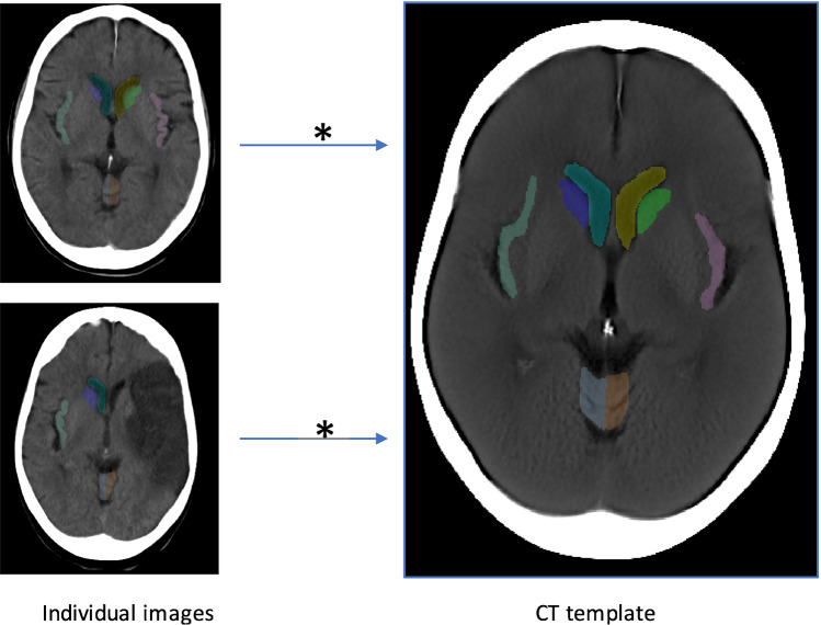
Methods: By reviewing CT scans in suspected stroke patients and filtering the AIBL MRI database, respectively, we collected 50 normal-for-age CT and MRI scans to build a standard-resolution CT template and a high-resolution MRI template. The latter was manually segmented into anatomical brain regions. We then developed and validated a MRI to CT registration pipeline to align the MRI atlas onto the CT template. Finally, we developed a CT-to-CT-normalisation pipeline and tested its reliability by calculating Dice coefficient (Dice) and Average Hausdorff Distance (AHD) for predefined areas in 100 CT scans from ischaemic stroke patients.
Results: The resulting CT/MRI templates were age and sex matched to a general stroke population (median age 71.9 years (62.1-80.2), 60% male). Specifically, this accounts for relevant structural changes related to aging, which may affect registration. Applying the validated MRI to CT alignment (Dice > 0.78, Average Hausdorff Distance < 0.59 mm) resulted in our final CT-MRI atlas. The atlas has 52 manually segmented regions and covers the whole brain. The alignment of four cortical and subcortical brain regions with our CT-normalisation pipeline was reliable for small/medium/large infarct lesions (Dice coefficient > 0.5).
Conclusion: The newly created CT-MRI brain atlas has the potential to standardise stroke lesion segmentation. Together with the automated normalisation pipeline, it allows analysis of existing and new datasets to improve prediction tools for stroke patients (free download at https://forms.office.com/r/v4t3sWfbKs ).
Keywords: Computed tomography (CT); Magnetic resonance imaging (MRI); Neuroanatomical atlas; Population specific; Stroke; Template.
© 2021. The Author(s).
Conflict of interest statement
We declare that we have no conflict of interest.
Figures


References
MeSH terms
LinkOut - more resources
Full Text Sources
Other Literature Sources
Medical
