. 2022 Apr;53(2):437-448.
doi: 10.1007/s10735-021-10055-5. Epub 2022 Jan 30.

Endocrine secretory granule production is caused by a lack of REST and intragranular secretory content and accelerated by PROX1

Affiliations

Endocrine secretory granule production is caused by a lack of REST and intragranular secretory content and accelerated by PROX1

Jun Ishii et al. J Mol Histol. 2022 Apr.

Abstract

Endocrine secretory granules (ESGs) are morphological characteristics of endocrine/neuroendocrine cells and store peptide hormones/neurotransmitters. ESGs contain prohormones and ESG-related molecules, mainly chromogranin/secretogranin family proteins. However, the precise mechanism of ESG formation has not been elucidated. In this study, we experimentally induced ESGs in the non-neuroendocrine lung cancer cell line H1299. Since repressive element 1 silencing transcription factor (REST) and prospero homeobox 1 (PROX1) are closely associated with the expression of ESG-related molecules, we edited the REST gene and/or transfected PROX1 and then performed molecular biology, immunocytochemistry, and electron and immunoelectron microscopy assays to determine whether ESG-related molecules and ESGs were induced in H1299 cells. Although chromogranin/secretogranin family proteins were induced in H1299 cells by knockout of REST and the induction was accelerated by the PROX1 transgene, the ESGs could not be defined by electron microscopy. However, a small number of ESGs were detected in the H1299 cells lacking REST and expressing pro-opiomelanocortin (POMC) by electron microscopy. Furthermore, many ESGs were produced in the REST-lacking and PROX1- and POMC-expressing H1299 cells. These findings suggest that a lack of REST and the expression of genes related to ESG content are indispensable for ESG production and that PROX1 accelerates ESG production.Trial registration: Not applicable.

Keywords: Endocrine secretory granule; Non-neuroendocrine lung cancer; POMC; PROX1; REST.


Conflict of interest statement

The authors have no conflicts of interest to declare that are relevant to the content of this article.

Figures
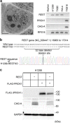
Fig. 1
Fig. 1
Establishment of REST gene-edited and/or PROX1-transfected H1299 cells. a (Left) Transmission electron micrograph of H1299 cells. The non-neuroendocrine lung cancer cell line H1299 does not express endocrine secretory granules (ESGs). The large globular structures with high electron density found in cells are secondary lysosomes. Scale bar, 5 μm. (Right) RT-PCR analyses of repressive element 1 silencing transcription factor (REST), prospero homeobox 1 (PROX1) and chromogranin A (CHG-A) in the lung cancer cell lines H1299 (large cell carcinoma), TKB5 (large cell carcinoma), and TKB16 (small cell carcinoma) and HeLa cells. H1299 cells, as well as TKB5 and HeLa cells, expressed REST but did not express PROX1 or CHG-A. PROX1 and CHG-A were expressed in the small cell carcinoma cell line TKB16. Ribosomal protein S18 (RPS18) served as an internal control. Ethidium bromide-stained gel, reverse images. b A schematic diagram of the REST gene editing status in H1299 cells. In REST knockout H1299 cells, the homozygous deletion of 32 bases in the REST gene (NG_029447.1, 16963 to 17014) was confirmed by Sanger sequencing. c Western blot analysis of FLAG-tagged PROX1 and CHG-A in the H1299 transfectants. FLAG signals were found in the FLAG-tagged PROX1-transfected H1299 cells. CHG-A was induced in the REST-deficient H1299 cells. GAPDH served as an internal control. "+" indicates that a specific gene was transfected, and "−" indicates that a specific gene was knocked out
Fig. 2
Fig. 2
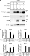
Effects of REST and PROX1 on ESG-related gene expression and ESG formation in H1299 cells. a Quantitative RT-PCR analyses were conducted to examine alterations in the expression of ESG-related genes induced by the knockout of REST and forced expression of FLAG-PROX1. "−" indicates that a specific gene was knocked out, and "+" indicates that a specific gene was transfected. The expression levels of CHG-A (chromogranin-A), CHG-B (chromogranin-B), SCG-2 (secretogranin-II) and SCG-3 (secretogranin-III) were highly induced by knockout of REST (− REST) and enhanced by PROX1 transfection (+ PROX1). Ribosomal protein S18 (RPS18) served as an internal control. The data are shown as the mean ± SD. b Transmission electron micrographs of wild-type H1299 cells and gene-transfected/gene-edited H1299 cells. "−" indicates that a specific gene was knocked out, and "+" indicates that a specific gene was transfected. No ESGs were observed in any of the cells. Scale bar, 2 μm
Fig. 3
Fig. 3
Effects of POMC on ESG-related gene expression in H1299 cells. a Western blot analysis of POMC gene products in H1299 transfectants. Signals of POMC gene products were observed in the POMC-transfected H1299 cells. CHG-A was induced in the REST-deficient H1299 cells. GAPDH served as an internal control. "+" indicates that a specific gene was transfected, and "−" indicates that a specific gene was knocked out. MW, molecular weight marker. b Quantitative RT-PCR analyses were conducted to examine alterations in the expression of ESG-related genes induced by the knockout of REST and forced expression of FLAG-PROX1/POMC. "−" indicates that a specific gene was knocked out, and "+" indicates that a specific gene was transfected. Sustained expression of ESG-related genes (CHG-A, CHG-B, SCG-2 and SCG-3) was found in the − REST H1299 cells and the − REST + PROX1 H1299 cells despite the additional transfection of POMC. Ribosomal protein S18 (RPS18) served as an internal control. The data are shown as the mean ± SD
Fig. 4
Fig. 4
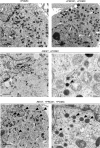
REST knockout and PROX1/POMC expression induce ESG-like structures in H1299 cells. Transmission electron micrographs of the gene-transfected/gene-edited H1299 cells. Many ESG-like structures were observed in the − REST + PROX1 + POMC H1299 cells (H1299-RESTKO-PROX1-POMC), and the same structures were observed in the − REST + POMC H1299 cells (H1299-RESTKO-POMC) but to a lesser extent. "+" indicates that a specific gene was transfected, and "−" indicates that a specific gene was knocked out. The arrowheads indicate representative ESG-like structures. Black scale bar, 2 μm. White scale bar, 0.5 μm
Fig. 5
Fig. 5
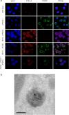
Immunofluorescence cytochemical and immunoelectron microscopic analyses of CHG-A and POMC in the REST-deficient H1299 cells transfected with or without PROX1 and/or POMC. a H1299 cells (wild-type), REST-deficient PROX1-transfected H1299 cells (− REST + PROX1), REST-deficient POMC-transfected H1299 cells (− REST + POMC), and REST-deficient PROX1- and POMC-transfected H1299 cells (− REST + PROX1 + POMC) were stained with a rabbit polyclonal anti-CHG-A antibody (Alexa Fluor 555-labelled), a mouse monoclonal anti-ACTH antibody (Alexa Fluor 488-labelled), which was used to detect POMC gene products, and DAPI (blue). Images were acquired by a confocal microscope. Red signals (CHG-A) and green signals (POMC) were colocalized in the − REST + PROX1 + POMC H1299 cells (H1299-RESTKO-PROX1-POMC). b PROX1- and POMC-transfected REST-deficient H1299 cells (H1299-RESTKO-PROX1-POMC) were processed for immunoelectron microscopy. Thin sections were treated with a rabbit polyclonal anti-CHG-A antibody and a mouse monoclonal anti-ACTH antibody, which was used to detect POMC gene products, and then incubated with an anti-rabbit secondary antibody coupled to 5 nm gold and an anti-mouse secondary antibody coupled to 15 nm gold. Gold colloids sized 5 nm and 15 nm were colocalized on the ESGs. Scale bar, 100 nm

Similar articles

Cited by

References

    1. Aoki H, Hara A, Era T, Kunisada T, Yamada Y. Genetic ablation of rest leads to in vitro-specific derepression of neuronal genes during neurogenesis. Development. 2012;139:667–677. doi: 10.1242/dev.072272. - DOI - PubMed
    1. Asadi F, Dhanvantari S. Stathmin-2 mediates glucagon secretion from pancreatic α-cells. Front Endocrinol. 2020;11:29. doi: 10.3389/fendo.2020.00029. - DOI - PMC - PubMed
    1. Asensio CS, Sirkis DW, Maas JW, Jr, Egami K, To T-L, Brodsky FM, Shu X, Cheng Y, Edwards RH. Self-assembly of VPS41 promotes sorting required for biogenesis of the regulated secretory pathway. Dev Cell. 2013;27:425–437. doi: 10.1016/j.devcel.2013.10.007. - DOI - PMC - PubMed
    1. Ballas N, Grunseich C, Lu DD, Speh JC, Mandel G. REST and its correpressors mediate plasticity of neuronal gene chromatin throughout neurogenesis. Cell. 2005;121:645–657. doi: 10.1016/j.cell.2005.03.013. - DOI - PubMed
    1. Beuret N, Stettler H, Renold A, Rutishauser J, Spiess M. Expression of regulated secretory proteins is sufficient to generate granule-like structures in constitutively secreting cells. J Biol Chem. 2004;279:20242–20249. doi: 10.1074/jbc.m310613200. - DOI - PubMed

MeSH terms