. 2022 Jan 14:12:724192.
doi: 10.3389/fphar.2021.724192. eCollection 2021.

Targeting HSP90 Inhibits Proliferation and Induces Apoptosis Through AKT1/ERK Pathway in Lung Cancer

Affiliations

Targeting HSP90 Inhibits Proliferation and Induces Apoptosis Through AKT1/ERK Pathway in Lung Cancer

Mengyuan Niu et al. Front Pharmacol. .

Abstract

Lung cancer is one of the most common malignant cancers worldwide. Searching for specific cancer targets and developing efficient therapies with lower toxicity is urgently needed. HPS90 is a key chaperon protein that has multiple client proteins involved in the development of cancer. In this study, we investigated the transcriptional levels of HSP90 isoforms in cancerous and normal tissues of lung cancer patients in multiple datasets. The higher expression of HSP90AA1 in cancer tissues correlated with poorer overall survival was observed. The higher levels of transcription and expression of HSP90AA1 and the activity of AKT1/ERK pathways were confirmed in lung cancer patient tissues. In both human and mouse lung cancer cell lines, knocking down HSP90AA1 promoted cell apoptosis through the inhibition of the pro-survival effect of AKT1 by decreasing the phosphorylation of itself and its downstream factors of mTOR and BAD, as well as downregulating Mcl1, Bcl-xl, and Survivin. The knockdown also suppressed lung cancer cell proliferation by inhibiting ERK activation and downregulating CyclinD1 expression. The treatment of 17-DMAG, an HSP90 inhibitor, recaptured these effects in vitro and inhibited tumor cell growth, and induced apoptosis without obvious side effects in lung tumor xenograft mouse models. This study suggests that targeting HSP90 by 17-DMAG could be a potential therapy for the treatment of lung cancer.

Keywords: 17-DMAG; AKT1; ERK; Hsp90; lung cancer.


Conflict of interest statement

The authors declare that the research was conducted in the absence of any commercial or financial relationships that could be construed as a potential conflict of interest.

Figures

FIGURE 1
FIGURE 1
Increased HSP90AA1 is correlated with decreased overall survival time in lung cancer patients. (A) HSP90AA1, HSP90AB1, HSP90B1 are all significantly up-regulated in lung cancer tissues in four different high throughput datasets. **p < 0.01, ****p < 0.0001. (B) Three HSP90 isoforms (AA1, AB1, B1) are positively correlated with each other. (C) HSP90AA1 is significantly correlated with a shortened overall survival time in three datasets.
FIGURE 2
FIGURE 2
The transcription profile of HSP90AA1 correlated genes was different between lung cancer and normal tissues. (A) The Venn plot indicated the overlapping and differences of the genes positively correlated with HSP90AA1 expression in normal (GTEX) and cancerous (TCGA LUAD) lungs. (B & C) Enrichment and correlations of HSP90 positively correlated genes in KEGG pathways. (D) The heatmap of indicated genes enriched in the corresponding pathways and their correlation with HSP90AA1 expression. (E) The correlation of BRCA1 and CDK1 with HSP90AA1 in the GTEX and TCGA databases. (F) The enrichment of GO terms of molecular function and biological process in HSP90AA1 correlated genes. (G) GSEA analysis of HSP90AA1 indicated the possible correlated genes enriched in KEGG pathways of cell cycle and DNA replication.
FIGURE 3
FIGURE 3
Transcription and western blot validation of AKT1/ERK signaling in lung cancer patients. (A) qPCR analysis of HSP90, AKT1, Bcl-xl, Survivin, Mcl1, and CyclinD1 in lung cancer patients’ tumors compared with Para cancerous tissue. Patients number, N = 40 The data represents the mean ± standard deviation (SD). ***p < 0.001 (B) Western blot analysis of HSP90, AKT1, Bcl-xl, Survivin, Mcl1, and CyclinD1 in lung cancer patient’s tumor compared with Para cancerous tissue. We randomly chose 6 patients and repeated the experiment three times.
FIGURE 4
FIGURE 4
17-DMAG inhibited lung cancer proliferation and induced cell apoptosis. (A) Molecular structural formula of 17-DMAG. (B) LLC and A549 lung cancer cells were treated with folded diluted 17-DMAG. Cell viability was determined by the MTS method. The IC50 value was determined to evaluate the inhibition of tumor cell growth by 17-DMAG. The value of each cell was the mean value of three independent experiments. (C) Time-course analysis of the growth inhibition effect of 17-DMAG in LLC and A549 cells. (D) BEAS-2B cells were treated with 17-DMAG at indicated concentrations for 24 h. The cell viability was determined by an MTS assay (n = 5). (E) LLC and A549 cells were treated with 0.2 or 1 μM 17-DMAG and then stained with PI and AnnexinV-FITC to detect apoptosis by flow cytometry. The columns showed the statistical FACS results of three independent experiments. ***p < 0.001.
FIGURE 5
FIGURE 5
Inhibiting HSP90 reversed AKT1/ERK activation in lung cancer cells. (A) LLC cells were treated with indicated concentrations of 17-DMAG and then subjected to western blot for measuring protein levels by indicated antibodies. (B) A549 cells were treated with indicated concentrations of 17-DMAG and then subjected to western blot for measuring protein levels by indicated antibodies. (C) The total RNA of 17-DMAG-treated LLC cells and A549 cells were extracted, and a qPCR method was adopted to test the transcription level of indicated genes. The result was obtained from three independent experiments. **p < 0.01, ***p < 0.001.
FIGURE 6
FIGURE 6
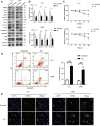
Knocking down of HSP90 inhibited lung cancer proliferation and induced cell apoptosis. LLC and A549 cells were transfected with HSP90 siRNA or control siRNA. (A) Western blotting with indicated antibodies was performed. (B) The total RNA of the cells was extracted, and a qPCR method was adopted to test the transcription level of indicated genes. (C) Time-course analysis of the cell growth detected by MTS assay. *p < 0.05, **p < 0.01. (D) The apoptosis level of the cells was evaluated by staining with PI and AnnexinV-FITC antibody and subjected by flow cytometry. The columns showed the statistical FACS results of three independent experiments. **p < 0.01, ***p < 0.001. (E) Analysis of apoptosis by TUNEL staining (blue fluorescence, DAPI staining for nuclei; green fluorescence, TUNEL-positive staining) in LLC and A549 cells. Scale bar = 30 μm.
FIGURE 7
FIGURE 7
17-DMAG inhibited tumor growth and induced cell death in vivo. Male C57/BL6 and Balb/c nude mice (n = 10) were injected with LLC and A549 cells, respectively (n = 10). After tumors reached approximately 100 mm3, mice were treated with or without 17-DMAG (15 mg/kg/day) intraperitoneally. (A) Tumor volumes of LLC and A549 bearing mice were calculated every day. ***p < 0.001. (B) The weight of each LLC or A549-bearing mouse was measured every other day before the treatment of 17-DMAG (n = 5 per group). (C) Tumor mass images of both LLC and A549 model mice at the end of the experiment (n = 5 per group). (D) LLC and A549 tumors treated with or without 17-DMAG were removed and weighed after the euthanasia of mice. ***p < 0.001. (E) Analysis of apoptosis by TUNEL staining (blue fluorescence, DAPI staining for nuclei; green fluorescence, TUNEL-positive staining) in LLC and A549 cell xenograft mouse-derived tissue from control, 17-DMAG–treated animals. Scale bar = 50 μm.
FIGURE 8
FIGURE 8
Inhibiting HSP90 reversed AKT1/ERK activation in vivo. (A) mRNA levels of indicated genes from LLC tumor tissue were tested by qPCR. *p < 0.05, **p < 0.01, ***p < 0.001. (B) mRNA levels of indicated genes from A549 tumor tissue were tested by qPCR. **p < 0.01, ***p < 0.001. (C) Lysates from LLC and A549 tumor tissue were analyzed by western blotting and probed with indicated antibodies. (D) Representative images of LLC tumor sections stained with CD8 antibody. The positive staining of CD8 was marked with arrows. Scale bar = 50 μm. (E) Expression of genes related to lymphocyte cytotoxicity. Relative mRNA expression levels were adjusted to the level of beta-actin mRNA for each sample. ***p < 0.001, ****p < 0.0001.

References

    1. Ambade A., Catalano D., Lim A., Mandrekar P. (2012). Inhibition of Heat Shock Protein (Molecular Weight 90 kDa) Attenuates Proinflammatory Cytokines and Prevents Lipopolysaccharide-Induced Liver Injury in Mice. Hepatology 55 (5), 1585–1595. 10.1002/hep.24802 - DOI - PMC - PubMed
    1. Basso A. D., Solit D. B., Chiosis G., Giri B., Tsichlis P., Rosen N. (2002). Akt Forms an Intracellular Complex with Heat Shock Protein 90 (Hsp90) and Cdc37 and Is Destabilized by Inhibitors of Hsp90 Function. J. Biol. Chem. 277 (42), 39858–39866. 10.1074/jbc.M206322200 - DOI - PubMed
    1. Brognard J., Clark A. S., Ni Y., Dennis P. A. (2001). Akt/protein Kinase B Is Constitutively Active in Non-small Cell Lung Cancer Cells and Promotes Cellular Survival and Resistance to Chemotherapy and Radiation. Cancer Res. 61 (10), 3986–3997. - PubMed
    1. Bucci M., Roviezzo F., Cicala C., Sessa W. C., Cirino G. (2000). Geldanamycin, an Inhibitor of Heat Shock Protein 90 (Hsp90) Mediated Signal Transduction Has Anti-inflammatory Effects and Interacts with Glucocorticoid Receptor In Vivo . Br. J. Pharmacol. 131 (1), 13–16. 10.1038/sj.bjp.0703549 - DOI - PMC - PubMed
    1. Chatterjee S., Bhattacharya S., Socinski M. A., Burns T. F. (2016). HSP90 Inhibitors in Lung Cancer: Promise Still Unfulfilled. Clin. Adv. Hematol. Oncol. 14 (5), 346–356. - PubMed