

HIV-Associated Structural and Functional Brain Alterations in Homosexual Males
- PMID: 35095719
- PMCID: PMC8796998
- DOI: 10.3389/fneur.2021.757374
HIV-Associated Structural and Functional Brain Alterations in Homosexual Males
Abstract
Purpose: Neuroimaging elucidations have shown structural and functional brain alterations in HIV-infected (HIV+) individuals when compared to HIV-negative (HIV-) controls. However, HIV- groups used in previous studies were not specifically considered for sexual orientation, which also affects the brain structures and functions. The current study aimed to characterize the brain alterations associated with HIV infection while controlling for sexual orientation.
Methods: Forty-three HIV+ and 40 HIV- homosexual men (HoM) were recruited and underwent resting-state MRI scanning. Group differences in gray matter volume (GMV) were assessed using a voxel-based morphometry analysis. Brain regions with the altered GMV in the HIV+ HoM group were then taken as regions of interest in a seed-based analysis to identify altered functional connectivity. Furthermore, the amplitude of low-frequency fluctuation (ALFF) and regional homogeneity values were compared between the two groups to evaluate the HIV-associated functional abnormalities in local brain regions.
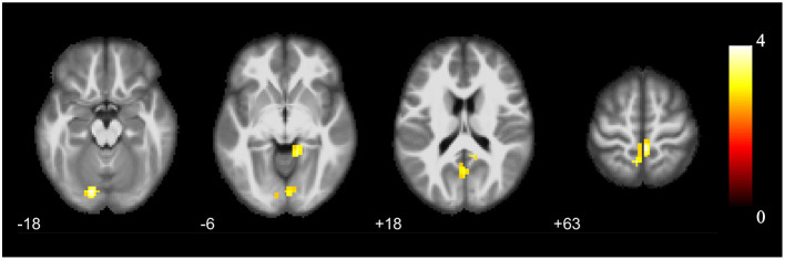
Results: HIV+ HoM showed significantly increased GMV in the bilateral parahippocampal gyrus and amygdala, and decreased GMV in the right inferior cerebellum, compared with the HIV- HoM. The brain regions with increased GMV were hyper-connected with the left superior cerebellum, right lingual gyrus, and left precuneus in the HIV+ HoM. Moreover, the ALFF values of the right fusiform gyrus, and left parahippocampal gyrus were increased in the HIV+ HoM. The regional homogeneity values of the right anterior cingulate and paracingulate gyri, and left superior cerebellum were decreased in the HIV+ HoM.
Conclusion: When the study population was restricted to HoM, HIV+ individuals exhibited structural alterations in the limbic system and cerebellum, and functional abnormalities in the limbic, cerebellum, and visual network. These findings complement the existing knowledge on the HIV-associated neurocognitive impairment from the previous neuroimaging studies by controlling for the potential confounding factor, sexual orientation. Future studies on brain alternations with the exclusion of related factors like sexual orientation are needed to understand the impact of HIV infection on neurocognitive function more accurately.
Keywords: HIV infection; amplitude of low frequency fluctuation; functional connectivity; gray matter volume; homosexual; regional homogeneity.
Copyright © 2022 Ma, Shi, Chen, Song, Liu, Zheng, Shi and Cai.
Conflict of interest statement
The authors declare that the research was conducted in the absence of any commercial or financial relationships that could be construed as a potential conflict of interest.
Figures


References
-
- Kallianpur KJ, Gerschenson M, Mitchell BI, LiButti DE, Umaki TM, Ndhlovu LC, et al. . Oxidative mitochondrial DNA damage in peripheral blood mononuclear cells is associated with reduced volumes of hippocampus and subcortical gray matter in chronically HIV-infected patients. Mitochondrion. (2016) 28:8–15. 10.1016/j.mito.2016.02.006 - DOI - PMC - PubMed
LinkOut - more resources
Full Text Sources