Chylothorax found in a patient with COVID-19
- PMID: 35096397
- PMCID: PMC8783950
- DOI: 10.1002/rcr2.836
Chylothorax found in a patient with COVID-19
Abstract
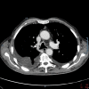
Coronavirus disease 2019 (COVID-19) is an infectious disease caused by severe acute respiratory syndrome coronavirus 2 (SARS-CoV-2) and its clinical spectrum ranges from mild to moderate or severe illness. A 78-year-old male was presented at emergency department with dyspnoea, dry cough and severe asthenia. The nasopharyngeal swab by real-time polymerase chain reaction confirmed a SARS-CoV-2 infection. The x-ray and the thoracic ultrasound revealed right pleural effusion. A diagnostic-therapeutic thoracentesis drained fluid identified as chylothorax. Subsequently, the patient underwent a chest computed tomography which showed the radiological hallmarks of COVID-19 and in the following weeks he underwent a chest magnetic resonance imaging to obtain a better view of mediastinal and lymphatic structures, which showed a partial thrombosis affecting the origin of superior vena cava and the distal tract of the right subclavian vein. For this reason, anticoagulant therapy was optimized and in the following weeks the patient was discharged for clinical and radiological improvement. This case demonstrates chylothorax as a possible and uncommon complication of COVID-19.
Keywords: COVID‐19; chylothorax; lymphatic system; thoracentesis; thrombosis.
© 2022 The Authors. Respirology Case Reports published by John Wiley & Sons Australia, Ltd on behalf of The Asian Pacific Society of Respirology.
Conflict of interest statement
None declared.
Figures
References
Publication types
LinkOut - more resources
Full Text Sources
Miscellaneous


