

Case Reports
doi: 10.1016/j.mmcr.2022.01.004.
eCollection 2022 Mar.
Disseminated Verruconis gallopava infection in a patient with systemic lupus erythematosus in Japan: A case report, literature review, and autopsy case
Affiliations
- PMID: 35096522
- PMCID: PMC8783064
- DOI: 10.1016/j.mmcr.2022.01.004
Item in Clipboard
Case Reports
Disseminated Verruconis gallopava infection in a patient with systemic lupus erythematosus in Japan: A case report, literature review, and autopsy case
Med Mycol Case Rep.
.
Abstract
Disseminated Verruconis gallopava infection is often reported in patients with severe immunodeficiency, such as those who have received an organ transplant or have hematological malignancies. The present report describes the first case of disseminated V. gallopava in a patient with systemic lupus erythematosus who used FK506 (Tacrolimus). In this case, β-D glucan was useful for diagnosis.
Keywords: Systemic lupus erythematosus; Verruconis gallopava.
© 2022 The Authors.
Conflict of interest statement
There are no conflicts of interests.
Figures
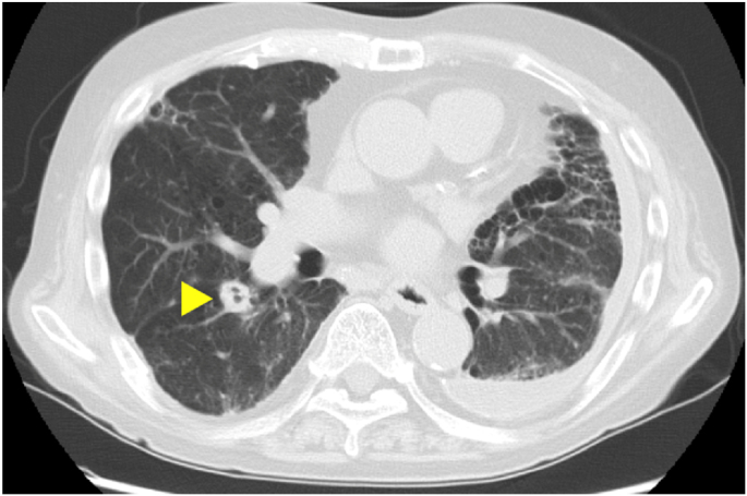
Computed tomography (CT) revealed a single nodule in his right lung(▶). Since it was a cavity lesion, we considered the possibility of fungal infection.
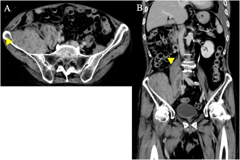
This figure is CT images of the inguinal lesion. A: Axial section of CT. B: Coronal section of CT. A huge low density area with contrast effect was observed on the wall in the right iliopsoas muscle(▶).
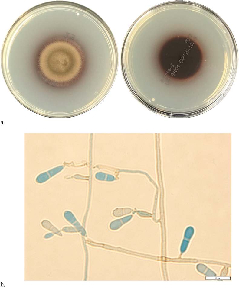
a. (left) Large colony (light olive green) grown on PDA (potato dextrose agar) at 28 °C on day 10 from a specimen obtained by a direct puncture. (right) The colony underside shows a reddish pigment. It has a two-celled cylindrical conidium at the tip. The shape of conidia is constricted at the septum. (For interpretation of the references to colour in this figure legend, the reader is referred to the Web version of this article.)
Similar articles
-
Successful treatment of disseminated Verruconis gallopava infection in a heart transplant recipient: A case report.Am J Health Syst Pharm. 2022 Jun 23;79(13):1066-1069. doi: 10.1093/ajhp/zxac063. Am J Health Syst Pharm. 2022. PMID: 35245929
-
Verruconis gallopava cardiac and endovascular infection with dissemination after renal transplantation: Case report and lessons learned.Med Mycol Case Rep. 2016 Dec 13;15:5-8. doi: 10.1016/j.mmcr.2016.12.006. eCollection 2017 Mar. Med Mycol Case Rep. 2016. PMID: 28053851 Free PMC article.
-
A case of Verruconis gallopava infection in a heart transplant recipient successfully treated with posaconazole.Transpl Infect Dis. 2019 Apr;21(2):e13044. doi: 10.1111/tid.13044. Epub 2019 Jan 10. Transpl Infect Dis. 2019. PMID: 30585691
-
Concomitant onset of systemic lupus erythematosus and disseminated histoplasmosis: a case-based review.Rheumatol Int. 2021 Sep;41(9):1673-1680. doi: 10.1007/s00296-020-04739-6. Epub 2020 Nov 4. Rheumatol Int. 2021. PMID: 33150492 Review.
-
Disseminated histoplasmosis in systemic lupus erythematosus: case report and review of the literature.Semin Arthritis Rheum. 1998 Dec;28(3):193-9. doi: 10.1016/s0049-0172(98)80036-x. Semin Arthritis Rheum. 1998. PMID: 9872480 Review.
Cited by
-
Recurrent Verruconis gallopava Infection at One Year after Excision of a Solitary Pulmonary Lesion.Intern Med. 2024 May 15;63(10):1499-1503. doi: 10.2169/internalmedicine.2263-23. Epub 2023 Sep 1. Intern Med. 2024. PMID: 37661451 Free PMC article.
-
RE-EVALUATION OF SYMPOVENTURIACEAE.Persoonia. 2022 Jul 12;48:219-260. doi: 10.3767/persoonia.2023.48.07. Epub 2022 Jun 17. Persoonia. 2022. PMID: 38234692 Free PMC article.
References
-
- Fukushiro K.R., Udagawa S., Kawashima Y., Kawamura Y. Subcutaneous abscesses caused by Ochroconis gallopavum. J. Med. Vet. Mycol. 1986;24:175–182. - PubMed
-
- Qureshi Z.A., Kwak E.J., Nguyen M.H., Silveira F.P. Ochroconis gallopava: a dematiaceous mold causing infections in transplant recipients. Clin. Transplant. 2012;26:E17–E23. - PubMed
-
- Wong J.S.J., Schousboe M.I., Metcalf S.S.L., et al. Ochroconis gallopava peritonitis in a cardiac transplant patient on continuous ambulatory peritoneal dialysis, Transpl. Inf. Disp. 2010;12:455–458. - PubMed
-
- Terreni A.A., Disalvo A.F., Barker A.S., Jr., Crymes W.B., Morris P.R., Dowda H., Jr. Disseminated Dactylaria gallopava infection in a diabetic patient with chronic lymphocytic leukemia of the T-cell type. Am. J. Clin. Pathol. 1990;94:104–107. - PubMed
Publication types
LinkOut - more resources
Full Text Sources
