

The Enhanced Expression of Cruzipain-Like Molecules in the Phytoflagellate Phytomonas serpens Recovered From the Invertebrate and Plant Hosts
- PMID: 35096661
- PMCID: PMC8793489
- DOI: 10.3389/fcimb.2021.819133
The Enhanced Expression of Cruzipain-Like Molecules in the Phytoflagellate Phytomonas serpens Recovered From the Invertebrate and Plant Hosts
Abstract
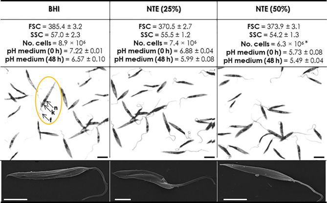
Phytomonas serpens is a protozoan parasite that alternates its life cycle between two hosts: an invertebrate vector and the tomato fruit. This phytoflagellate is able to synthesize proteins displaying similarity to the cysteine peptidase named cruzipain, an important virulence factor from Trypanosoma cruzi, the etiologic agent of Chagas disease. Herein, the growth of P. serpens in complex medium (BHI) supplemented with natural tomato extract (NTE) resulted in the increased expression of cysteine peptidases, as verified by the hydrolysis of the fluorogenic substrate Z-Phe-Arg-AMC and by gelatin-SDS-PAGE. Phytoflagellates showed no changes in morphology, morphometry and viability, but the proliferation was slightly reduced when cultivated in the presence of NTE. The enhanced proteolytic activity was accompanied by a significant increase in the expression of cruzipain-like molecules, as verified by flow cytometry using anti-cruzipain antibodies. In parallel, parasites incubated under chemically defined conditions (PBS supplemented with glucose) and added of different concentration of NTE revealed an augmentation in the production of cruzipain-like molecules in a typically dose-dependent way. Similarly, P. serpens recovered from the infection of mature tomatoes showed an increase in the expression of molecules homologous to cruzipain; however, cells showed a smaller size compared to parasites grown in BHI medium. Furthermore, phytoflagellates incubated with dissected salivary glands from Oncopeltus fasciatus or recovered from the hemolymph of infected insects also showed a strong enhance in the expression of cruzipain-like molecules that is more relevant in the hemolymph. Collectively, our results showed that cysteine peptidases displaying similarities to cruzipain are more expressed during the life cycle of the phytoflagellate P. serpens both in the invertebrate and plant hosts.
Keywords: Oncopeltus fasciatus; Phytomonas serpens; cruzipain; interaction; invertebrate vector; tomato.
Copyright © 2022 Oliveira, Elias, Dias, Lopes, d’Avila-Levy, Santos and Branquinha.
Conflict of interest statement
The authors declare that the research was conducted in the absence of any commercial or financial relationships that could be construed as a potential conflict of interest.
Figures


Similar articles
-
Phytomonas serpens: cysteine peptidase inhibitors interfere with growth, ultrastructure and host adhesion.Int J Parasitol. 2006 Jan;36(1):47-56. doi: 10.1016/j.ijpara.2005.09.004. Epub 2005 Oct 5. Int J Parasitol. 2006. PMID: 16310789
-
Cysteine peptidases in the tomato trypanosomatid Phytomonas serpens: influence of growth conditions, similarities with cruzipain and secretion to the extracellular environment.Exp Parasitol. 2008 Dec;120(4):343-52. doi: 10.1016/j.exppara.2008.08.011. Epub 2008 Aug 31. Exp Parasitol. 2008. PMID: 18793639
-
Cysteine peptidases from Phytomonas serpens: biochemical and immunological approaches.FEMS Immunol Med Microbiol. 2009 Dec;57(3):247-56. doi: 10.1111/j.1574-695X.2009.00604.x. Epub 2009 Sep 1. FEMS Immunol Med Microbiol. 2009. PMID: 19780820
-
Phytomonas serpens: immunological similarities with the human trypanosomatid pathogens.Microbes Infect. 2007 Jul;9(8):915-21. doi: 10.1016/j.micinf.2007.03.018. Epub 2007 Apr 12. Microbes Infect. 2007. PMID: 17556002 Review.
-
Cruzipain, the major cysteine proteinase from the protozoan parasite Trypanosoma cruzi.Biol Chem. 1997 Jan;378(1):1-10. doi: 10.1515/bchm.1997.378.1.1. Biol Chem. 1997. PMID: 9049059 Review.
Cited by
-
Differential expression of peptidases in Strigomonas culicis wild-type and aposymbiotic strains: from proteomic data to proteolytic activity.Mem Inst Oswaldo Cruz. 2024 Dec 9;119:e240110. doi: 10.1590/0074-02760240110. eCollection 2024. Mem Inst Oswaldo Cruz. 2024. PMID: 39661825 Free PMC article.
-
Silver(I) and Copper(II) 1,10-Phenanthroline-5,6-dione Complexes as Promising Antivirulence Strategy against Leishmania: Focus on Gp63 (Leishmanolysin).Trop Med Infect Dis. 2023 Jun 30;8(7):348. doi: 10.3390/tropicalmed8070348. Trop Med Infect Dis. 2023. PMID: 37505644 Free PMC article.
References
-
- Branquinha M. H., Oliveira S. S., Sangenito L. S., Sodre C. L., Kneipp L. F., d'Avila-Levy C. M., et al. . (2015). Cruzipain: An Update on Its Potential as Chemotherapy Target Against the Human Pathogen Trypanosoma cruzi . Curr. Med. Chem. 22 (18), 2225–2235. doi: 10.2174/0929867322666150521091652 - DOI - PubMed
-
- Breganó J. W., Picão R. C., Graça V. K., Menolli R. A., Itow Jankevicius S., Pinge Filho P., et al. . (2003). Phytomonas serpens, a Tomato Parasite, Shares Antigens With Trypanosoma cruzi That Are Recognized by Human Sera and Induce Protective Immunity in Mice. FEMS Immunol. Med. Microbiol. 39 (3), 257–264. doi: 10.1016/S0928-8244(03)00256-6 - DOI - PubMed
-
- Camargo E., Wallace F. (1994). Vectors of Plant Parasites of the Genus Phytomonas (Protozoa, Zoomastigophorea, Kinetoplastida). Adv. Dis. Vector Res. 10, 333–359. doi: 10.1007/978-1-4612-2590-4_12 - DOI
Publication types
MeSH terms
Substances
LinkOut - more resources
Full Text Sources
Research Materials