Review
. 2022 Jan 14:9:794069.
doi: 10.3389/fped.2021.794069. eCollection 2021.

Diagnostic Approach to Macrocephaly in Children

Affiliations
Review

Diagnostic Approach to Macrocephaly in Children

Andrea Accogli et al. Front Pediatr. .

Abstract

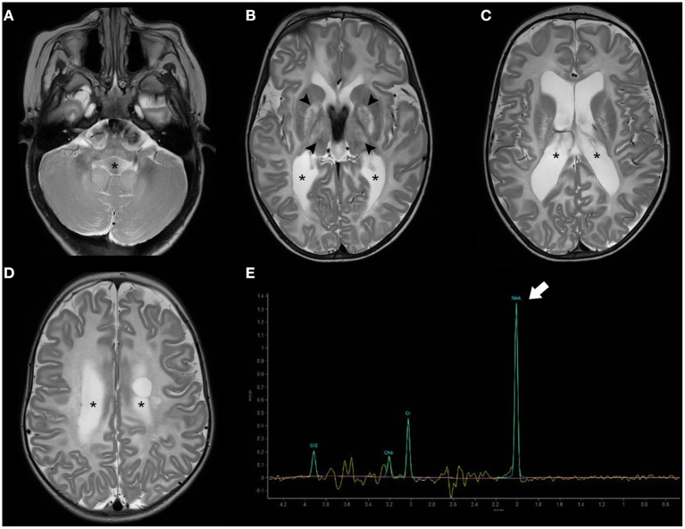
Macrocephaly affects up to 5% of the pediatric population and is defined as an abnormally large head with an occipitofrontal circumference (OFC) >2 standard deviations (SD) above the mean for a given age and sex. Taking into account that about 2-3% of the healthy population has an OFC between 2 and 3 SD, macrocephaly is considered as "clinically relevant" when OFC is above 3 SD. This implies the urgent need for a diagnostic workflow to use in the clinical setting to dissect the several causes of increased OFC, from the benign form of familial macrocephaly and the Benign enlargement of subarachnoid spaces (BESS) to many pathological conditions, including genetic disorders. Moreover, macrocephaly should be differentiated by megalencephaly (MEG), which refers exclusively to brain overgrowth, exceeding twice the SD (3SD-"clinically relevant" megalencephaly). While macrocephaly can be isolated and benign or may be the first indication of an underlying congenital, genetic, or acquired disorder, megalencephaly is most likely due to a genetic cause. Apart from the head size evaluation, a detailed family and personal history, neuroimaging, and a careful clinical evaluation are crucial to reach the correct diagnosis. In this review, we seek to underline the clinical aspects of macrocephaly and megalencephaly, emphasizing the main differential diagnosis with a major focus on common genetic disorders. We thus provide a clinico-radiological algorithm to guide pediatricians in the assessment of children with macrocephaly.

Keywords: brain MRI; genetic diagnosis; head circumference; macrocephaly; megalencephaly.


Conflict of interest statement

The authors declare that the research was conducted in the absence of any commercial or financial relationships that could be construed as a potential conflict of interest.

Figures

Figure 1
Figure 1
Brain MRI findings in Benign enlargement of subarachnoid spaces (BESS) of infancy. Coronal Head US image (A) of a 7-months-old girl with macrocephaly shows bilateral enlargement of the frontal subarachnoid spaces and anterior interhemispheric fissure (thick white arrows). Note the presence of a few vessels (white arrow) on US Doppler (B) crossing the enlarged subarachnoid space on the right side, corresponding to the “cortical vein sign.” Coronal (C) and axial (D–F) T2WI performed a few days after better depict these findings, including presence of multiple small vessels bilaterally within these enlarged subarachnoid spaces (black arrowheads). In addition, there is a small left frontal T2 hyperintense subdural collection (black thick arrows) as well as mild enlargement of the ventricular system (black asterisks). Follow-up brain MRI of the same patient performed at 2.3 years of age including axial T2WI (G–I) depict expected spontaneous interval reduction of the aforementioned enlarged subarachnoid spaces and resolution of the thin left subdural collection.
Figure 2
Figure 2
Longitudinal brain MRI findings in Glutaric Aciduria type I. Axial (A–C) and coronal (D) T2TWI of an affected 1.3-years-old boy demonstrates enlargement of the anterior temporal subarachnoid spaces (black arrowheads) and widening of the Sylvian fissures (thick black arrows) due to incomplete opercularization. There are also bilateral, symmetric hyperintensities in the globus pallidus/posterior putamen, substantia nigra and central tegmental tracts (white arrows). Axial b1000 images (E,F) also depict hyperintensity in the globus pallidus and posterior putamen bilaterally (white arrows). There was corresponding mild hypointensity in the ADC maps (not shown), in keeping with restricted diffusion.
Figure 3
Figure 3
Brain MRI findings in L2-Hydroxyglutaric Aciduria. Axial T2WI (A–E) of an affected 11-year-old boy show diffuse, bilateral, and symmetric hyperintensity involving mainly the subcortical and deep cerebral white-matter, while the periventricular white-matter, corpus callosum (asterisks), and internal capsules (white arrowheads) are relatively spared. There is also mild, symmetric hyperintensity of the caudate nuclei and putamina (black arrows) and dentate nuclei (thick white arrows). Axial T1WI (F) demonstrate bilateral, symmetric hypointensity involving mainly the subcortical cerebral white matter/U fibers (white arrows).
Figure 4
Figure 4
Imaging characteristics of Mucopolyssacaridosis. Sagittal T1WI (A) and axial T2WI (B–D) of a boy with Hurler syndrome performed at 4 years of age depict frontal bossing (white arrowhead), mild diffuse enlargement of the ventricular system (asterisks) as well as multiple dilated perivascular spaces (white thick arrows). Also note platybasia of the cervical vertebrae (black thick arrows), J-shaped sella (black arrowhead), flat nasal bridge (open arrow), and mild hypertrophy of the occipital squama (curved arrow), at this time point without significant stenosis of the cranio-cervical junction.
Figure 5
Figure 5
Brain MRI findings of Alexander Disease (infantile form). Axial T2WI (A–D) of a 1.2-years-old girl exhibit diffuse, symmetric, and confluent areas of white-matter hyperintensity with a postero-anterior gradient. Also, note areas of hyperintensity in the medulla oblongata (thick black arrows) as well as in the caudate nuclei and putamina (asterisks). Axial b1000 images (E,F) show focal areas of mild symmetric hyperintensity involving the globus pallidus and the head of the caudate bilaterally (white arrows). There was corresponding hypointensity in the ADC maps (not shown) in keeping with restricted diffusion. Axial post-gadolinium T1WI (G,H) depict small “caps” of contrast enhancement around the frontal horns bilaterally (thick white arrows).
Figure 6
Figure 6
Brain MRI findings of Canavan disease. Axial T2WI (A–D) of a 2.3-years-old female infant with Canavan disease demonstrate diffuse, bilateral, and symmetric supratentorial and infratentorial white-matter hyperintensity associated with enlargement of the ventricular system (asterisks). There is also hyperintensity of the central gray-matter, mainly the globus pallidus and thalami bilaterally (black arrowheads). MR proton spectroscopy (intermediate TE) (E) depict increased NAA peak (thick white arrow) relatively to the other peaks, a feature near pathognomonic of this disease.
Figure 7
Figure 7
Imaging characteristics of Megalencephalic Leukoencephalopathy With Subcortical Cysts (classic form). Axial T2WI (A–D), axial (E,F), and coronal (G) T1WI, and coronal FLAIR (H) performed in an affected boy at 1.8 years of age show diffuse, bilateral, and symmetric cerebral white matter hyperintensity including the subcortical white-matter/U-fibers, leading to brain swelling and effacement of the cortical sulci. There is a characteristic double-line signal abnormality in the posterior limb of the internal capsule (white arrows), i.e., a residual central dark line surrounded by two strands of hyperintensity in this location. Also note the typical areas of cystic-like white-matter rarefaction in the temporal-polar regions on T1WI and FLAIR (thick white arrows).
Figure 8
Figure 8
Imaging characteristics of Childhood Ataxia with Central Hypomyelination/Vanishing White Matter Disease (CACH/VWMD). Axial T2WI (A–D) and FLAIR (E–H) of an 11-year-old boy with CACH/VWMD demonstrates bilateral, confluent, symmetric periventricular, and deep white matter hyperintensity, with relative sparing of the anterior limb of the internal capsule (black arrowheads) and the subcortical white-matter/U-fibers. There are areas of cystic degeneration around the frontal horns (white arrows) and in the centrum semi-oval bilaterally (thick black arrows), with some radiation stripes depicted within the latter location.
Figure 9
Figure 9
Imaging characteristics of Neurofibromatosis Type 1 (NF1). Axial FLAIR (A–C) and sagittal T1 WI (D) of an 18-year-old boy with NF1 reveals multiple focal areas of abnormal hyperintensity (FASI) (white arrows) distributed in the dentate nuclei, middle cerebellar peduncles, mesencephalic tegment, hippocampi, and pulvinar bilaterally as well as left globus pallidus. There is also diffuse thickening of the corpus callosum (thick white arrows). Coronal contrast-enhanced T1WI FAT SAT of the orbits (E) of the same patient show thickening, tortuosity, and enhancement of the intra-orbital segment of the right optic nerve (open arrow), in keeping with an ipsilateral optic nerve glioma.
Figure 10
Figure 10
Imaging characteristics of Tuberous Sclerosis (TS). Axial (A) and coronal (B) FLAIR images, sagittal T2WI (C) and axial post-gadolinium TWI (D) of a 14-years-old boy with TS demonstrate a contrast-enhanced mass larger than 10 mm in the region of the right foramen of Monro (thick white arrow) and with progressive growth in comparison with previous studies (not shown), compatible with a giant cell astrocytoma. There is the contralateral deviation of the septum pellucidum and asymmetric dilatation of the ventricular system (asterisks) including the temporal horns with incipient signs of periventricular interstitial edema and effacement of the cortical sulci in keeping with decompensated hydrocephalus. Also note a small contralateral, contrast-enhancing subependymal nodule (black arrow). Axial T1WI (E–G) and coronal T2WI (H) in another patient with TS at the age of 11 years reveals right hemimegalencephaly associated with marked abnormal cortex and white matter as well as enlargement of the ipsilateral ventricular system (asterisks). Also, note small subependymal nodules (white arrowheads) as well as enlargement of the right cerebellar hemisphere (thick white arrows).
Figure 11
Figure 11
Imaging characteristics of brain overgrowth disorders (continuation). Axial T2WI (A–C) of a 16-years-old boy demonstrate right unilateral megalencephaly/hemimegalencephaly with abnormal cortex (black arrows) and abnormal white-matter signal intensity (asterisks). There is also enlargement of the right mesencephalon (white arrowhead) and medulla oblongata (black arrowhead) as well as enlarged and dysplastic right cerebellar hemisphere (thick white arrow). These findings were previously designated as “total hemimegalencephaly.” Coronal (D) and axial (E) T1WE and axial T2WI (F) of another child performed at 1.8 years of age show a right anterior form of unilateral megalencephaly/hemimegalencephaly with abnormal cortex (white arrowheads) and abnormal white-matter signal intensity (asterisks) within the affected region. Coronal (G) and axial (H,I) T2WI acquired in a third child at 2 months of age depict instead a right posterior form of unilateral megalencephaly/hemimegalencephaly, also with abnormal cortex (white arrowheads) and white-matter (asterisks) within the affected region, with enlargement of the ipsilateral occipital horn of the lateral ventricle.
Figure 12
Figure 12
Photos of affected patients with either somatic or germline mutations in genes of the PI3K-AKT-mTOR pathway. (A–C) Photos of the face (A), occipital region (B), and left foot (C) of a subject with MCAP (somatic PIK3CA mutation p.Pro104Leu) showing MEG, occipital capillary malformation, and syndactyly of the second, third, and fourth toes. (D,E) Photos of the face (D) and left foot (E) of a subject with MCAP (somatic PIK3CA mutation p.Glu545Asp) showing MEG, capillary malformation of the philtrum, skin laxity of the forehead, and syndactyly of the second, third, and fourth toes. (F,G) Photos of lower limbs with pigmentary defects in a patient with Megalencephaly-Polymicrogyria-Pigmentary Mosaicism Syndrome (somatic MTOR mutation p.Thr1977Ile). (H) Diffuse vascular anomaly in the left lower leg in a subject with somatic mutation in PIK3CA (p.Gly914Arg). (I–L) Photos of the face (I) and lower extremities (L) of a subject with MCAP (somatic PIK3CA p.Met1043Ile) showing facial and body asymmetry, MEG with a prominent forehead, and capillary malformations on the face and body. (M) Photo of a subject with MEG, frontal bossing, nevus flammenus and retrognathia, harboring a germline variant in MTOR (p.Glu1799Lys). (N) Photos of a female with MEG and broad forehead harboring a germline variant in PIK3CA (p.Pro104Leu). Part of these photos are re-printed with permission from ref (182).
Figure 13
Figure 13
Imaging characteristics of brain overgrowth disorders. Sagittal (A) and axial (B) T1WI and axial T2WI (C) of a 3.5-years-old boy with macrocephaly due to a pathogenic PTEN mutation reveals signs of bilateral, symmetric megalencephaly with regular cortex and normal signal-intensity white matter. Note presence of frontal bossing (arrowhead), mega corpus callosum (open arrows), thickened anterior commissure (arrow), mild tonsillar herniation (thick white arrow), and peri-atrial dilated perivascular spaces, mainly on the left side (black thick arrow). Axial T1 WI (D–F) of a different patient with macrocephaly at 3.4 years of age show signs of unilateral megalencephaly/hemimegalencephaly with normal cortex and normal white matter. Note the “occipital sign” (black arrows) as well as anterior pointing of the left frontal horn (thick black arrow) and thickening of the midline structures (black arrowheads). Axial T1WI (G), axial T2WI (H), and sagittal T1WI (I) in a child with megalencephaly capillary syndrome performed at 1.6 years of age depict bilateral, asymmetric dysplastic megalencephaly with abnormal perisylvian polymicrogyric cortex (black arrows) and white matter signal changes (asterisks) as well as bilateral cerebellar dysplasia (thick white arrows). Also, note bilateral facial lipomatous lesions (white arrowheads).
Figure 14
Figure 14
Diagnostic algorithm of macrocephaly. BESS, Benign enlargement of subarachnoid spaces; BRRS, Bannayan-Riley-Ruvalcaba syndrome; CS, Cowden syndrome; CNS, central nervous system; D2HA, D2-hydroxyglutaric aciduria; L2HA, L2-hydroxyglutaric aciduria; DD, developmental delay; KTS, Klippel-Trenaunay syndrome; ICP, increased intracranial pressure; GA1, glutaric aciduria type 1; MAV, arteriovenous malformations; MPS, mucopolysaccharidosis; AD, Alexander disease; CD, Canavan disease; MLC, megalencephalic leukoencephalopathy with subcortical cysts; MCAP, megalencephaly-capillary malformation syndrome; MPPH, megalencephaly-polymicrogyria-polydactyly-hydrocephalus syndrome; MPPM, megalencephaly-polymicrogyria and pigmentary mosaicism; CACH/VWMD, childhood ataxia with central hypomyelination/vanishing white matter disease.

References

    1. Medina LS, Frawley K, Zurakowski D, Buttros D, DeGrauw AJC, Crone KR. Children with macrocrania: clinical and imaging predictors of disorders requiring surgery. Am J Neuroradiol. (2001) 22:564–70. - PMC - PubMed
    1. Tan AP, Mankad K, Gonçalves FG, Talenti G, Alexia E. Macrocephaly: solving the diagnostic dilemma. Top Magn Reson Imaging. (2018) 27:197–217. 10.1097/RMR.0000000000000170 - DOI - PubMed
    1. Nellhaus G. Head circumference from birth to eighteen years. Pediatrics. (1968) 41:106–14. - PubMed
    1. DeMyer W. Megalencephaly: types, clinical syndromes, and management. Pediatr Neurol. (1986) 2:321–8. 10.1016/0887-8994(86)90072-X - DOI - PubMed
    1. Severino M, Geraldo AF, Utz N, Tortora D, Pogledic I, Klonowski W, et al. . Definitions and classification of malformations of cortical development: practical guidelines. Brain. (2020) 143:2874–94. 10.1093/brain/awaa174 - DOI - PMC - PubMed

LinkOut - more resources