Malignant solitary fibrous tumor in the central nervous system treated with surgery, radiotherapy and anlotinib: A case report
- PMID: 35097089
- PMCID: PMC8771389
- DOI: 10.12998/wjcc.v10.i2.631
Malignant solitary fibrous tumor in the central nervous system treated with surgery, radiotherapy and anlotinib: A case report
Abstract
Background: Solitary fibrous tumor (SFT) of the central nervous system is rare. It is predominantly benign and rarely malignant. There is no established standardized treatment regimen for malignant intracranial SFTs.
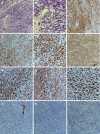
Case summary: We present a rare case of SFT in a 9-year-old girl with a space-occupying effect in the frontal-parietal lobes. She underwent craniotomy, and the mass was resected. Immunohistochemistry examination of the specimen showed that Ki-67 proliferation index staining was highly positive in 80% of tumor cells. Whole exome sequencing of the surgical tissue showed 38 somatic gene mutations and 1 gene amplification such as fibroblast growth factor receptor 4 or TP53. At 1.5 mo after surgery, head magnetic resonance imaging revealed that the tumor had recurred. The patient received 60 Gy and 30 fractions of intensity modulated radiotherapy. The patient then received anlotinib 8 mg po qd for 1-14 d of a 21 d cycle. Following this regimen, the patient achieved stable disease for > 17 mo. Magnetic resonance imaging at 1.5 year after surgery showed that the tumor had not progressed.
Conclusion: This is the first reported case of SFT of the central nervous system treated with surgery, radiotherapy and anlotinib. This regimen may be an effective treatment option for malignant intracranial SFT patients.
Keywords: Anlotinib; Biological therapy; Case report; Mutation; Recurrence; Sequence analysis.
©The Author(s) 2022. Published by Baishideng Publishing Group Inc. All rights reserved.
Conflict of interest statement
Conflict-of-interest statement: The authors declare that they have no competing interest.
Figures


Similar articles
-
Intracranial Primary Malignant Solitary Fibrous Tumor/Hemangiopericytoma Masquerading as Meningioma: Report of a Rare Case.Int J Gen Med. 2020 Oct 29;13:963-967. doi: 10.2147/IJGM.S279483. eCollection 2020. Int J Gen Med. 2020. PMID: 33149660 Free PMC article.
-
Malignant solitary fibrous tumor of the greater omentum: A case report and review of literature.World J Clin Cases. 2021 Jan 16;9(2):445-456. doi: 10.12998/wjcc.v9.i2.445. World J Clin Cases. 2021. PMID: 33521114 Free PMC article.
-
Systemic metastasis in malignant solitary fibrous tumor of the liver: two case reports and literature review.Front Oncol. 2024 Oct 2;14:1418547. doi: 10.3389/fonc.2024.1418547. eCollection 2024. Front Oncol. 2024. PMID: 39416460 Free PMC article.
-
Solitary fibrous tumor arising from Cranial Nerve VI in the prepontine cistern: case report and review of a tumor subpopulation mimicking schwannoma.Neurosurgery. 2006 Oct;59(4):E939-40; discussion E940. doi: 10.1227/01.NEU.0000232660.21537.60. Neurosurgery. 2006. PMID: 17038929 Review.
-
Spontaneous Tumor Regression of Intracranial Solitary Fibrous Tumor Originating From the Medulla Oblongata: A Case Report and Literature Review.World Neurosurg. 2019 Oct;130:400-404. doi: 10.1016/j.wneu.2019.07.052. Epub 2019 Jul 18. World Neurosurg. 2019. PMID: 31326640 Review.
Cited by
-
A clinical investigation of extracranial metastases in 17 cases of intracranial solitary fibrous tumors.World J Surg Oncol. 2025 Jul 1;23(1):257. doi: 10.1186/s12957-025-03902-2. World J Surg Oncol. 2025. PMID: 40597169 Free PMC article.
-
Solitary fibrous tumor of the central nervous system invading and penetrating the skull: A case report.Oncol Lett. 2023 Jan 10;25(2):81. doi: 10.3892/ol.2023.13667. eCollection 2023 Feb. Oncol Lett. 2023. PMID: 36742362 Free PMC article.
References
-
- Louis DN, Perry A, Reifenberger G, von Deimling A, Figarella-Branger D, Cavenee WK, Ohgaki H, Wiestler OD, Kleihues P, Ellison DW. The 2016 World Health Organization Classification of Tumors of the Central Nervous System: a summary. Acta Neuropathol. 2016;131:803–820. - PubMed
-
- Carneiro SS, Scheithauer BW, Nascimento AG, Hirose T, Davis DH. Solitary fibrous tumor of the meninges: a lesion distinct from fibrous meningioma. A clinicopathologic and immunohistochemical study. Am J Clin Pathol. 1996;106:217–224. - PubMed
-
- Giordan E, Marton E, Wennberg AM, Guerriero A, Canova G. A review of solitary fibrous tumor/hemangiopericytoma tumor and a comparison of risk factors for recurrence, metastases, and death among patients with spinal and intracranial tumors. Neurosurg Rev. 2021;44:1299–1312. - PubMed
-
- Tani E, Wejde J, Åström K, Wingmo IL, Larsson O, Haglund F. FNA cytology of solitary fibrous tumors and the diagnostic value of STAT6 immunocytochemistry. Cancer Cytopathol. 2018;126:36–43. - PubMed
-
- Fritchie K, Jensch K, Moskalev EA, Caron A, Jenkins S, Link M, Brown PD, Rodriguez FJ, Guajardo A, Brat D, Velázquez Vega JE, Perry A, Wu A, Raleigh DR, Santagata S, Louis DN, Brastianos PK, Kaplan A, Alexander BM, Rossi S, Ferrarese F, Haller F, Giannini C. The impact of histopathology and NAB2-STAT6 fusion subtype in classification and grading of meningeal solitary fibrous tumor/hemangiopericytoma. Acta Neuropathol. 2019;137:307–319. - PMC - PubMed
Publication types
LinkOut - more resources
Full Text Sources
Research Materials
Miscellaneous

