Review
. 2019 Jul 19;4(3):2473011419852931.
doi: 10.1177/2473011419852931. eCollection 2019 Jul.

Ankle Arthritis

Affiliations
Review

Ankle Arthritis

Vu Le et al. Foot Ankle Orthop. .

Abstract

Ankle arthritis is a major source of morbidity impacting a younger working age population than hip and knee arthritis. Unlike the hip and knee, more than 70% of ankle arthritis cases are post-traumatic, with the remainder being inflammatory or primary arthritis. Nonoperative treatment begins with lifestyle and shoe-wear modifications and progresses to bracing, physical therapy, anti-inflammatory medications, and intra-articular injections. Ankle arthrodesis and total ankle arthroplasty are the 2 main surgical options for end-stage ankle arthritis, with debridement, realignment osteotomy, and distraction arthroplasty being appropriate for limited indications.

Level of evidence: Level V, expert opinion.

Keywords: ankle; ankle arthritis; ankle arthrodesis; arthritis; total ankle replacement.


Conflict of interest statement

Declaration of Conflicting Interests: The author(s) declared the following potential conflicts of interest with respect to the research, authorship, and/or publication of this article: Vu Le, MD, FRCSC, reports personal fees from Purdue Pharma (Canada), outside the submitted work. Andrea Veljkovic, MD, MPH, FRCSC, reports grants from Acumed, from AIC, from Therapia, personal fees from Arthrex, grants from Zimmer, outside the submitted work. Kevin Wing, MD, FRCSC, reports grants from Acumed, grants and personal fees from Wright medical, grants from Ferring, grants from Zimmer, grants from Synthes, grants from Bioventus, outside the submitted work. Murray Penner, MD, FRCSC, reports grants and personal fees from Wright medical, grants from Zimmer, grants from Synthes, grants and personal fees from Springer, grants from Arthrex, other from Cdn. Orthop. Foot & Ankle society, other from Intl. Federation of Foot & Ankle societies, outside the submitted work. Alastair Younger, MB, ChB, MSc, ChM, FRCSC, reports grants and personal fees from Acumed, grants and personal fees from Wright medical, grants and personal fees from Ferring, grants and personal fees from Zimmer, grants from Synthes, grants and personal fees from Bioventus, outside the submitted work. ICMJE forms for all authors are available online.

Figures

Figure 1.
Figure 1.
Arthroscopic view of the medial gutter of a right ankle from the anteromedial portal. A shaver is debriding articular cartilage and fibrous tissue via an accessory inferior medial portal.
Figure 2.
Figure 2.
Arthroscopic view of the medial gutter of a right ankle from the anteromedial portal after debridement.
Figure 3.
Figure 3.
Arthroscopic view from the anteromedial portal of a right ankle looking laterally over the talar dome at the fibula and lateral gutter. Articular cartilage has been removed.
Figure 4.
Figure 4.
Arthroscopic view from the anteromedial portal of a right ankle looking laterally over the talar dome at the fibula and lateral gutter. Further debridement of fibrous tissue has been performed and bare subchondral bone has been fenestrated with an arthroscopic burr.
Figure 5.
Figure 5.
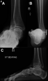
Case 1. Varus nonconcentric post-traumatic ankle arthritis weightbearing preoperative radiographs. Note the previous bimalleolar ankle fracture fixation. (A) Anteroposterior view; (B) hindfoot alignment view; (C) lateral view.
Figure 6.
Figure 6.
Case 1. Varus nonconcentric ankle arthritis weightbearing radiographs after arthroscopic fusion with percutaneous screw fixation. Note that the previous medial malleolar fracture fixation was removed. The lateral screw was inserted first under compression to reduce the varus talar tilt, followed by the medial screw, and finally the posterior screw. (A) Anteroposterior view; (B) hindfoot alignment view; (C) lateral view.
Figure 7.
Figure 7.
Case 2. Valgus concentric ankle arthritis with ball-and-socket deformity and planovalgus foot. Weightbearing preoperative radiographs. (A) Anteroposterior view; (B) hindfoot alignment view; (C) lateral view.
Figure 8.
Figure 8.
Case 2. Intraoperative fluoroscopic images of arthroscopic ankle fusion of valgus concentric ankle arthritis with ball-and-socket deformity after joint preparation and gastrocnemius recession to enhance deformity reduction. The ball-and-socket articulation allows simple angular reduction via screw placement. (A) Reduction of valgus alignment performed manually followed by K-wire placement to hold. Medial screw inserted first under compression to further reduce out of valgus. (B, C) Anteroposterior view of second screw inserted as full-threaded strut to prevent collapse back into valgus. (D, E) Third screw inserted anterolaterally under compression to obtain appropriate ankle dorsiflexion.
Figure 9.
Figure 9.
Case 2. Valgus concentric ankle arthritis with ball-and-socket deformity. Weightbearing postoperative radiographs after arthroscopic fusion with percutaneous screw fixation. Also note the concomitant medial cuneiform dorsal opening wedge osteotomy for correction of a dorsiflexed first ray. (A) Anteroposterior view; (B) hindfoot alignment view; (C) lateral view.
Figure 10.
Figure 10.
Case 3. Anteriorly translated varus nonconcentric ankle arthritis preoperative weightbearing radiographs. Note the previous medial malleolar fracture screw fixation. (A) Anteroposterior view. (B) Lateral view.
Figure 11.
Figure 11.
Case 3. Intraoperative fluoroscopic images of arthroscopic ankle fusion of case 3 after joint preparation and gastrocnemius recession to enhance posterior translation. (A) The posterior plafond is burred to allow more posterior translation. (B-C) Platelet-derived growth factor bone graft substitute is inserted into the anterior void (panel b) followed by demineralized bone matrix (panel c). (D) Ankle is manually reduced and provisionally fixed with K-wires. (E) First screw is inserted posterolaterally to reduce the anterior translation and varus talar tilt. (F, G) Second screw is inserted medially to compress medial side of joint. (H, I) Third screw is inserted anterolaterally as a fully threaded strut to avoid drifting back anteriorly. A fourth posteromedial screw is inserted under compression as well (not shown).
Figure 12.
Figure 12.
Case 3. Postoperative weightbearing radiographs of case 3. A circumferential cast is applied due to patient noncompliance. (A) Anteroposterior view. (B) Hindfoot alignment view. (C) Lateral view. Note that a concomitant first metatarsal dorsiflexion osteotomy was performed to help create a plantigrade foot.

References

    1. Agel J, Coetzee JC, Sangeorzan BJ, Roberts MM, Hansen ST. Functional limitations of patients with end-stage ankle arthrosis. Foot Ankle Int. 2005;26(7):537–539. - PubMed
    1. Al-Ashhab ME. Primary ankle arthrodesis for severely comminuted tibial pilon fractures. Orthopedics. 2017;40(2):e378–e381. - PubMed
    1. Albert E. Zur resection des kniegelenkes [For the resection of the knee joint]. Wien Med Press. 1879;20:705–708.
    1. Amendola A, Petrik J, Webster-Bogaert S. Ankle arthroscopy: outcome in 79 consecutive patients. Arthroscopy. 1996;12(5):565–573. - PubMed
    1. Balaji S, Selvaraj V, Devadoss S, Devadoss A. Transfibular ankle arthrodesis: a novel method for ankle fusion—a short term retrospective study. Indian J Orthop. 2017;51(1):75. - PMC - PubMed