Review
. 2020 Feb 13;5(1):2473011419896763.
doi: 10.1177/2473011419896763. eCollection 2020 Jan.

Evaluation and Treatment of Chronic Plantar Fasciitis

Affiliations
Review

Evaluation and Treatment of Chronic Plantar Fasciitis

L Daniel Latt et al. Foot Ankle Orthop. .

Abstract

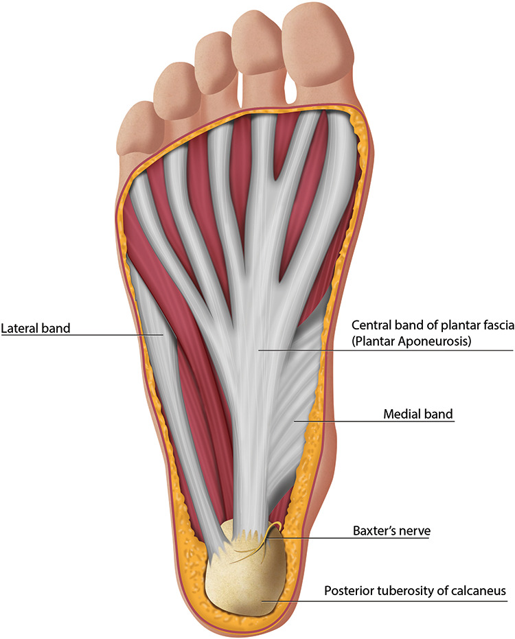
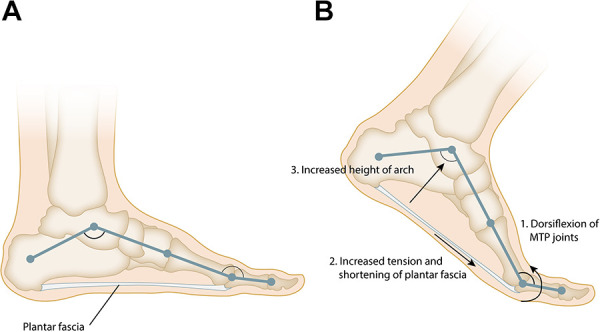
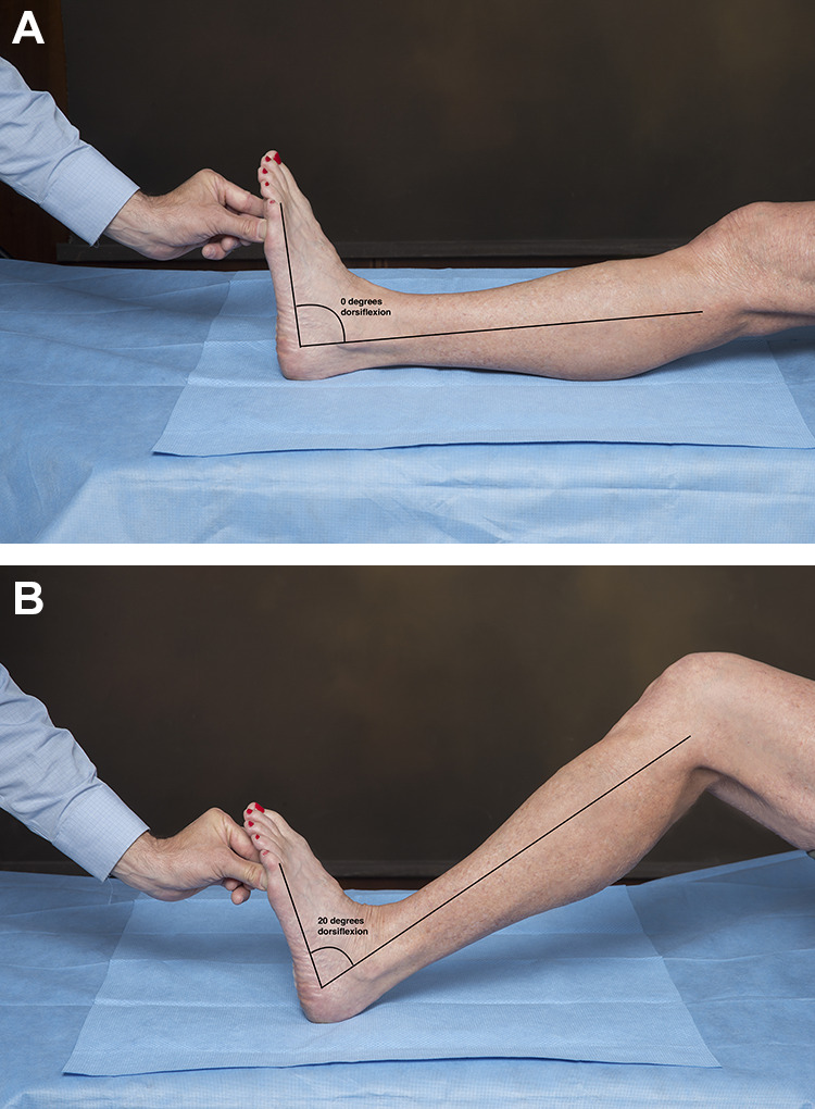
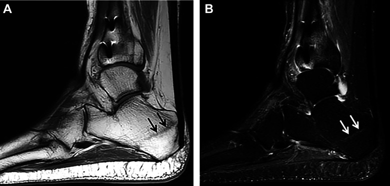
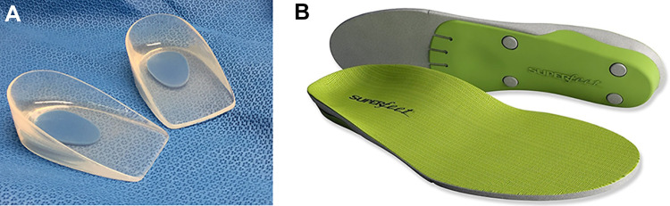
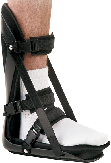
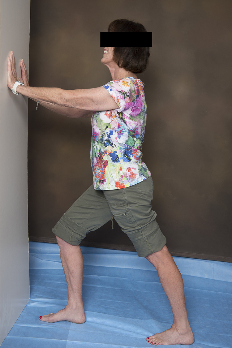
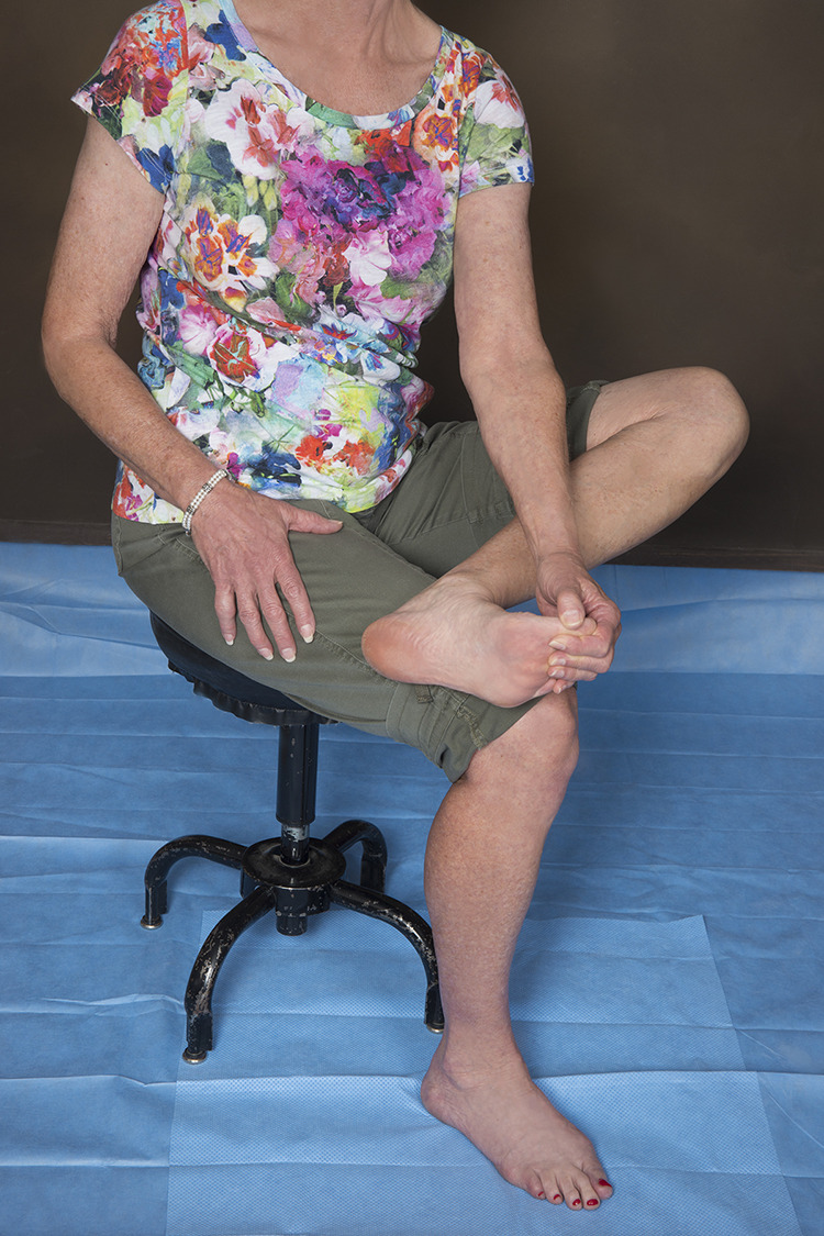
Plantar fasciitis is the most common cause of chronic heel pain in adults, affecting both young active patients and older sedentary individuals. It results from repetitive stress to the plantar fascia at its origin on the medial tubercle of the calcaneus and is often associated with gastrocnemius tightness. The diagnosis can be made clinically with a focused history and physical examination; imaging is reserved for atypical presentations and those that do not respond to initial treatment. The most common presenting symptom is aching plantar heel pain, which is worst with first step in the morning or after periods of rest. Diagnosis is confirmed with point tenderness at the origin of the plantar fascia on the medial tubercle of the calcaneus. Initial treatment consists of activity modification, anti-inflammatory medication, gastrocnemius and plantar fascia stretching, and an in-shoe orthosis that lifts and cushions the heel. These nonoperative treatments lead to complete resolution of pain in 90% of patients but can take 3-6 months. Patients who remain symptomatic despite a 6-month trial of nonoperative therapy may be considered for minimally invasive treatment or surgery. Platelet-rich plasma injections and therapeutic ultrasound are among a number of minimally invasive treatments that stimulate the body's healing response. Corticosteroid injections temporarily relieve pain, but may increase the risk of plantar fascia rupture and fat pad atrophy. Botulinum toxin injections relax the calf muscles, which decreases the stress in the plantar fascia. Operative treatments include gastrocnemius recession and medial head of gastrocnemius release, which decrease the stress on the plantar fascia and partial planter fasciotomy, which stimulates a healing response.

Level of evidence: Level V, expert opinion.

Keywords: fasciosis; plantar fascia; tendinosis; tendon disorders.


Conflict of interest statement

Declaration of Conflicting Interests: The author(s) declared no potential conflicts of interest with respect to the research, authorship, and/or publication of this article. ICMJE forms for all authors are available online.

Figures

Figure 1.
Figure 1.
Anatomy of the plantar aspect of the foot demonstrating the bands of the plantar fascia.
Figure 2.
Figure 2.
(A) The plantar fascia and the longitudinal arch of the foot form a truss. (B) Dorsiflexion of the toes during the late stance phase of gait tensions the plantar fascia around the metatarsal heads leading to an increase in the height and stability of the longitudinal arch of the foot, this effect is known as the “Windlass” mechanism.
Figure 3.
Figure 3.
The Silverskiold test is used to assess for gastrocnemius equinus. The maximum passive ankle dorsiflexion is compared with the knee (A) extended to (B) flexed. The difference between dorsiflexion in these 2 positions is the contribution of the gastrocnemius to the equinus contracture because the gastrocnemius crosses both the ankle and the knee joints whereas the other plantarflexors of the ankle do not. Dorsiflexion of less than 10 degrees with the knee extended or a difference of greater than 10 degrees confirms the presence of gastrocnemius equinus contracture.
Figure 4.
Figure 4.
In the calcaneus squeeze test, the examiner’s fingers are interlaced, the thenar or hypothenar eminences are then squeezed together compressing the posterior tuberosity of the calcaneus. The test is positive if this elicits the patient’s pain and is more painful than pressing plantarly at the insertion of the plantar fascia.
Figure 5.
Figure 5.
Weightbearing lateral radiograph of the foot demonstrating a large plantar calcaneal spur consistent with chronic heel cord tightness.
Figure 6.
Figure 6.
Long axis ultrasound imaging of the plantar fascia demonstrates thickening (arrows) of the proximal medial/central band, the right plantar fascia measuring up to 6 mm. There is a small region of hypoechogenicity (asterisk) within the medial aspect of the central band consistent with a moderate-grade partial-thickness tear.
Figure 7.
Figure 7.
(A) Sagittal T1-weighted MRI showing intermediate signal intensity in the region of the origin of the central band of the plantar fascia (arrows). (B) Sagittal STIR MRI showing heterogeneous increased signal intensity involving the proximal central limb of the plantar fascia adjacent to the vitamin E marker (arrows) consistent with plantar fasciitis. A full-thickness partial-width tear of the proximal central band of the plantar fascia is seen (arrowhead) as well as associated perifascial subcutaneous and deep soft tissue edema and a small focus of mild focal bone marrow edema in the adjacent calcaneal tuberosity (open arrowhead). MRI, magnetic resonance imaging; STIR, short tau inversion recovery.
Figure 8.
Figure 8.
(A) Sagittal, (B) coronal, and (C) axial T1-weighted MRI showing severe atrophy of the abductor digiti minimi muscle (arrows) consistent with Baxter neuropathy with entrapment of the inferior calcaneal nerve. MRI, magnetic resonance imaging.
Figure 9.
Figure 9.
(A) Sagittal T1-weighted MRI of the ankle demonstrating an oblique curvilinear hypointense marrow signal in the posterior tuberosity of the calcaneus (arrows) consistent with a stress fracture (B) Sagittal STIR MRI showing high signal calcaneal fracture line (arrows) with minimal surrounding bone marrow edema, consistent with a healing/healed stress/insufficiency fracture. MRI, magnetic resonance imaging; STIR, short tau inversion recovery.
Figure 10.
Figure 10.
In-shoe orthoses used in the treatment of plantar fasciitis: (A) gel heel cups, (B) over-the-counter arch supports. Image used with permission of Superfeet Worldwide, Inc.
Figure 11.
Figure 11.
Night splint. Image used with permission of Breg, Inc.
Figure 12.
Figure 12.
In the gastrocnemius stretch, the hands are placed against the wall, the leg being stretched is slid posteriorly with the knee bent as far as it will go and then the knee is straightened.
Figure 13.
Figure 13.
In the plantar fascia stretch, the foot to be stretched is placed on top of the contralateral knee, the ankle is maximally dorsiflexed, and then the toes are pulled up, tensioning the plantar fascia. The plantar fascia is then gently massaged along its length.

References

    1. Abbassian A, Kohls-Gatzoulis J, Solan MC. Proximal medial gastrocnemius release in the treatment of recalcitrant plantar fasciitis. Foot Ankle Int. 2012;33(1):14–19. - PubMed
    1. Ahmad J, Ahmad SH, Jones K. Treatment of plantar fasciitis with botulinum toxin: a randomized, controlled study. Foot Ankle Int. 2017;38(1):1–7. - PubMed
    1. Alshami AM, Babri AS, Souvlis T, Coppieters MW. Biomechanical evaluation of two clinical tests for plantar heel pain: the dorsiflexion-eversion test for tarsal tunnel syndrome and the windlass test for plantar fasciitis. Foot Ankle Int. 2007;28(4):499–505. - PubMed
    1. Barton JK, Rice PS, Howard CC, et al. Structural and functional assessment of intense therapeutic ultrasound effects on partial Achilles tendon transection. In: Advanced Biomedical and Clinical Diagnostic and Surgical Guidance Systems XVI 2018, San Francisco, United States, January 28-30, 2018. Bellingham, WA: SPIE; 2018.
    1. Bazaz R, Ferkel RD. Results of endoscopic plantar fascia release. Foot Ankle Int. 2007;28(5):549–556. - PubMed

LinkOut - more resources