

High Incidence of Hippocampal Abnormalities in Pediatric Patients with Congenital Cytomegalovirus Infection
- PMID: 35098496
- PMCID: PMC9444318
- DOI: 10.1055/a-1754-1142
High Incidence of Hippocampal Abnormalities in Pediatric Patients with Congenital Cytomegalovirus Infection
Abstract
Background: Congenital cytomegalovirus (CMV) infection exhibits polymicrogyria, intracranial calcification, white matter lesions, and several types of intracranial lesions on magnetic resonance imaging (MRI), in addition to various developmental disorders and epilepsies. However, little is known on the presence of hippocampal abnormality in this affliction. The aim of this study is to clarify the incidence of hippocampal abnormality in congenital CMV infection.
Methods: Seventeen children diagnosed as having congenital CMV infection along with 17 age-matched pediatric controls were retrospectively evaluated by brain MRI and clinical review. The measurement data were obtained from conventional coronal sections in this retrospective study. Hippocampal malrotation (HIMAL) was defined as a hippocampal diameter ratio (i.e., the ratio of the height and width of the hippocampus) of >0.92.
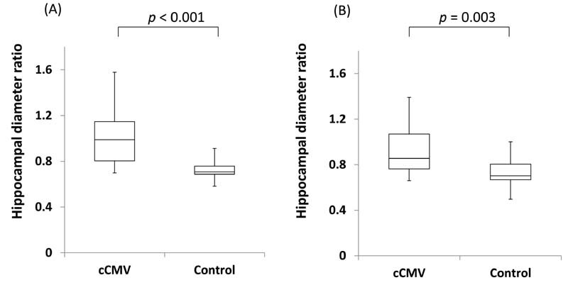
Results: Hippocampal diameter ratios were significantly higher in the congenital CMV infection group (0.99 [range: 0.70-1.58] on the right side and 0.85 [range: 0.66-1.39] on the left side) than in controls (0.71 [range: 0.58-0.91] and 0.70 [range: 0.50-1.00], respectively). HIMAL was present in 17 of 34 hippocampi (50%) in the congenital CMV infection group and 1 of 34 hippocampi (2.9%) in controls. No correlations were detected between HIMAL and intelligence quotient/developmental quotient or the occurrences of autism spectrum disorder or epilepsy.
Conclusion: This study is the first to demonstrate the incidence of hippocampal abnormality to be significantly higher in congenital CMV infection patients than in age-matched controls. Further study is necessary to clarify the associations of HIMAL with other clinical and developmental features.
The Author(s). This is an open access article published by Thieme under the terms of the Creative Commons Attribution-NonDerivative-NonCommercial License, permitting copying and reproduction so long as the original work is given appropriate credit. Contents may not be used for commercial purposes, or adapted, remixed, transformed or built upon. (https://creativecommons.org/licenses/by-nc-nd/4.0/).
Conflict of interest statement
None declared.
Figures

References
-
- Boppana S B, Pass R F, Britt W J, Stagno S, Alford C A. Symptomatic congenital cytomegalovirus infection: neonatal morbidity and mortality. Pediatr Infect Dis J. 1992;11(02):93–99. - PubMed
-
- Ivarsson S A, Lernmark B, Svanberg L. Ten-year clinical, developmental, and intellectual follow-up of children with congenital cytomegalovirus infection without neurologic symptoms at one year of age. Pediatrics. 1997;99(06):800–803. - PubMed
-
- Pass R F, Stagno S, Myers G J, Alford C A. Outcome of symptomatic congenital cytomegalovirus infection: results of long-term longitudinal follow-up. Pediatrics. 1980;66(05):758–762. - PubMed
-
- Zhang X W, Li F, Yu X W, Shi X W, Shi J, Zhang J P. Physical and intellectual development in children with asymptomatic congenital cytomegalovirus infection: a longitudinal cohort study in Qinba mountain area, China. J Clin Virol. 2007;40(03):180–185. - PubMed
-
- Barbi M, Binda S, Caroppo S, Primache V. Neonatal screening for congenital cytomegalovirus infection and hearing loss. J Clin Virol. 2006;35(02):206–209. - PubMed