Type A thymoma in a pet rabbit
- PMID: 35098805
- PMCID: PMC8921795
- DOI: 10.1177/10406387221077086
Type A thymoma in a pet rabbit
Abstract
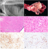
A 4-y-old, spayed female, mixed-breed domesticated rabbit (Oryctolagus cuniculus domesticus) was presented because of progressive bilateral exophthalmos, with a large mediastinal mass in the cranial thorax. Palliative radiation therapy was elected, and 4 fractions of 5 Gy were delivered twice weekly under general anesthesia using 3-dimensional conformal radiation therapy for a total dose of 20 Gy, guided by an on-board cone beam CT scan. Quality-of-life and respiratory rate improved before sudden death that followed an episode of dyspnea. The overall survival time following initial diagnosis was 93 d, with 68 d after the first dose of radiation. An autopsy was performed, and the mass was diagnosed as a type A thymoma. The diagnosis was confirmed with positive immunohistochemical labeling of the neoplastic cells for cytokeratin 5/6 and cytokeratin 7.
Keywords: World Health Organization; domesticated rabbits; immunohistochemistry; radiation therapy; thymoma; type A thymoma.
Conflict of interest statement
Figures

References
-
- Bertram CA, et al.. Neoplasia and tumor-like lesions in pet rabbits (Oryctolagus cuniculus): a retrospective analysis of cases between 1995 and 2019. Vet Pathol 2021;58:901–911. - PubMed
-
- Detterbeck FC. Clinical value of the WHO classification system of thymoma. Ann Thorac Surg 2006;81:2328–2334. - PubMed
-
- Jekl V. Respiratory disorders in rabbits. Vet Clin North Am Exot Anim Pract 2021;24:459–482. - PubMed
-
- Morrisey JK, McEntee M. Therapeutic options for thymoma in the rabbit. Sem Avian Exot Pet Med 2005;14:175–181.
-
- Sanchez-Migallon DG, et al.. Radiation therapy for the treatment of thymoma in rabbits (Oryctolagus cuniculus). J Exot Pet Med 2006;15:138–144.
MeSH terms
LinkOut - more resources
Full Text Sources
Medical
Research Materials

