Clinical Trial
. 2022 Jan 31;6(1):5.
doi: 10.1186/s41747-021-00256-8.

Myocardial CT perfusion imaging for the detection of obstructive coronary artery disease: multisegment reconstruction does not improve diagnostic performance

Affiliations
Clinical Trial

Myocardial CT perfusion imaging for the detection of obstructive coronary artery disease: multisegment reconstruction does not improve diagnostic performance

Daniel Preuß et al. Eur Radiol Exp. .

Abstract

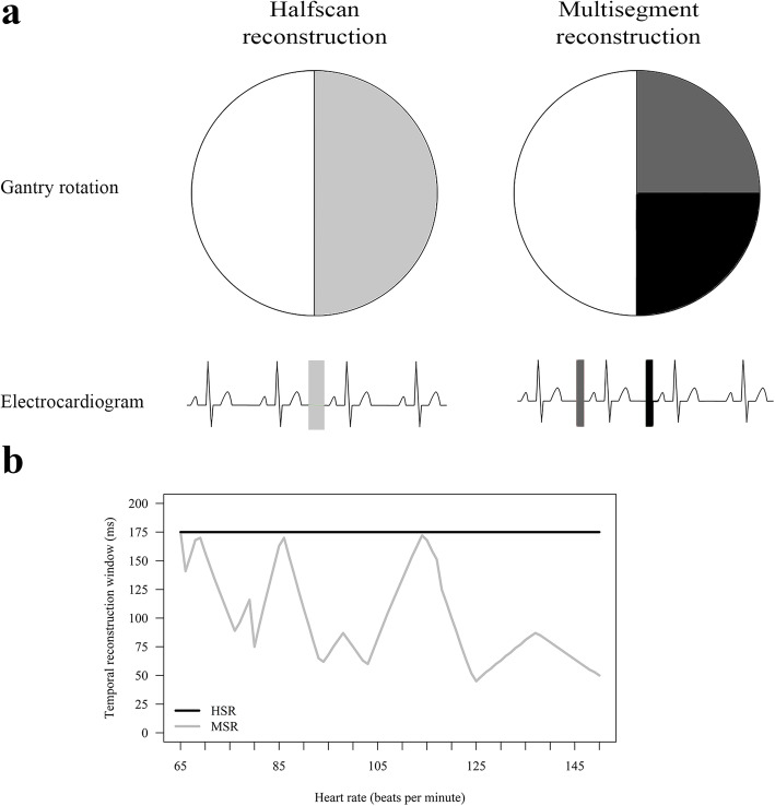
Background: Multisegment reconstruction (MSR) was introduced to shorten the temporal reconstruction window of computed tomography (CT) and thereby reduce motion artefacts. We investigated whether MSR of myocardial CT perfusion (CTP) can improve diagnostic performance in detecting obstructive coronary artery disease (CAD) compared with halfscan reconstruction (HSR).

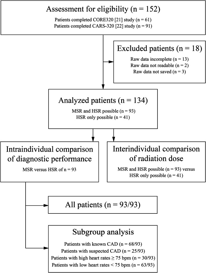
Methods: A total of 134 patients (median age 65.7 years) with clinical indication for invasive coronary angiography and without cardiac surgery prospectively underwent static CTP. In 93 patients with multisegment acquisition, we retrospectively performed both MSR and HSR and searched both reconstructions for perfusion defects. Subgroups with known (n = 68) or suspected CAD (n = 25) and high heart rate (n = 30) were analysed. The area under the curve (AUC) was compared applying DeLong approach using ≥ 50% stenosis on invasive coronary angiography as reference standard.

Results: Per-patient analysis revealed the overall AUC of MSR (0.65 [95% confidence interval 0.53, 0.78]) to be inferior to that of HSR (0.79 [0.69, 0.88]; p = 0.011). AUCs of MSR and HSR were similar in all subgroups analysed (known CAD 0.62 [0.45, 0.79] versus 0.72 [0.57, 0.86]; p = 0.157; suspected CAD 0.80 [0.63, 0.97] versus 0.89 [0.77, 1.00]; p = 0.243; high heart rate 0.46 [0.19, 0.73] versus 0.55 [0.33, 0.77]; p = 0.389). Median stress radiation dose was higher for MSR than for HSR (6.67 mSv versus 3.64 mSv, p < 0.001).

Conclusions: MSR did not improve diagnostic performance of myocardial CTP imaging while increasing radiation dose compared with HSR.

Trial registration: CORE320: clinicaltrials.gov NCT00934037, CARS-320: NCT00967876.

Keywords: Coronary angiography; Coronary artery disease; Multidetector computed tomography; Myocardial perfusion imaging; Sensitivity and specificity.


Conflict of interest statement

Institutional master research agreements exist with Siemens, General Electric, Philips, and Canon Medical Systems (former Toshiba Medical Systems). The terms of these arrangements are managed by the legal department of Charité – Universitätsmedizin Berlin.

Prof. Dewey has received grant support from the FP7 Program of the European Commission for the randomized multicenter DISCHARGE trial (603266-2, HEALTH-2012.2.4.-2). He also received grant support from German Research Foundation (DFG) in the Heisenberg Program (DE 1361/14-1), graduate programme on quantitative biomedical imaging (BIOQIC, GRK 2260/1), for fractal analysis of myocardial perfusion (DE 1361/18-1), the Priority Programme Radiomics for the investigation of coronary plaque and coronary flow (DE 1361/19-1 [428222922] and 20-1 [428223139] in SPP 2177/1). He also received funding from the Berlin University Alliance (GC_SC_PC 27) and from the Digital Health Accelerator of the Berlin Institute of Health. Prof. Dewey has received lecture fees from Canon Medical Systems (former Toshiba Medical Systems), Guerbet. Prof. Dewey is European Society of Radiology (ESR) Research Chair (2019–2022) and the opinions expressed in this article are the author’s own and do not represent the view of ESR. Per the guiding principles of ESR, the work as Research Chair is on a voluntary basis and only travel expenses are remunerated. Prof. Dewey is also the editor of Cardiac CT, published by Springer Nature, and offers hands-on courses on CT imaging (www.ct-kurs.de). Prof. Dewey holds a joint patent with Florian Michallek on dynamic perfusion analysis using fractal analysis (PCT/EP2016/071551).

Dr. Rief received grant support for the investigation of coronary plaque from the ‘Radiomics’ Priority Programme of the German Research Foundation (DFG).

Other authors declared no conflicts of interest.

Figures

Fig. 1
Fig. 1
Flow chart showing the patient selection strategy for comparison of diagnostic performance and radiation dose of MSR and HSR of myocardial CTP imaging datasets in patients with suspected or known CAD. The study population for intraindividual comparison of diagnostic performance consisted of 93 of 134 patients with multisegment acquisition of stress CTP allowing both HSR and MSR. The remaining 41 of 134 patients had predefined single-segment acquisition due to heart rates < 65 bpm, and only HSR was possible. These patients served as a control group for interindividual radiation dose comparison. bpm Beats per minute, CAD Coronary artery disease, CTP Computed tomography perfusion, HSR Halfscan reconstruction, MSR Multisegment reconstruction
Fig. 2
Fig. 2
Technical background for generating temporal reconstruction windows of MSR and HSR (a) and their dependency on patient’s heart rate (b). a The minimum partial scan raw data needed to reconstruct a volume require half a gantry rotation (+ fan angle). HSR uses partial scan raw data acquired within one heart beat (illustrated on the left in light grey) and consequently generates a per-segment temporal reconstruction window of half a gantry rotation. MSR uses partial scan raw data of approximately half a gantry rotation acquired in at least two successive heart beats, illustrated on the right for two segments in dark grey (segment one) and black (successive segment two). The resulting minimum per-segment temporal reconstruction window of MSR can be calculated as follows: Temporal reconstruction window = gantry rotation time / (2 × number of acquired segments). Thereby, acquiring more segments improves the per (heart)-segment temporal reconstruction window of MSR compared with HSR by up to the same factor as the number of segments acquired [see references [–11]] at the cost of a higher radiation dose [see references [8, 12]]. b Line graphs showing the per-segment temporal reconstruction window of MSR and HSR depending on individual patient’s heart rate for a 350-ms gantry rotation time and acquisition of up to four segments. The temporal reconstruction window of HSR is always 175 ms, whereas the temporal reconstruction window of MSR ranges from 44 to 175 ms depending on heart rate. Data by courtesy of the equipment vendor. HSR Halfscan reconstruction, MSR Multisegment reconstruction
Fig. 3
Fig. 3
HSR and two-segment MSR of stress myocardial CTP in a 55-year-old man with suspected coronary artery disease and typical angina pectoris in comparison with invasive coronary angiography. a HSR shows a moderate perfusion defect in the left anterior descending artery territory (arrow), which is also suggested by moderate hypoattenuation in the corresponding area (circle) of the polar myocardial attenuation map; CTP with HSR was considered positive. b MSR shows only very slightly reduced perfusion (arrowhead) and only weak hypoattenuation in the corresponding area (dotted circle) of the polar myocardial attenuation map; CTP with MSR was considered negative. c Invasive coronary angiography reveals visually high-grade diameter stenosis (*) of the left anterior descending artery with 61% stenosis in quantitative invasive coronary angiography (**), corresponding to a true-positive CTP with HSR (a) and a false-negative CTP with MSR (b). Contrast-enhanced CTP in mid-heart short-axis view with 8-mm slice thickness and rainbow-red colour preset using a predefined window level/window width of 200/400. CTP Computed tomography perfusion, HSR Halfscan reconstruction, MSR Multisegment reconstruction
Fig. 4
Fig. 4
All patients: receiver operating characteristic areas under the curve for comparison of MSR and HSR of myocardial competed tomography perfusion in per-patient (a) and per-territory analysis (b) using 50% diameter stenosis detected on quantitative coronary angiography as a reference standard. In all 93 patients, the area under the curve of MSR was inferior to that of HSR for both levels of analysis (p < 0.001). HSR Halfscan reconstruction, MSR Multisegment reconstruction
Fig. 5
Fig. 5
Boxplots showing estimated radiation dose in mSv of stress computed tomography perfusion in patients with multisegment acquisition, which allowed both MSR and HSR (93 patients), and in patients with HSR only (41 patients). Boundaries of boxes represent the lower and upper quartiles and horizontal lines in boxes the medians. Outliers are depicted as individual open circles. In the HSR-only group, MSR was not possible in 9 patients despite multisegment acquisition of two segments. Mean radiation dose of patients with both MSR and HSR was higher than that of patients with HSR (p < 0.001). HSR Halfscan reconstruction, MSR Multisegment reconstruction

References

    1. Fihn SD, Gardin JM, Abrams J, et al. 2012 ACCF/AHA/ACP/AATS/PCNA/SCAI/STS Guideline for the diagnosis and management of patients with stable ischemic heart disease: a report of the American College of Cardiology Foundation/American Heart Association Task Force on Practice Guidelines, and the American College of Physicians, American Association for Thoracic Surgery, Preventive Cardiovascular Nurses Association, Society for Cardiovascular Angiography and Interventions, and Society of Thoracic Surgeons. J Am Coll Cardiol. 2012;60:e44–e164. doi: 10.1016/j.jacc.2012.07.013. - DOI - PubMed
    1. Montalescot G, Sechtem U, Achenbach S, et al. 2013 ESC guidelines on the management of stable coronary artery disease: the Task Force on the management of stable coronary artery disease of the European Society of Cardiology. Eur Heart J. 2013;34:2949–3003. doi: 10.1093/eurheartj/eht310.P4876. - DOI - PubMed
    1. George RT, Mehra VC, Chen MY, et al. (2014) Myocardial CT perfusion imaging and SPECT for the diagnosis of coronary artery disease: a head-to-head comparison from the CORE320 multicenter diagnostic performance study. Radiology 272:407–416 10.1148/radiol.14140806 - PMC - PubMed
    1. Rief M, Chen MY, Vavere AL, et al. (2018) Coronary artery disease: analysis of diagnostic performance of CT perfusion and MR perfusion imaging in comparison with quantitative coronary angiography and SPECT-multicenter prospective trial. Radiology 286:461–470 10.1148/radiol.2017162447 - PMC - PubMed
    1. Sorgaard MH, Kofoed KF, Linde JJ, et al. Diagnostic accuracy of static CT perfusion for the detection of myocardial ischemia. A systematic review and meta-analysis. J Cardiovasc Comput Tomogr. 2016;10:450–457. doi: 10.1016/j.jcct.2016.09.003. - DOI - PubMed

Publication types

MeSH terms

Associated data