. 2022 Feb 4;12(2):jkab451.
doi: 10.1093/g3journal/jkab451.

A nonfunctional copy of the salmonid sex-determining gene (sdY) is responsible for the "apparent" XY females in Chinook salmon, Oncorhynchus tshawytscha

Affiliations

A nonfunctional copy of the salmonid sex-determining gene (sdY) is responsible for the "apparent" XY females in Chinook salmon, Oncorhynchus tshawytscha

Sylvain Bertho et al. G3 (Bethesda). .

Abstract

Many salmonids have a male heterogametic (XX/XY) sex determination system, and they are supposed to have a conserved master sex-determining gene (sdY) that interacts at the protein level with Foxl2 leading to the blockage of the synergistic induction of Foxl2 and Nr5a1 of the cyp19a1a promoter. However, this hypothesis of a conserved master sex-determining role of sdY in salmonids is challenged by a few exceptions, one of them being the presence of naturally occurring "apparent" XY Chinook salmon, Oncorhynchus tshawytscha, females. Here, we show that some XY Chinook salmon females have a sdY gene (sdY-N183), with 1 missense mutation leading to a substitution of a conserved isoleucine to an asparagine (I183N). In contrast, Chinook salmon males have both a nonmutated sdY-I183 gene and the missense mutation sdY-N183 gene. The 3-dimensional model of SdY-I183N predicts that the I183N hydrophobic to hydrophilic amino acid change leads to a modification in the SdY β-sandwich structure. Using in vitro cell transfection assays, we found that SdY-I183N, like the wild-type SdY, is preferentially localized in the cytoplasm. However, compared to wild-type SdY, SdY-I183N is more prone to degradation, its nuclear translocation by Foxl2 is reduced, and SdY-I183N is unable to significantly repress the synergistic Foxl2/Nr5a1 induction of the cyp19a1a promoter. Altogether, our results suggest that the sdY-N183 gene of XY Chinook females is nonfunctional and that SdY-I183N is no longer able to promote testicular differentiation by impairing the synthesis of estrogens in the early differentiating gonads of wild Chinook salmon XY females.

Keywords: sdY; XY females; salmonids; sex determination; sex reversal.


Figures

Fig. 1.
Fig. 1.
XY Chinook salmon females have a missense mutation in a conserved position of the sdY coding sequence. a) Schematic representation of Chinook salmon SdY sequence with its 4 exons depicted as square boxes (E1–E4) and the introns as broken lines with intron sizes (bp). b) Remapping of transcriptome reads (N = number of raw remapped reads) from a chinook male testis revealed 2 SNVs (A/G and A/T) in the coding region of the sdY gene. Representative sequencing chromatograms of parts of the genomic sdY coding sequencing containing SNVs in XY females c) and XY males c′) leading to a synonymous mutation in exon 2 (A/G) and a missense mutation in exon 3 (A/T). d) Alignment of Irf9a, Irf9b, and SdY protein sequences in different salmonid species showing the conservation of isoleucine 183 (I) highlighted in gray color and its modification to asparagine (N) only in XY Chinook salmon females (SdY-I183N).
Fig. 2.
Fig. 2.
Sex chromosomes, sex genotypes and sex phenotypes in Chinook salmon. a) Schematic representation of sex chromosomes and the hypothetical relation between sex genotypes and sex phenotypes in Chinook salmon. According to our model, phenotypic females can be normal XX females or XY females (XY−) bearing a Y-chromosome (Y−) with a single copy sdY-N183 gene. Phenotypic males can be XY males (XY+) bearing a Y-chromosome (Y+) with 2 copies of the sdY gene, i.e. sdY-I183 and sdY-N183, or Y−Y+ resulting from the crossing of an XY+ male with an XY− female. In turn, a Y−Y+ males crossed with an XY− female can also generate Y−Y− phenotypic females. b) Representative chromatograms of the sequences around the sdY I183N mutation (exon 3) in Chinook salmon. XY females (XY−) are homozygotes A/A for the I183N mutation and males are heterozygotes (A/T). Y−Y− females cannot be discriminated from XY− females based on the chromatogram analysis (single A peak of homozygosity in both cases), but XY+ and Y−Y+ could be in theory identified based on the relative peak height of the A/T “pseudo” alleles. With a 1:1 ratio of sdY-I183 and sdY-N183, XY+ males should have an equal A/T peak height and Y−Y+ with a 1:2 ratio of sdY-I183 and sdY-N183 should have an A peak height double from the T peak at the same position. Such chromatogram examples are shown in (b) but due to potential variability of the sequencing reactions this genotyping approach was not retained as an accurate approach to discriminate XY− males from Y−Y+ males in our analyses.
Fig. 3.
Fig. 3.
The I183N SdY mutation affects locally the structure of SdY. a) Model of SdY-I183N (green) deduced from the protein-protein interaction domain template of IRF5 (PDB ID 3DSH) embedded in the surface representation (gray). b) Magnification around the asparagine residue (N183) in cyan indicated by a black arrow. The mutation is located at the beginning of the β7-strand embedded in a hydrophobic pocket leading to a local misfolding.
Fig. 4.
Fig. 4.
SdY-I183N is localized predominantly in the cytoplasm and is only slightly translocated in the nucleus after cotransfection with Foxl2b2. SdY-I183N alone is mainly detected in the cytoplasm a−a″) with some transfected cells, however, showing a nucleo-cytoplasmic localization [see (e) for quantification of the different localization percentage] and even in some cells a restricted localization in the nucleus b–b″). After cotransfection with Foxl2b2, SdY-I183N remains also mostly cytoplasmic c–c″) with more transfected cells showing a nucleo-cytoplasmic localization [(d−d″) and panel (e) for quantification of the different localization percentage] and a complete localization in the nucleus. e) Quantification of the percentage of transfected cells (measured on 50 transfected cells) with an SdY-I183N localization in the cytoplasm (white bar), in the nucleus (black bar) or with a nucleo-cytoplasmic localization (gray bar) with (Foxl2b2+) or without (Foxl2b2−) cotransfection with Foxl2b2. Human Embryonic Kidney cells (HEK 293T) were transiently cotransfected with rainbow trout SdY-I183N in fusion with 3× Flag tag either with a nucleus marker, i.e. Histone H2B-mCherry (H2B), or a rainbow trout Foxl2b2-mCherry expression construct. Rainbow trout SdY-I183N was detected with an FLAG antibody and the nucleus was stain in red for the H2B construct a′ and b′) or in blue with Hoechst c″ and d″). Scale bar = 5 μm a″–d″).
Fig. 5.
Fig. 5.
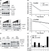
SdY-I183N is unstable even in the presence of Foxl2b2. Cycloheximide (CHX) time course was performed to assess SdY or SdY-I183N stability in the presence or absence of Foxl2b2. HEK cells were transiently transfected with SdY, SdY-I183N, or Foxl2b2 a–a′) alone or with SdY or SdY-I183N in combination with Foxl2b2 b–b′). Cells were treated with 50 μm of CHX and harvest at 4 and 8 h a′ and b′). Lysates were standardized for total protein concentration and expression levels of SdY, SdY-I183N, or Foxl2b2 were detected by Western blotting. Tubulin was blotted as a loading control. Foxl2b2 increased SdY but not SdY-I183N stability b and d). e) Western blot analysis of SdY, SdY-I183N alone, or in combination with Foxl2b2 protein levels following 8 h treatment with proteasome inhibitor MG132. Cells were treated with DMSO [vehicle (control), indicated by a − sign] and MG132 (20 µM, indicated by a + sign). Tubulin was blotted as a loading control f). Quantification of (e).
Fig. 6
Fig. 6
SdY-I183N does not prevent Foxl2/Nr5a1-positive regulation of the cyp19a1a promoter (see also Supplementary Fig. 1). The cyp19a1a promoter activity (cyp19a1a promoter coupled to firefly luciferase) was measured in HEK 293 cells using a luciferase reporter assay and cotransfection of fixed quantities of nr5a1 (100 ng), foxl2 (200 ng), and variable quantities (25–300 ng) of sdY-N183. Results are calculated from the mean ± SEM of 3 biological replicates in 1 experiment. Statistics were calculated with a one-way ANOVA with post hoc Dunnett tests. N.S: not statistically significant. Empty vector control (pGL3).

References

    1. Ayllon F, Solberg MF, Besnier F, Fjelldal PG, Hansen TJ, Wargelius A, Edvardsen RB, Glover KA.. Autosomal sdY pseudogenes explain discordances between phenotypic sex and DNA marker for sex identification in Atlantic salmon. Front Genet. 2020;11:544207. - PMC - PubMed
    1. Bachtrog D, Mank JE, Peichel CL, Kirkpatrick M, Otto SP, Ashman T-L, Hahn MW, Kitano J, Mayrose I, Ming R, et al.; Tree of Sex Consortium. Sex determination: why so many ways of doing it? PLoS Biol. 2014;12(7):e1001899. - PMC - PubMed
    1. Baroiller J-F, D'Cotta H.. The reversible sex of gonochoristic fish: insights and consequences. Sex Dev. 2016;10(5–6):242–266. - PubMed
    1. Bertho S, Herpin A, Branthonne A, Jouanno E, Yano A, Nicol B, Muller T, Pannetier M, Pailhoux E, Miwa M, et al.. The unusual rainbow trout sex determination gene hijacked the canonical vertebrate gonadal differentiation pathway. Proc Natl Acad Sci U S A. 2018;115:12781–12786. - PMC - PubMed
    1. Bertho S, Herpin A, Schartl M, Guiguen Y.. Lessons from an unusual vertebrate sex-determining gene. Philos Trans R Soc Lond B Biol Sci. 2021;376(1832):20200092. - PMC - PubMed

Publication types

LinkOut - more resources