Review
. 2022 Feb 4;220(2):iyab197.
doi: 10.1093/genetics/iyab197.

Mechanisms of sex determination and X-chromosome dosage compensation

Affiliations
Review

Mechanisms of sex determination and X-chromosome dosage compensation

Barbara J Meyer. Genetics. .

Abstract

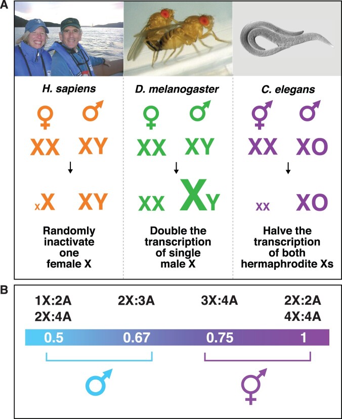
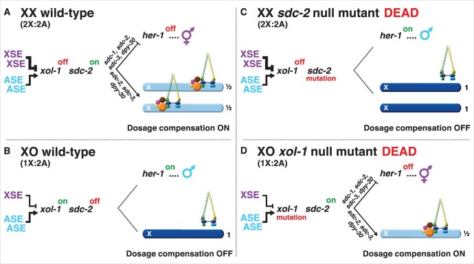
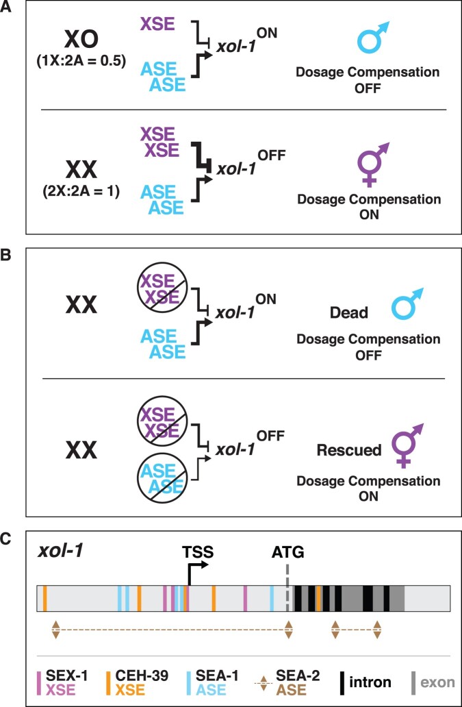
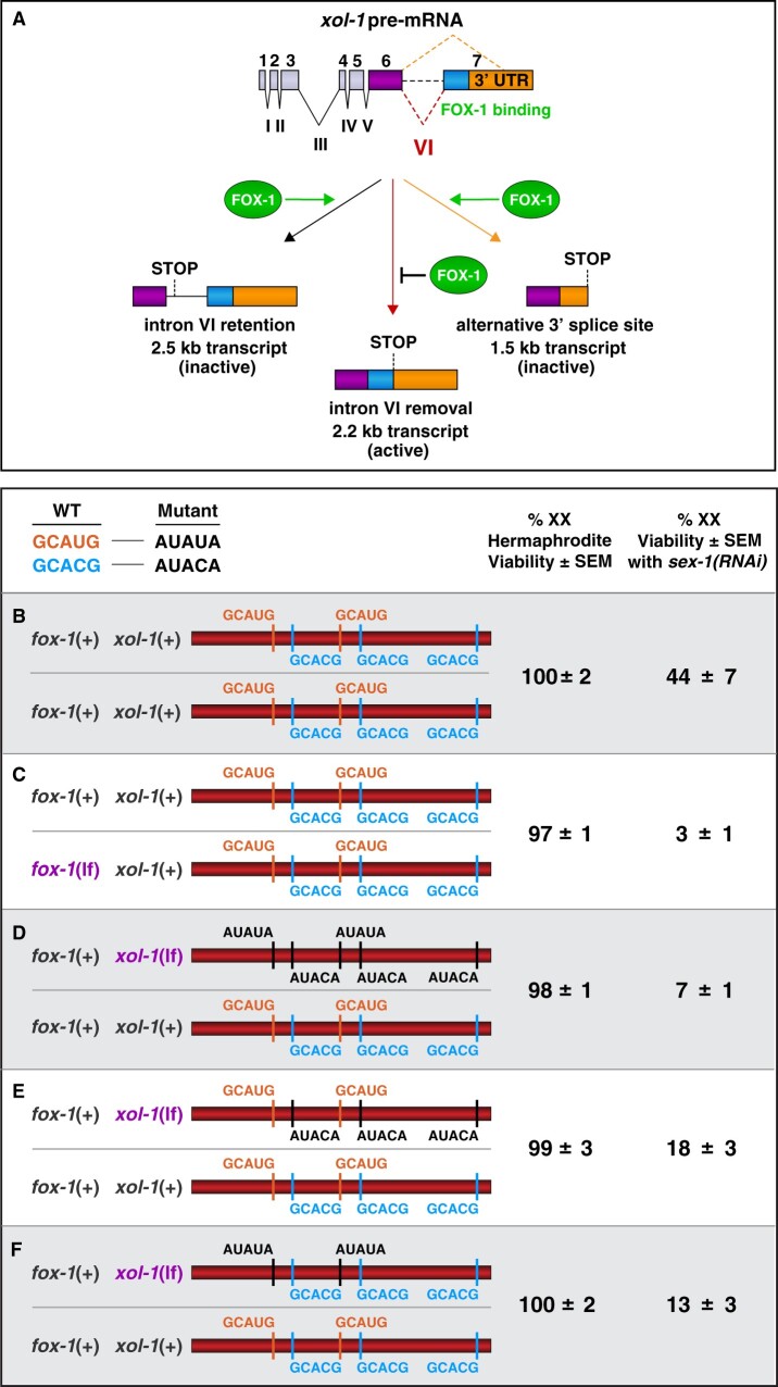
Abnormalities in chromosome number have the potential to disrupt the balance of gene expression and thereby decrease organismal fitness and viability. Such abnormalities occur in most solid tumors and also cause severe developmental defects and spontaneous abortions. In contrast to the imbalances in chromosome dose that cause pathologies, the difference in X-chromosome dose used to determine sexual fate across diverse species is well tolerated. Dosage compensation mechanisms have evolved in such species to balance X-chromosome gene expression between the sexes, allowing them to tolerate the difference in X-chromosome dose. This review analyzes the chromosome counting mechanism that tallies X-chromosome number to determine sex (XO male and XX hermaphrodite) in the nematode Caenorhabditis elegans and the associated dosage compensation mechanism that balances X-chromosome gene expression between the sexes. Dissecting the molecular mechanisms underlying X-chromosome counting has revealed how small quantitative differences in intracellular signals can be translated into dramatically different fates. Dissecting the process of X-chromosome dosage compensation has revealed the interplay between chromatin modification and chromosome structure in regulating gene expression over vast chromosomal territories.

Keywords: Caenorhabditis elegans; WormBook; X-chromosome counting; X-chromosome dosage compensation; cell fate determination; chromosome segregation; chromosome structure; histone modification; mRNA splicing regulation; sex determination; transcriptional regulation.


Figures

Figure 1
Figure 1
Diverse strategies for X-chromosome dosage compensation. (A) Organisms use different strategies to ensure that males and females or hermaphrodites produce comparable levels of X-linked gene products, despite the twofold difference in X dose between the sexes. Female human and Mus musculus mammals (XX) randomly inactivate most genes on one X chromosome. Male D. melanogaster fruit flies (XY) double transcription of their singe X chromosome. Hermaphrodite C. elegans worms (XX) reduce transcription of both X chromosomes by half. (B) The nematode calculates the ratio of X chromosomes to sets of autosomes to determine sexual fate. Chromosome counting is executed with remarkable precision in the nematode such that diploid animals with one X chromosome (1X:2A, ratio 0.5) and triploid animals with two X chromosomes (2X:3A, ratio of 0.67) become fertile males, while diploid animals with two X chromosomes (2X:2A, ratio 1.0) and tetraploid animals with three X chromosomes (3X:4A, ratio of 0.75) become fertile hermaphrodites. Other organisms like fruit flies discriminate less well such that only a ratio of 0.5 results in fertile males, and a ratio of 1.0 results in fertile females, with intermediate ratios generating sterile intersexes.
Figure 2
Figure 2
Overview of the X:A signal and the regulatory hierarchy that controls nematode sex determination and dosage compensation. (A, B) In wild-type animals, the X:A signal that determines sexual fate is a competition between a set of genes on X called XSEs that represses their direct gene target xol-1 (XO lethal) in a cumulative dose-dependent manner via transcriptional and post-transcriptional mechanisms and a set of genes on autosomes called ASEs that stimulate xol-1 transcription in a cumulative dose-dependent manner. xol-1 is the master sex-determination switch gene that must be activated in XO animals to set the male fate and must be repressed in XX animals to permit the hermaphrodite fate. (A) Two doses of XSEs in diploid XX animals win out and repress xol-1, but (B) the single dose of XSEs in diploid XO animals does not turn xol-1 off. (B) xol-1 triggers male sexual development in wild-type XO animals by repressing the feminizing switch gene sdc-2 (sex determination and dosage compensation). (A) Together with sdc-1, sdc-3, and dpy-30, the sdc-2 gene induces hermaphrodite sexual development in XX animals by repressing the male sex-determining gene her-1. Together with sdc-3 and dpy-30, sdc-2 triggers binding of a dosage compensation complex (DCC) onto both hermaphrodite X chromosomes to repress gene expression by half. sdc-1 is essential for DCC activity, but not for loading of the DCC onto X. The DCC is a condensin complex that restructures the topology of X. (C) sdc-2 mutations kill XX animals by prevent the DCC from binding to X chromosomes, resulting in overexpression of X-linked genes. The mutations also masculinize XX animals, because her-1 is not repressed. (D) Loss-of-function xol-1 mutations enable sdc-2 to be active and permit the DCC to bind the single male X, thereby killing XO animals from reduced X-chromosome expression. The dying xol-1 XO mutant animals are feminized because her-1 is repressed. Hence, mutations that disrupt elements of the X:A signal itself transform sexual fate, but also kill due to altered X-chromosome gene expression.
Figure 3
Figure 3
Dissecting the X:A sex determination signal. (A) XSE regulate xol-1 in a dose-dependent manner in the context of two doses of ASE. Two doses of XSEs win out and repress xol-1 in diploid animals with two doses of ASE, which stimulate xol-1 expression. One XSE dose does not prevail in repressing. When xol-1 is activated in 1X:2A animals, the dosage compensation machinery is turned off. XO animals are viable and develop as males. When xol-1 is repressed in 2X:2A animals, the dosage compensation machinery is activated, thereby reducing X-linked gene expression by half. XX animals are viable and develop as hermaphrodites. (B) Loss-of-function mutations in XSEs were identified in genetic screens because they caused a xol-1 reporter transgene to be activated in XX animals, resulting in the masculinization and death of XX animals. XSEs were also discovered as suppressors of the male lethality caused by duplication of large regions of X. Loss-of-function mutations in ASEs were identified in genetic screens because they suppressed the lethality of mutations in XSEs and prevented the transformation of sexual fate caused by them. (C) Locations of binding sites in the 5′ xol-1 regulator regions for the XSEs (SEX-1 and CEH-39) that repress xol-1 transcription and the ASEs (SEA-1 and SEA-2) that activate xol-1 transcription. The general regions of SEA-2 binding were defined but not yet the precise binding sites. SEX-1 is a nuclear hormone receptor; CEH-39 is a ONECUT homeodomain protein; SEA-1 is a T-box protein; SEA-2 is a zinc-finger protein. After the molecular tug-of-war to control xol-1 transcription, a second tier of regulation occurs to control xol-1 pre-mRNA splicing by the XSE FOX-1 (see Figure  5).
Figure 4
Figure 4
Dose-dependent pre-mRNA splicing regulation of xol-1 by the RNA binding protein FOX-1, an XSE. (A) Summary of xol-1 splicing regulation by FOX-1. By binding to multiple GCAUG and GCACG motifs in intron VI of xol-1, FOX-1 reduces formation of the male-determining 2.2 kb transcript by causing intron VI retention (2.5 kb transcript) or by directing use of an alternative 3′ splice acceptor site, causing deletion of essential exon 7 coding sequences (blue) and part of the 3′ UTR (orange) (1.5 kb transcript). (B–F) Two copies of fox-1(+) and multiple high-affinity GCAUG and GCACG motifs in both copies of intron VI are essential for FOX-1-mediated alternative splicing of xol-1 in XX animals. DNA sequences in the upper left compare the wild-type vs mutant versions of FOX-1 binding motifs in xol-1 intron VI that were used to assess regulation by FOX-1. Diagrams on the left show sequences of FOX-1 binding motifs in either a fox-1 mutant or in animals carrying heterozygous combinations of mutant motifs in xol-1 intron VI. Viability of XX progeny were assayed in each cohort of sex-1(+) or sex-1(RNAi) XX animals to assess the dose-dependence of FOX-1 action in regulating xol-1 splicing. Percentages on the right reflect the viability of sex-1(+) or sex-1(RNAi) XX animals with different heterozygous combinations of intron VI or fox-1 mutations. None of the heterozygous mutant combinations in intron VI or fox-1 affects the viability of sex-1(+) animals, but they have strong effects on sex-1(RNAi) animals. (B, C) Mutating one copy of fox-1 reduced viability of sex-1(RNAi) XX animals from 44% to 3%. (D) Similarly, mutating one copy of all GCAUG and GCACG motifs in one intron reduced viability to 7%. Mutating only one copy of the three GCACG motifs (E) or one copy of the two GCAUG motifs (F) in one intron had an intermediate effect, resulting in 18% or 13% viability. Thus, FOX-1 acts in a dose-dependent manner to regulate xol-1 splicing in XX vs XO animals.
Figure 5
Figure 5
Model for X:A signal assessment: two tiers of xol-1 repression. XSEs and ASEs bind directly to numerous nonoverlapping sites in the 5′ regulatory region of xol-1 to antagonize each other’s opposing transcriptional activities and thereby control xol-1 transcription. Molecular rivalry at the xol-1 promoter between the XSE transcriptional repressors and ASE transcriptional activators causes high xol-1 transcript levels in 1X:2A embryos with one dose of XSEs and low levels in 2X:2A embryos with two doses of XSE. In a second tier of xol-1 repression, the XSE RNA binding protein FOX-1 then enhances the fidelity of X-chromosome counting by binding to numerous GCAUG and GCAUG motifs in intron VI (yellow) of the residual xol-1 pre-mRNA, thereby causing nonproductive alternative splicing and hence xol-1 mRNA variants that have in-frame stop codons or lack essential exons. High XOL-1 protein induces the male fate and low XOL-1 permits the hermaphrodite fate. Light gray rectangles represent 5′ and 3′ xol-1 regulatory regions, dark gray rectangles represent xol-1 exons, black rectangles represent unregulated xol-1 introns, and the yellow rectangle represents the alternatively spliced intron VI regulated by FOX-1. The orange rectangle that represents a CEH-39 binding site in the gene body is located in an exon.
Figure 6
Figure 6
Targeting the dosage compensation complex to X chromosomes. (A) The DCC contains 10 identified subunits, including five condensin-like subunits (DPY-27, MIX-1, DPY-26, DPY-28, and CAPG-1) that are homologous to canonical condensin subunits SMC2, SMC4, CAP-H, CAP-D2, and CAP-G1, respectively, which are conserved from yeast to human. The DCC also includes the XX-specific novel protein SDC-2 that is expressed specifically in XX animals and triggers assembly of the DCC onto X. Two DCC subunits aid SDC-2 in recruiting the complex to X: SDC-3 (a zinc-finger protein) and DPY-30 (also a subunit of the MLL/COMPASS H3K4me3 methyltransferase complex). Two additional subunits, SDC-1 (a zinc-finger protein) and DPY-21 (Jumonji C H4K20me2 demethylase), are required for DCC activity but not for assembly of the DCC onto X. (B) Possible models for the mechanism by which the DCC is targeted to X. A single site on X could recruit the DCC and nucleate spreading across X (1). A limited number of sites could recruit the DCC and either nucleate DCC spreading (arrows) (2) or not (3). If no spreading occurs, the DCC would act over long distance to repress gene expression (3). A high density of sites could recruit the DCC but no spreading would occur, implying direct, short-range gene regulation by the DCC (4). Model 2 representing DCC recruitment to specific sites on X followed by spreading is the mechanism supported by all available data. (C) Enlargement of the DNA section from the 4.37- to 4.40-Mb region on the left end of X showing adjacent rex and dox DCC binding sites mapped by ChIP-chip (shown) and ChIP-seq experiments and assayed for autonomous DCC recruitment ability in vivo. Sites were classified into two categories based on their ability to bind the complex when detached from X chromosomes. rex sites (recruitment elements on X) bind the complex robustly in vivo when they are detached from X and are present either in multiple copies on extrachromosomal arrays or in low copy number integrated onto an autosome. dox sites (dependent on X) fail to bind the DCC when detached from X and require the X-chromosome context of rex sites for their DCC binding ability. DCC binding at rex sites facilitates binding at dox sites nearby, but the mechanism of spreading is not known. (D) A 12 base pair consensus motif identified by motif searches is enriched at rex sites relative to dox sites and on X chromosomes relative to autosomes. It recruits the DCC to X but cannot be the sole X-enriched motif to do so. Mutations within the motif disrupt the ability of rex sites to bind the DCC. (E) DCC binding to chromosome V is facilitated by proximity to rex sites located on the X part of an X:V fusion chromosome. DCC binding on X is able to spread into the 2 Mb region of chromosome V adjacent to the fusion break point. Chromosomes X (17.7 Mb) and V (20.9 Mb) are drawn to scale.
Figure 7
Figure 7
Three condensin complexes carry out distinct functions in C. elegans. (A) Comparison of the DCC condensin complex compared with the two other independent condensin complexes in C. elegans. The DCC condensin binds to X chromosomes and reduces X expression in XX embryos (B). It shares four subunits with condensin I as shown; DPY-27 replaces SMC-2 as the fifth subunit. Condensin I plays minor roles in chromosome segregation during mitosis and meiosis. Condensin II is the prime condensin complex responsible for mitotic and meiotic chromosome compaction, resolution, and segregation. It shares one subunit with the DCC (MIX-1) and two subunits (SMC-4 and MIX) with condensin I. (B) SDC-2 is bound to both X chromosomes. Shown is an XX embryo expressing SDC-2::mNeonGreen (green) and RNA Polymerase II::mRuby (red), which is dispersed throughout the nucleus. (C) Condensin II binding on holocentric mitotic chromosomes. Shown is a two-cell embryo with one cell in metaphase (left) and one in prophase (right). Condensin II (magenta) colocalizes with holocentric chromosome binding proteins all along the outer edge of each chromosome (blue), adjacent to where the mitotic spindle (green) attaches. (D) Disruption of condensin II causes severe defects in mitotic chromosome segregation. Shown is a progression of images, from fertilization through the first cell division, of a single wild-type or hcp-6 mutant embryo carrying GFP::H2B histone-tagged chromosomes. In hcp-6 mutants, prophase chromosomes are not properly condensed, chromosomes fail to align properly on the metaphase plate, and chromatin bridges occur between separating homologous chromosomes in anaphase, thereby preventing chromosome segregation, as seen by the fully connected sperm and oocyte chromosomes in telophase. o, oocyte pronucleus; s, sperm pronucleus; p, polar bodies. (E) Axis lengths of meiotic pachytene chromosomes are extended in mutants depleted of condensin I or condensin II. Shown are images of computationally straightened X-chromosome axes in pachytene nuclei of wild-type animals or heterozygous condensin mutants that were labeled for the cohesin axis protein COH-3/4 (red), a center X FISH probe (green), and a right end X FISH probe (blue). Chromosomes are displayed horizontally. Genotypes of animals and average total chromosome axis length with SEM are shown adjacent to each image.
Figure 8
Figure 8
Plausible strategy for dosage compensation and for balancing gene expression between X chromosomes and autosomes. (A) Evidence that the DCC controls gene expression by limiting RNA polymerase recruitment to promoters. A uniform increase in transcriptionally engaged RNA polymerase (1.7-fold) across the length of X-linked genes, from promoters to 3′ ends, in response to disruption of dosage compensation implicates reduction of RNA polymerase recruitment to X-linked promoters as a plausible mechanism of dosage compensation. Levels of transcriptionally engaged RNA polymerase were measured by global run-on sequencing experiments. The figure shows metagene analysis comparing levels of transcriptionally engaged RNA polymerase from wild-type control embryos and sdc-2 mutant embryos. All genes are depicted by the convention that 5′ ends (−1 kb to + 500 bp of the transcript start sites) and 3′ ends (500 bp upstream to 1 kb downstream of the 3′ end) are not scaled but all gene bodies are scaled to 2 kb. (B) Levels of transcriptionally engaged RNA polymerase on genes of autosomes are slightly decreased in sdc-2 mutant vs wild-type control embryos, potentially because the limited amount of RNA polymerase in the cell is recruited to the numerous nondosage-compensated X-linked genes in the sdc-2 mutant. Analysis was conducted and depicted as in (A). Average levels of transcriptionally engaged polymerase are similar between X and autosomal genes (Kruesi et al. 2013). (B) Balancing gene expression between X chromosomes and autosomes. Recognizing that the reduction of X-chromosome gene expression in XX females (or hermaphrodites) as a mechanism for dosage compensation between sexes might create a deleterious reduction in X-chromosome products for both sexes, Susumo Ohno proposed a two-step mechanism for the recruitment of autosomal genes to X chromosomes and the concomitant regulation of X-linked gene expression (Ohno 1967). During the evolution of X chromosomes from autosomes and the connected establishment of X-chromosome dosage compensation, a mechanism would arise to increase the expression level of autosomal genes translocating to X by twofold in both sexes (step 1). This upregulation of X expression would make expression from the male X equal to that of the ancestral autosomes but would cause a twofold overexpression of X-linked genes in females (or hermaphrodites) relative to the ancestral autosomes. The overexpression in females (or hermaphrodites) would then be offset by an X-chromosome dosage compensation process that reduced X expression in females (or hermaphrodites), thereby balancing X expression between sexes, as well as balancing expression between female (or hermaphrodite) X chromosomes and the ancestral autosomes (step 2). Evidence from gene expression studies supports this model for C. elegans.
Figure 9
Figure 9
X-chromosome domain architecture established by DCC binding to rex sites regulates C. elegans lifespan but not dosage compensation. (A) DCC binding at each of eight high-occupancy rex sites (red rectangles) results in a TAD boundary on hermaphrodite X chromosomes. Median lifespan of wild-type XX hermaphrodites is 23 days. (B) The sdc-2 XX mutant animals lack all DCC-dependent TAD boundaries on X, and embryos exhibit overexpression of X-linked genes and die. X-chromosome volume is expanded. (C, D) A single rex deletion at each boundary disrupts the boundary (C) and a single rex insertion (D) creates a new boundary, demonstrating that a high-occupancy rex site on X can be both necessary and sufficient to define a DCC-dependent boundary location. In contrast to sdc-2 mutant embryos, 8rexΔ mutant embryos exhibited no changes in X volume or X expression, and 8rexΔ adults lack dosage-compensation mutant phenotypes. Hence, TAD boundaries are neither the cause nor consequence of DCC-mediated gene repression. Abrogating TAD structure did, however, reduce thermotolerance, accelerate aging, and shorten lifespan by 20% (C), implicating chromosome architecture in stress responses and aging. Inserting a rex site in a new location in 8rexΔ mutants failed to suppress the reduced lifespan or reduced thermotolerance (D).
Figure 10
Figure 10
Loop extrusion model for TAD formation by the DCC. (A) The popular model about TAD boundary formation called loop extrusion is supported by available data for the induction of X-chromosome structure by the DCC. In this model, the DCC condensin (blue) loads onto chromatin with SDC proteins (magenta) and extrudes loops of increasing size until the extrusion is halted by binding to a high-occupancy rex site with multiple X-enriched motifs (red). Because DCC-mediated loops do not cross high-occupancy rex sites, the rex sites define the locations of TAD boundaries. The SDC loading factors could travel with condensin subunits from loading sites on X to the highest-affinity rex sites, where they bind stably and block extrusion. Alternatively, condensin alone could bind at low levels to some X sites without SDC proteins and extrude loops until encountering SDC proteins bound at a rex site. Only boundary rex sites are drawn, even though numerous rex sites with a range of DCC binding affinities act as loading sites and confer X specificity. (B) When high-occupancy rex sites are deleted (orange), TAD boundaries are lost, but other DCC-mediated DNA interactions remain, most notably those in the 0.1–1 Mb length scale. The 8rexΔ X maintains the same level of compaction as the wild-type X.
Figure 11
Figure 11
Control of X-chromosome histone modification, topology, and repression by a histone H4K20 demethylase DCC subunit that catalyzes formation of H4K20me1. (A) During the establishment and maintenance of dosage compensation, the DCC enriches the histone modification H4K20me1 on both hermaphrodite X chromosomes. H4K20me1 is also enriched on the inactive X chromosome of female mammals, revealing a common feature of diverse dosage compensation strategies. (B) The DPY-21 H4K20me2 histone demethylase regulates 3D X-chromosome structure and gene expression by catalyzing enrichment of H4K20me1. The 1.8 Å crystal structure of DPY-21 and biochemical assays in vitro identified a novel, highly conserved H4K20me2 JmjC demethylase subfamily that converts H4K20me2 to H4K20me1 in an Fe2+ and α-ketoglutarate-dependent manner. In somatic cells, DPY-21 binds to X chromosomes via the DCC and enriches H4K20me1 to repress gene expression. The H4K20me1 enrichment controls the higher-order structure of X chromosomes by facilitating compaction and TAD formation. In germ cells, DPY-21 enriches HK20me1 on autosomes, but not X, in a DCC-independent manner to promote chromosome compaction.

References

    1. Akerib CC, Meyer BJ.. 1994. Identification of X chromosome regions in Caenorhabditis elegans that contain sex-determination signal elements. Genetics. 138:1105–1125. - PMC - PubMed
    1. Albritton SE, Kranz AL, Winterkorn LH, Street LA, Ercan S.. 2017. Cooperation between a hierarchical set of recruitment sites targets the X chromosome for dosage compensation. Elife. 6:e23645. doi:10.7554/eLife.23645. - PMC - PubMed
    1. Anderson EC, Frankino PA, Higuchi-Sanabria R, Yang Q, Bian Q, et al.2019. X chromosome domain architecture regulates Caenorhabditis elegans lifespan but not dosage compensation. Dev Cell. 51:192–207.e6. doi:10.1016/j.devcel.2019.08.004. - PMC - PubMed
    1. Arda HE, Taubert S, MacNeil LT, Conine CC, Tsuda B, et al.2010. Functional modularity of nuclear hormone receptors in a Caenorhabditis elegans metabolic gene regulatory network. Mol Syst Biol. 6:367.doi:10.1038/msb.2010.23. - PMC - PubMed
    1. Beagan JA, Phillips-Cremins JE.. 2020. On the existence and functionality of topologically associating domains. Nat Genet. 52:8–16. doi:10.1038/s41588-019-0561-1. - PMC - PubMed

Publication types

Substances